Kaplan Cynthia G., MD Color Atlas of Gross Placental Pathology
Подождите немного. Документ загружается.


Placenta Previa
When implantation is low in the uterus the point of membrane rupture
will be near the placental edge in a vaginal delivery. True placenta previa
develops with a low implantation when placental villous tissue covers the
cervical opening (Figure 2.9). Complete previas are usually delivered by
cesarean. This process is often difficult to confirm on placental exami-
nation, particularly if there has not been significant clinical bleeding. The
maternal surface may show old or fresh hemorrhage (Figure 2.10) or
merely a 1-cm to 2-cm circular deposition of fibrin in the region of the
cervix.
18 Chapter 2 Basic Placental Anatomy and Development
Figure 2.9. This gravid supracervical hysterectomy was done for placenta previa
with excessive maternal bleeding at a nonviable gestational age. Note the pale
villous tissue completely covering the region of the cervical os at 6 o’clock
cervical os and extending around the entire lower uterine segment. The placenta
could not be manually removed from the uterus and there was extensive
placenta accreta.

Placenta Accreta
Invasion of the placenta into the uterine wall should stop before the
myometrium is reached, leaving a layer of decidua separating the anchor-
ing villi from the muscle. Decidual tissue apparently limits placental
growth. If such limitation does not occur the placenta will adhere abnor-
mally to the uterus and may extend into the myometrium. Placenta
acreta, increta or percreta results if there is invasion to, into, or through
the myometrium respectively (Figure 2.11 to Figure 2.13). These
processes often occur in the setting of damage to the endometrium
by previous cesarean sections or other uterine scarring and low
Placenta Accreta 19
Figure 2.10. This near term placenta with complete placenta previa recapitulates
the shape of the lower portion of the uterus, being folded back on itself at the
cervix.There is brown, old hemorrhage in the area of the os due to placental sep-
aration. No accreta was present.
20 Chapter 2 Basic Placental Anatomy and Development
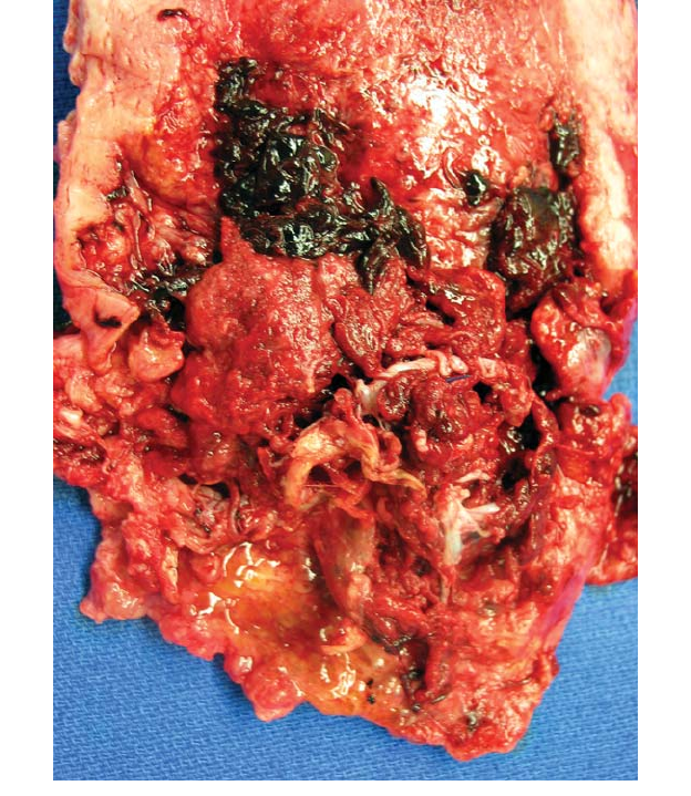
Figure 2.11. This opened post-partum uterus shows extensively invasive adher-
ent placental tissue in the lower segment. Thus this was a placenta previa that
had partially separated as well as invaded into the muscle, an increta. Accreta,
previa, and placental separation are frequently seen together.
Placenta Accreta 21
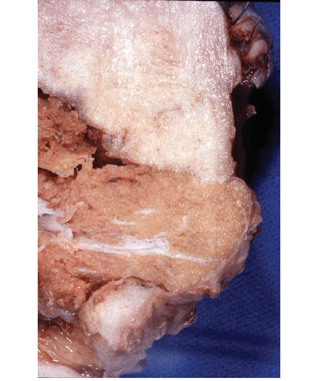
Figure 2.12. This fixed postpartum hysterectomy specimen reveals retained pale
placental tissue invading focally nearly through the wall of the uterus, with thin-
ning to less than 1 mm of serosal tissue (placenta increta).
22 Chapter 2 Basic Placental Anatomy and Development
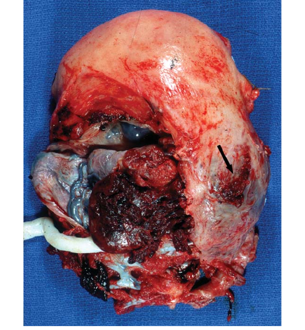
Figure 2.13. This uterus still contains the placenta as it could not be removed
readily at delivery and focally the placental tissue has perforated the
myometrium (arrow). Fibrin and organization of the clot were present on his-
tology. The adjacent thinned lower uterine area appears somewhat blue from the
closely underlying placental. An unsutured vertical cesarean section incision is
present.
implantation. Accreta is most easily identified in hysterectomy speci-
mens. It is usually not possible to make the gross diagnosis of sympto-
matic accreta and its more invasive forms in a delivered placenta, and
only rarely on placental microscopy. Accreta may be found both grossly
and microscopically in post-partum currettings for bleeding (Figure
2.14). Partial myomectomies of regions with increta are occasionally per-
formed (Figure 2.15).
Abnormal adherence of the placenta is not the only cause of life
threatening postpartum hemorrhage. Uterine atony is another frequent
cause of postpartum hysterectomies (Figure 2.16).
Placenta Accreta 23
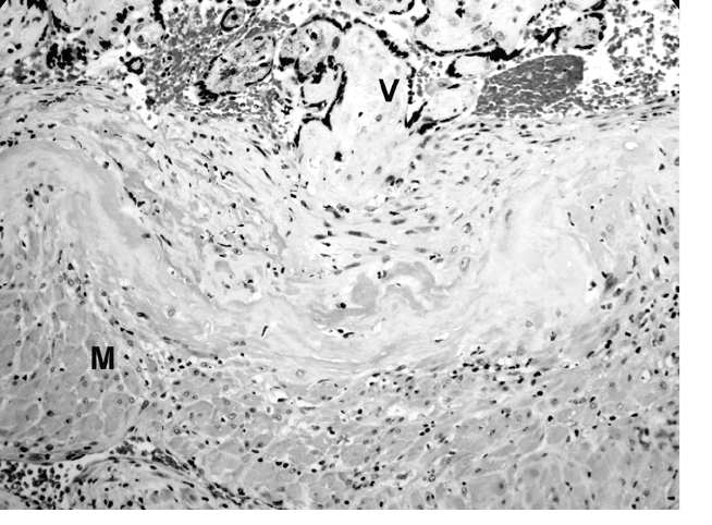
Figure 2.14. This is from the post-partum currettings in a woman with bleeding
3 weeks after delivery. Fibrotic avascular villi (V) can be seen directly adjacent
to myometrium (M) without intervening decidua.
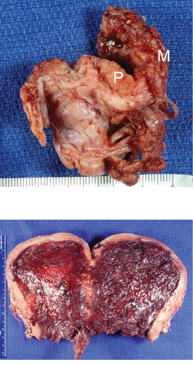
Figure 2.15. This piece of myometrium was resected in a woman with a local-
ized area of accreta. The placental tissue invades well into the muscle.
Figure 2.16. This postpartum supracervical hysterectomy was done for uncon-
trollable bleeding after delivery. The myometrial wall is thin and the fresh uterus
was floppy. No adherent placenta was identified on thorough sectioning.
3
Umbilical Cord
25
The umbilical cord is the lifeline of the fetus. Complete cord occlusion
often leads to fetal demise while intermittent obstruction has been asso-
ciated with intrauterine brain damage. Cord compression and vasospasm
are important factors in fetal distress. Careful umbilical cord examina-
tion often reveals significant lesions which may be associated with these
processes.
Development
The umbilical cord forms in the region of the body stalk where the
embryo is attached to the chorion. This area contains the allantois,
omphalomesenteric duct, vitelline vessels and evolving umbilical arter-
ies and vein. The expanding amnion surrounds these structures and
covers the umbilical cord. Eventually most of the embryonic elements as
well as the right umbilical vein disappear, leaving two arteries and one
vein (Figure 3.1). Embryologic remnants are frequent on microscopy, but
are rarely visible grossly. Allantoic remnants show a transitional-type
epithelium and occur most often near the fetal end, between the arter-
ies. Omphalomesenteric remnants may be ductal and lined by gastroin-
testinal epithelium or vascular (Figure 3.2).
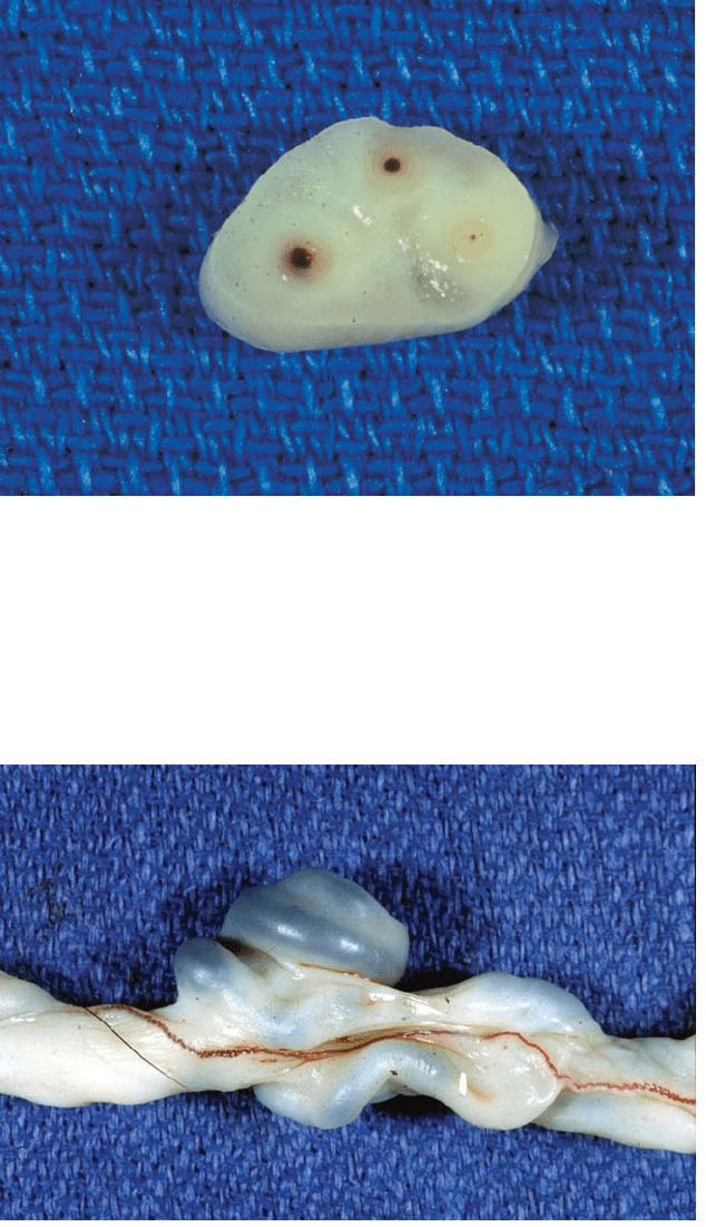
Single Umbilical Artery
The absence of one umbilical artery is a common anomaly, occurring in
about 1% of deliveries (Figure 3.3). It is more frequently seen with twins
and velamentous cord insertions. About 20% of infants missing one
artery will have other major congenital anomalies which may involve any
organ system. Many are of chromosomal etiology. The abnormalities
are generally apparent in the neonatal period, except for the increased
incidence of inguinal hernias. The “nonmalformed” infants missing one
umbilical artery are slightly growth-retarded overall and have increased
perinatal mortality. Cord accidents have been unusually frequent in this
group.
26 Chapter 3 Umbilical Cord
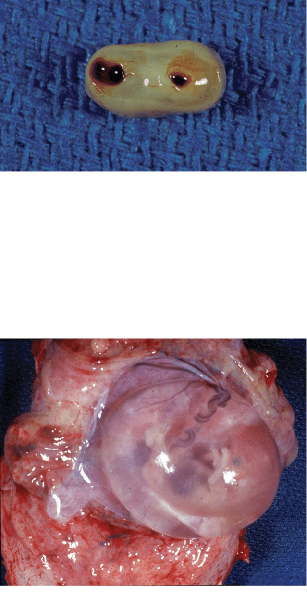
Figure 3.1. A normal three-vessel cord contains two arteries and one vein. The
arteries are often more contracted than the vein, but it is not always possible to
identify the type of vessel grossly. Most embryologic remnants are too small to
be seen by eye.
Figure 3.2. The small zigzag vessel on the cord surface is a vitelline vascular
remnant. Under the microscope these have no muscular wall and are sometimes
multiple suggesting a hemangioma. The dilated blue area is a small “false knot,”
an area of redundant length of the umbilical vein.
Twist
The spiral twisting of the cord is established early in development
(Figure 3.4). Most commonly it is counterclockwise, a so-called left twist
(Figure 3.5, Figure 3.6).The etiology of twisting is unknown. It does allow
the arteries to surround and help protect the vein from compression.
The number of twists in the cord can be counted, as excessive twisting is
Twist 27
Figure 3.3. An
umbilical cord with a
single umbilical
artery shows only
two vascular lumens.
Frequently the two
arteries fuse in the
last few centimeters
of cord above the
fetal surface, thus
multiple cuts along
the cord should be
made to confirm the
number of vessels.
Figure 3.4. Umbilical
cord twist is
established early
in development,
as shown in this
10-week gestation.
It usually twists
in a left or
counterclockwise
direction (7 : 1).