Kaplan Cynthia G., MD Color Atlas of Gross Placental Pathology
Подождите немного. Документ загружается.

8 Chapter 1 Examination Procedures
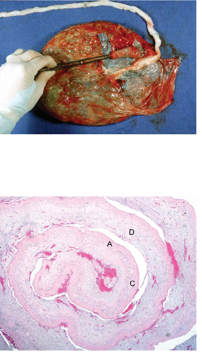
Figure 1.7. The membranes of this normal, term placenta have been placed in
their in situ uterine position. With a vaginal delivery, the minimal distance from
the hole of rupture to the edge of the placental disk indicates the site of the pla-
centa in the uterus. Shorter lengths indicate low-lying placentas. This shows a
membrane roll being made from the rupture point to the margin of the placenta.
It is then pinned, cut, and fixed. A larger length of membranes can be rolled and
two sections cut from different areas.
Figure 1.8. Histologic section of a cross section of a membrane roll shows the
numerous layers visible by this technique.Amnion (A), chorion (C), and attached
decidua (D) with small blood vessels are present.
Technique of Gross Examination 9
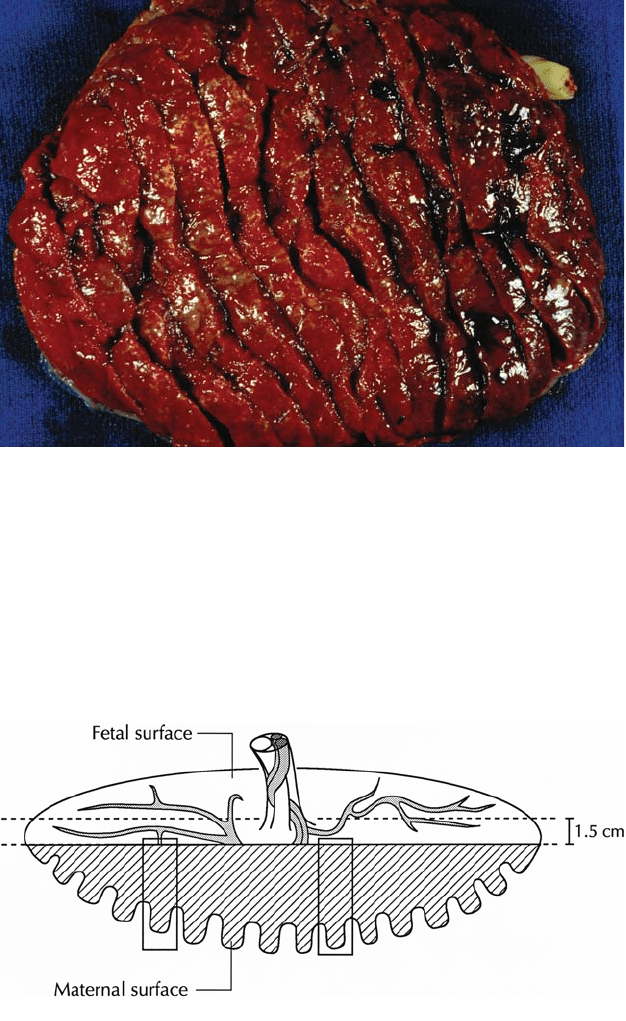
Figure 1.9. Mature placenta after transverse cuts (1.5 cm to 2 cm) have been
made on the maternal surface in order to examine the villous tissue. The knife
has a tendency to skip over firmer areas and simultaneous palpation of the villous
tissue is necessary. The fetal surface is not usually cut and keeps the placenta
somewhat intact.
Figure 1.10. A transverse strip of placental tissue from the central region includ-
ing the cord is routinely saved. This piece should be thin enough to adequate fix.
Histologic blocks of villi including small surface vessels are taken from at least
two separate areas in the placental midzone (boxes). These should not be from
areas with thick subchorionic fibrin or hemorrhage as this masks inflammation.
The placental margin has substantial artifact and is not ideal for assessing villous
configuration. It may show more inflammation or decidual vascular change and
can be submitted in addition. Placentas with significant pathologic processes
require extra blocks to sample these.
Placental Weight
Placental weight is not a precise measurement and will vary with the
methodology of examination. It is affected by fixation, the presence of
cord, membranes, and loose clot, the amount of blood retained, and the
intactness of the maternal surface. Fresh refrigerated placentas lose a
small amount of weight with storage, whereas formalin fixation leads to
an increase, no more than 10% in either case. The value of placental
weight is largely at the extremes, taking into account the gestational age
and weight of the baby. A relatively heavy or light placenta often indi-
cates an abnormal pregnancy. At term, the infant usually weighs about 7
to 8 times the placental weight. The ratio decreases earlier in gestation.
Most term placentas weighing more than 750 grams or less than 350
grams will warrant histology. There are standard tables for placental
weight by gestational age and by fetal weight as well as those with fetal-
placental ratios by gestational age (Appendices B-1, B-2, and B-3).
Histologic Sectioning
Although it is possible to cut blocks from fresh placental tissue, this is
far easier after some fixation has occurred. Sharp blades are important
to keep the amnion on the placental surface intact. On most placentas
cord (2 pieces from different sites), membrane roll, and two to three full
thickness pieces of villous tissue including fetal and maternal surfaces
are an adequate sample. The pieces of placental villous tissue should be
from separate areas (different cotyledons), and not from the margin of
the placenta, which frequently shows changes of diminished blood flow
(Figure 1.10). The fetal surface of the section should include small blood
vessels, and be free of substantial subchorionic clot or fibrin. Early
changes of ascending infection are often masked in areas with thick sub-
chorionic deposits. If the placental sections are too large to fit in the cas-
sette, they will need to be divided. Additional representative sections of
significant lesions or differences in villous character are also taken. En
face blocks of the basal plate may be useful for evaluating maternal vas-
culature. It is not necessary to section every infarct, hemorrhagic lesion,
and so forth, as long as they are clearly identifiable grossly and ade-
quately described. Blocking can be done by a trained technician.The spe-
cific type of fixation, processing, cutting, and staining may greatly alter
the histology of the placental villous tissue. This is particularly important
in the assessment of villous structure and maturation. Anyone looking
at even a few placentas needs to become familiar with the appearance
of villous tissue at different points in gestation as prepared in their his-
tology lab.
Reports
For reports, the form on which the original gross information is recorded
can often serve as the actual report or a master for rapid typing of
reports. These forms can readily incorporate the microscopic exam and
10 Chapter 1 Examination Procedures
diagnoses. Some hospitals use placental check lists while in others reports
are narrative. The special requirements of twin placentas should be
either a separate form or incorporated into the singleton worksheet.
(Appendix A1,2)
Reports 11
2
Basic Placental Anatomy
and Development
12
Some appreciation of placental development and structure is necessary
to understand its examination and certain pathology. While the placenta
shows extensive growth and histologic change in the second and third
trimesters, the basic gross morphology is established early in pregnancy,
before the end of the first trimester.
Development
Trophoblastic tissue is the major component of the placenta. By 4 to 5
days after fertilization, trophoblasts differentiate from the external cells
of the morula as it becomes a blastocyst.The trophoblastic cells prolifer-
ate rapidly and surround the inner cell mass, covering the entire surface
of the blastocyst. Attachment to the endometrial surface and implanta-
tion occur at 5 to 6 days, usually in the upper part of the uterus. Implan-
tation is interstitial and the blastocyst becomes totally embedded in the
endometrium. As the developing conception grows, it protrudes into the
endometrial cavity. The endometrial stroma undergoes decidual change.
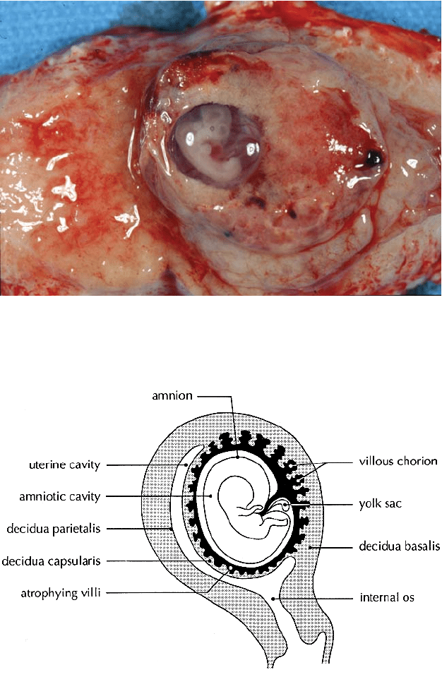
At first the entire gestational sac is covered by chorionic villi (Figure 2.1,
Figure 2.2). As the sac enlarges, its surface thins, forming the peripheral
membranes which are composed of decidua capsularis, atrophied
chorion, and amnion.The definitive placenta is left at the base. With con-
tinued growth of the conception there is apposition of the membranes
with the decidua vera of the opposite side of the uterus, but no true fusion.
The fetal-placental circulation begins at about 9 days when lacunae
form in the syncytial trophoblast. By days 10 to 12 these lacunae link
with maternal blood vessels which have been eroded by trophoblastic
invasion. The intermediate trophoblastic cells are responsible for inva-
sion into the uterine wall and maternal vasculature. The primary fetal
chorionic villi have formed by 14 days and consist of cords of cytotro-
phoblast covered by syncytial trophoblast. Shortly after there is invasion
of avascular extraembryonic mesenchyme from the embryonic body
stalk into these columns forming secondary villi. Capillaries develop
within the villous stroma and form networks by 20 days (tertiary villi).
These vessels communicate with the fetus through vessels differentiat-
ing from the chorion and the connecting stalk, the large surface vessels
and umbilical cord. The circulation is functional by the end of the third
developmental week. The placenta grows through branching of the
villous tree. Primary stem villi break up below the chorionic plate to form
Development 13
Figure 2.1. This embryo of 6 developmental weeks was removed in situ during a
hysterectomy for cervical carcinoma. The decidua has been partially removed to
reveal the chorionic villi which cover the entire early gestational sac. Part of the
chorion has also been dissected showing the amniotic sac containing the embryo.
Figure 2.2. The embryo lies within the chorionic and amniotic sacs. Note the yolk
sac between them. The capsular chorionic villi associated with the evolving
peripheral membranes are undergoing atrophy creating the discoid placenta at
the base into which the cord inserts.
14 Chapter 2 Basic Placental Anatomy and Development
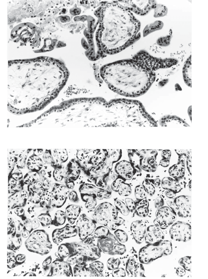
A
B
Figure 2.3. Histologic maturation of villi (A) Very early first trimester villi show
abundant stroma without vessels. Two layers of trophoblast (cyto and syncy-
tiotrophoblast) are present, without syncytial knots. (B) At the same magnifica-
tion, term chorionic villi are much smaller and show little stroma. There are
numerous blood vessels and only syncytial trophoblast is visible on the surface
with numerous knots.

secondary and tertiary stem villi and finally distal terminal villi.“Anchor-
ing” villi are present at the base of the placenta. Normal maturation of
villi entails several features. There is progressive diminution in villous
size and stromal content with an increasing portion of the villus com-
posed of blood vessels. The syncytiotrophoblast nuclei become aggre-
gated into “knots,” and the cytoplasm thins over vessels forming
vasculosyncytial membranes. The originally prominent cytotrophoblastic
layer disappears and by term few cytotrophoblasts are recognized on
light microscopy (Figures 2.3A,B).
Placental Shape
The shape of the placenta is quite variable. Generally it is round to ovoid
and about 18-cm to 20-cm diameter by 1.5-cm to 2.5-cm thick at term.
Failure of atrophy of capsular villi leads to succenturiate lobes (Figure
2.4, Figure 2.5). Bilobate placentas result from uterine sulcal implanta-
tion (Figure 2.6), while unusually shaped often multilobate placentas
may be due to uterine cavity abnormalities (Figure 2.7). A diffuse thin
placenta without free membranes is extremely rare and known as pla-
centa membranacea. (Figure 2.8). This may represent a shallow implan-
tation with persistence of virtually all the capsular villi. While these
alterations should be described, they are of little significance except for
potential problems related to the velamentous vessels that often accom-
pany them and placenta previa.
Placental Shape 15
Figure 2.4. Succenturiate lobes are formed if some of the capsular villous tissue
fails to atrophy during development. Such tissue can potentially be left behind
at delivery leading to bleeding from retained placenta. True succenturiate lobes
are connected to the main placental mass by velamentous vessels which can be
damaged. This slightly immature placenta shows at least four such lobes, one
large and three small. Succenturiate lobes often become infarcted or fibrinous.
One of the small lobes is yellow and atrophic (arrow).

16 Chapter 2 Basic Placental Anatomy and Development
Figure 2.5. The term “partial” lobes can be used to help describe some of the
irregularities of outline. These are lobe-like marginal placental areas which are
connected by bridges of villous tissue and do not show velamentous vessels.
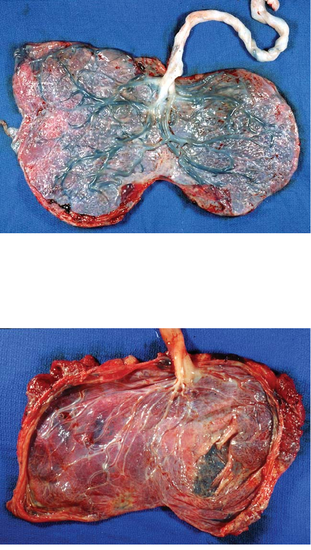
Figure 2.6. This mature bilobate placenta has two distinct lobes of roughly equal
proportions. The umbilical cord inserts between them, velamentously into the
membranes. Although this resembles a placenta with a large succenturiate lobe,
this configuration more likely arises through a different mechanism. Implanta-
tion in a lateral uterine sulcus will lead to relatively equal growth along the ante-
rior and posterior walls.
Placental Shape 17
Figure 2.7. An unusual uterine shape, scarring, or intracavitary lesions may be
reflected in the placental outline. Such abnormalities impede placental growth
in certain areas and the remaining tissue extends into other regions. This large
irregular placenta suggests an abnormal uterine cavity.
Figure 2.8. This is a very large thin immature placenta with villi covering the
entire sac except for small areas of membranes including an enclosed window. It
likely covered the cervical os. The placenta was cut through at Cesarean section
and was extensively disrupted.