Kaplan Cynthia G., MD Color Atlas of Gross Placental Pathology
Подождите немного. Документ загружается.


58 Chapter 4 Fetal Membrances and Surface
Figure 4.22. Histology of chorioamnionitis reveals neutrophils from the mater-
nal intervillous space (ivs) extending into the chorion (C) and amnion (A).
Meconium
Meconium in the amniotic fluid commonly causes green discolored mem-
branes particularly in late gestation. An exposed placenta can have
several gross appearances (Figure 4.23 to Figure 4.25). The entire time
course of histologic meconium change is not clearly established. In vitro
studies suggest meconium rapidly reaches macrophages in the amnion
(one hour) (Figure 4.26) and is in the chorion within three hours.
Whether this corresponds to the time course in vivo is unknown, but
alterations occur within hours, not days. The passage of meconium has
long been taken as a sign of fetal stress. Current thinking regarding the
significance of meconium in the amniotic fluid is less defined. Some, but
not all, infants in distress pass meconium, and many infants with meco-
nium have not had hypoxic events. Many term placentas may show a

Meconium 59
Figure 4.23. Fetal
passage of meconium
leads to green
coloration of the
placenta. This is
recent meconium,
which is in the
amnion but does not
stain the chorion as
revealed by reflection
of the amnion.
Experimental studies
suggest amniotic
staining occurs within
one hour and
chorionic staining in
approximately three
hours. Meconium
has a variety of
appearances. It may
be thick or thin and
color ranges from
yellow to dark green.
Figure 4.24. This
meconium-stained
placenta shows
yellow-green
coloration of the
amnion and chorion,
suggesting a longer
duration of passage.
There is an amniotic
web (arrow) and
the adjacent
amnion is retracted,
revealing the stained
chorion (c).

60 Chapter 4 Fetal Membrances and Surface
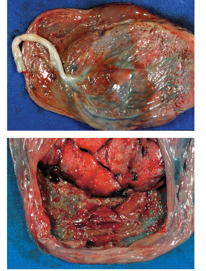
Figure 4.25. This is a near term placenta is from an intrauterine fetal demise and
shows severe, longstanding meconium exposure.The cause of death here was the
tight cord knot (arrow). There is cord congestion on the placental side. On
microscopy, inflammation will often accompany meconium.
Figure 4.26. Histology of membranes stained with meconium reveals fresh, free
meconium containing squames and hair (arrow) as well as vacuolated pigmented
macrophages in the amniotic connective tissue (arrowhead). The pigment does
not stain for iron.

Retromembranous Hemorrhage 61
vaguely green color with a few pigmented macrophages in the mem-
branes. Subsequent to affecting the membranes, meconium discolors the
umbilical cord. All green appearing placentas do not have meconium
pigment. Extensive old hemorrhage or severe ascending infection can
lead to similar coloration (Figure 4.19 to Figure 4.21). These are impor-
tant considerations in preterm pregnancies when passage of meconium
is less likely.
Retromembranous Hemorrhage
Red-brown thickenings and yellow areas mark old hemorrhages
behind the membranes (Figure 4.27, Figure 4.28). These are quite
common, particularly in multiple gestations, and result from confined
regions of hemorrhage in areas of decidual necrosis. Problems related to
these are rare. Other thickenings in the membranes may represent
compressed fetuses (Figure 4.29, Figure 4.30) and rarely retained IUD’s
(Figure 4.31).
Figure 4.27. This very immature placenta shows marked discoloration and
opacity of the fetal surface. This is most likely to be from old bleeding
and ascending infection which are common together in extremely premature
deliveries.
62 Chapter 4 Fetal Membrances and Surface
A
B
Figure 4.28. (A) Brown or yellow discolorations on the membranes usually
reflect old, retromembranous hemorrhages. These may be associated with other
hemorrhage in the placenta, but are frequently isolated. Such lesions are quite
common. A clinical history of mild bleeding can sometimes be elicited. (B) The
maternal surface better shows the old brown-red clotted blood on the
membranes.
Retromembranous Hemorrhage 63
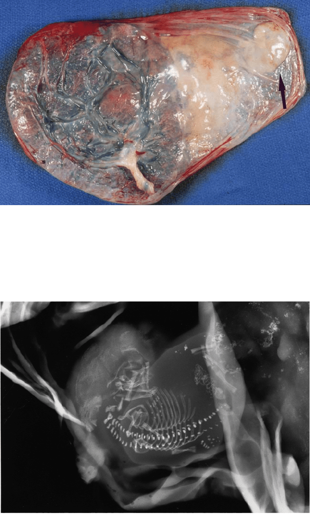
Figure 4.29. Careful examination of the membranes may reveal the presence of
an atrophied twin (arrow). These are usually firm ovoid nodules with a smooth
outline, as distinct from old hemorrhage or decidual necrosis. Eye pigment can
often be identified. Examination of the dividing membranes revealed this pla-
centa to be monochorionic and its size suggested 12 weeks gestation.
Figure 4.30. Specimen radiograph of the patient in Figure 4-29 confirms the fetal
presence. Skeletal examination will reveal gestational age and some anomalies.

64 Chapter 4 Fetal Membrances and Surface
Figure 4.32. Old
thrombosis is present
in several of the
veins on the fetal
surface. Veins are the
most common vessels
to find thrombi. The
vessels on the
placental surface can
be distinguished
grossly since fetal
arteries cross over
veins. Identification
of the vessel type is
not possible
histologically. This
placenta was
associated with an
unusually long and
highly twisted cord.
The infant did not
have problems in the
newborn period.
Figure 4.31. Intrauterine contraceptive devices are not always effective in
preventing pregnancies. A “copper T” was embedded on the maternal side of the
membranes, the characteristic location. There was both old (arrow) and recent
hemorrhage, with discoloration visible from the fetal side. The pregnancy in this
case proceeded normally with a healthy full-term infant. Velamentous cord
insertions are common in pregnancies with IUDs in place, perhaps due to the
effects on implantation.
Thrombosis
Thrombosis of the fetal surface vessels is an important observation.
These occur most commonly in fetal veins (Figure 4.32 to Figure 4.34).
Thrombosis is sometimes is associated with inflammation, meconium, or

Thrombosis 65
Figure 4.33. Two regions of thrombosed arteries and veins are present on the
surface of this slightly immature placenta. Hemolytic coloration of the sur-
rounding membranes is seen in these areas and the variations in color suggest
they are of different ages. In one area, several of the thrombosed vessels connect
to succenturiate lobes. Since vessels run through thinned placental tissue, it is
possible mechanical obstruction was a factor. There was extensive associated
villous change.
Figure 4.34. Cross-sectional view of surface vessels similar to those in Figure 4.30
shows the nonocclusive nature of many of these lesions which are largely
calcified.

vascular obstruction, but frequently there is no apparent causation. Cal-
cification of vessel walls represents old thrombosis, and most thrombi are
nonocclusive. Many more thrombi will be identified on microscopy
(Figure 4.35).
66 Chapter 4 Fetal Membrances and Surface
Figure 4.35. Histologic view of a partially occluded large fetal vessel shows fib-
rinous material on one side.
5
Lesions of the Villous Tissue
67
The general gross morphology of the placenta is established before the
end of the first trimester, and further change is largely limited to growth
and histologic maturation of villi. During placental examination the
villous tissue is examined from the maternal side before and after trans-
verse cuts have been made. While visual inspection is important, palpa-
tion of the placenta may be even more revealing of pathologic processes.
Most villous lesions show diagnostic gross morphology. The common
abnormalities are predominantly related to placental circulation (Figure
5.1).Alterations in the fetal and maternal components can be recognized
and distinguished.
Calcification
Calcification may be a striking feature of the maternal surface and villous
tissue (Figure 5.2). The degree is quite variable and the etiology is
unknown. Even very large amounts have no recognized pathologic
sequelae. Generally, calcification increases with gestational age, but is
quite variable.
Color
The color of the villous tissue tends to become darken with advancing
gestational age. Color is largely determined by fetal hemoglobin content
including the level of hematocrit and total blood volume. The placentas
of immature infants, who characteristically have lower hematocrits, are
paler than those of term infants (Figure 5.3, Figure 5.4). Unusual fetal
vascular congestion or fetal blood loss will lead to dark or light villous
color (Figure 5.5). In hydrops fetalis the placenta is very pale and coarse
(Figure 5.6 to Figure 5.8). There are many etiologies for hydrops includ-
ing isoimmunization, infection, cytogenetic abnormalities, malforma-
tions, and metabolic diseases. Some of these are readily diagnosed
through placental histology.