Kaplan Cynthia G., MD Color Atlas of Gross Placental Pathology
Подождите немного. Документ загружается.

68 Chapter 5 Lesions of the Villous Tissue
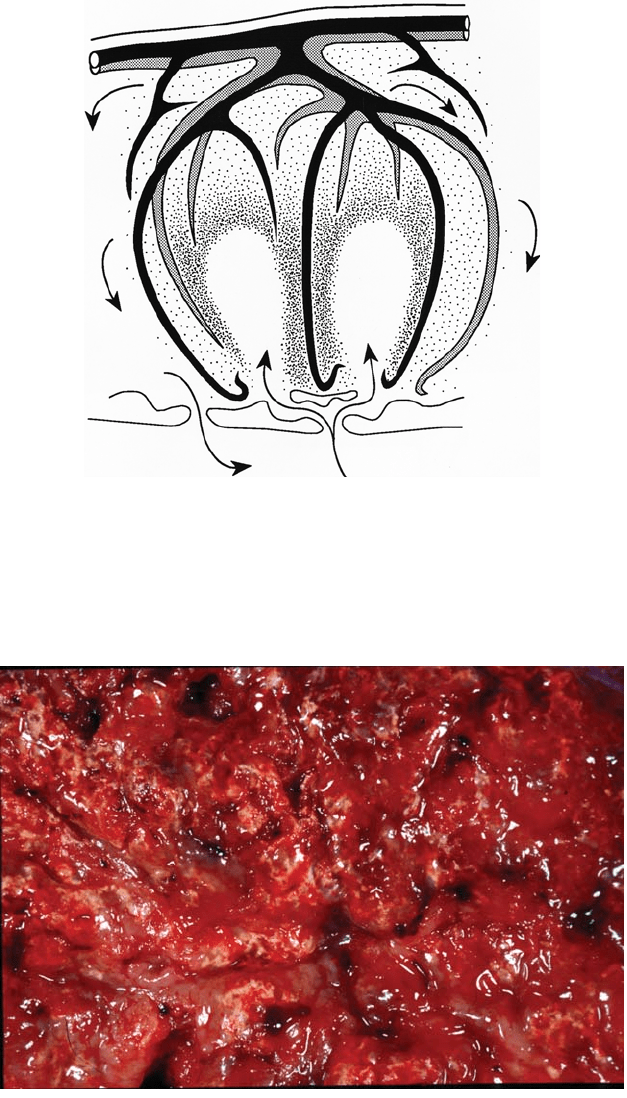
Figure 5.1. In this schematic of a placental district, maternal blood is shown
being injected from an altered decidual spiral arteriole (bottom) into the central
intervillous space where it often leaves a “hole” (see Figure 5.4). The blood flows
toward the fetal surface, and drains back passively to decidual veins. The midzone
of the placenta is best perfused, with poorer flow at the base, under the fetal
surface, and at the margins. The fetal arteries (solid) run over fetal veins
(hatched) and both branch to capillaries at the villous level.
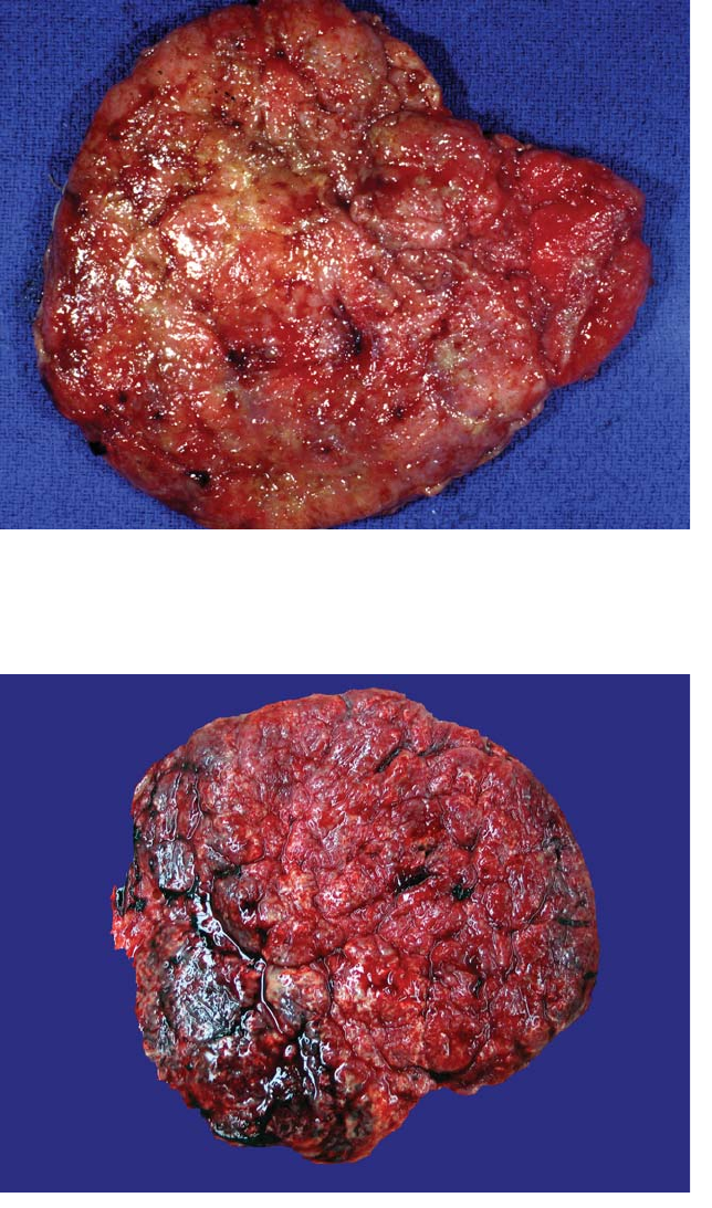
Figure 5.2. Yellow-white areas of abundant calcification can be seen on the red
maternal surface of this normal term placenta. It will also be present within the
parenchyma, giving a gritty sensation and sound on cutting. Grossly visible cal-
cification is quite variable, but tends to increase with advancing gestation. It is
not associated with disease, even if large amounts are present.
Color 69
Figure 5.3. The parenchyma of this immature (24-week) placenta shows no cal-
cification and is relatively pale red. Color of the villous tissue is largely related
to hemoglobin content. Thus mature placentas have a darker coloration than
immature ones (compare also with Figure 1.6).
Figure 5.4. The maternal surface of this term placenta shows a rough line from
11 o’clock to 5 o’clock demarcating two zones of differing color. This is an arti-
fact, created by the positioning of the fresh placenta in the container. The darker
portion was folded under the slightly paler area. Some of the color difference
may be related to dependant congestion. Additionally, the paler red portion was
also more exposed to the air and has more oxygenated hemoglobin, an effect
common in packaged ground meat.

70 Chapter 5 Lesions of the Villous Tissue
Figure 5.5. In the central portion of the villous tissue there is a rounded depres-
sion.This is a site of maternal blood injection into the intervillous space, a normal
finding. The placenta is also a very deep red color, which is usually due to con-
gestion of fetal villous vessels with blood. This is commonly caused by early cord
clamping, and the infant may be hypovolemic. Dark placentas also occur in some
cases of maternal diabetes and with certain microscopic villous vascular abnor-
malities (e.g., chorangiosis). Such villous tissue may feel unusually soft, particu-
larly in the abnormal deep red placentas.
Figure 5.6. A transverse
slice of a hydropic placenta
(middle) is compared with
an age matched preterm
(top) and normal term
placenta (bottom). Extreme
pallor is usually present in
hydropic placentas, due
to factors including fetal
anemia, villous edema, and
inappropriately immature
villous histology. Such
placentas are large and thick
with coarse villous structure.
Color 71
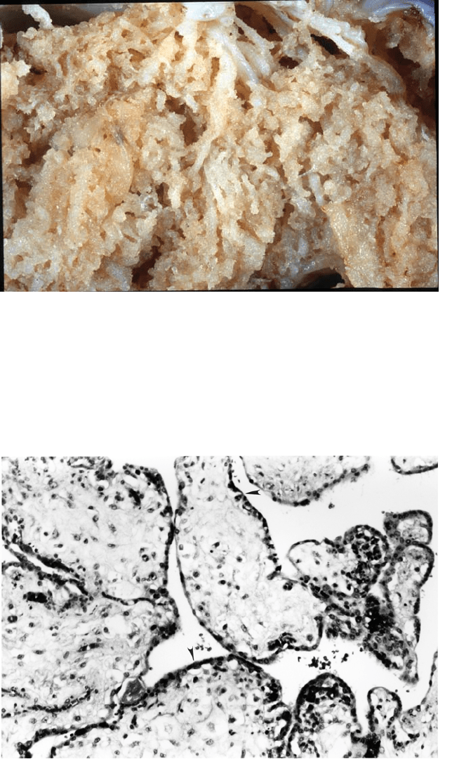
Figure 5.7. A close view of a fixed piece of a hydropic placenta better shows the
coarse villous structure. Such placentas may be extremely friable.The pallor here
is marked. Placentas also appear pale if they have lost most of their fetal blood
either before, during or after delivery (draining the cord, villous disruption). In
contrast to hydrops, the gross villous size is normal in such cases.
Figure 5.8. Microscopically, these hydropic villi show unusually large size for this
near full-term gestation (compare with Figure 2.11, same magnification). The
stroma is abundant and edematous. The covering trophoblastic layer shows few
syncytial knots, and cytotrophoblasts are easily identified (arrowheads), features
of abnormal immaturity. There are no specific diagnostic features. The infant
showed premature closure of the ductus arteriosus.
Infarcts
True villous infarcts are quite common and usually distinctive on gross
exam. These are villous regions that have lost their maternal blood
supply. They are based on the maternal surface and have rather linear
defined margins (Figure 5.9, Figure 5.10). Infarcts are more solid and feel
firmer than the adjacent tissue. They appear granular due to the remain-
ing collapsed villi in varying stages of degeneration. Over time the color
changes from red to white. Cystic change and hemorrhagic regions may
be seen in infarcts. Infarction is seen most commonly at the placental
margin where there is less blood flow.A small (1-cm) lesion of this nature
is usually insignificant (Figure 5.11, Figure 5.12). Central and large mar-
ginal infarcts suggest maternal vascular disease, particularly if they are
extensive or in placentas from preterm births. Examination of the entire
sliced placenta is often a good means to assess the extent of villous
damage in such cases (Figure 5.13). Fetal problems such as growth
restriction are often present with 15% or more infarction. Since infarcts
collapse and shrink over time, they actually represent a greater portion
of villous tissue than their dimensions would imply.The fetus can survive
the loss of more than 50% of its placenta if the increase is gradual.
Histologically infarcts are relatively uninteresting.The early ones show
villous congestion and collapse with loss of the intervillous space,
accounting for the gross firmness and red color. There is subsequent loss
72 Chapter 5 Lesions of the Villous Tissue
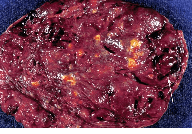
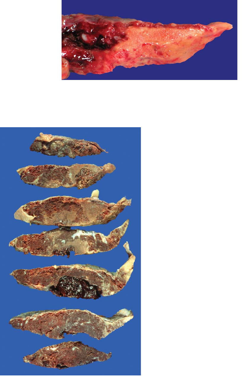
Figure 5.9. The maternal surface of this 30-week placenta from a mother with
severe preeclampsia reveals many infarcts, yellow polygonal areas which feel
quite firm. The villous tissue is dark and mature appearing (see Figure 5.2), and
the placenta was quite small. Areas of fresh retroplacental blood clot are also
identifiable at 2 (arrow), 4, and 11 o’clock. Placentas with substantial infarction
frequently show premature separation, both processes reflecting maternal
vascular disease.

Infarcts 73
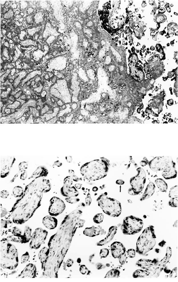
Figure 5.10. Cross-section of a multiply infarcted placenta highlights two central
lesions. Infarcts are usually well demarcated, square and based on the maternal
surface. There is a spared region under the fetal surface due to cross-circulation
between districts. Villous collapse leads to the granular appearance, while color
reflects the age of the lesion and degeneration of blood. The dark red lesion is
relatively recent. The smaller, paler one has been present longer. The exact time
course of these changes is unknown.
Figure 5.11. A fresh marginal infarct is present at the edge of this placenta (*s).
It is minimally different in color, but is more solid and compact. Infarcts at the
margin are triangular and often extend to the fetal surface. Such very recent
infarcts are often better palpated than seen.
74 Chapter 5 Lesions of the Villous Tissue
Figure 5.12. Old marginal infarcts are very common, reflecting the poor perfu-
sion near the edge of the placenta.A small lesion is of little significance.This older
lesion has lost much of its color. It can be seen to extend on the maternal surface.
The placenta shown is thin and the infracted region occurred in a partial lobe.
Infarction is particularly common in the periphery of irregularly shaped placen-
tas and relatively large lesions may be seen in otherwise normal placentas.
Figure 5.13. The extent of infarction in a
placenta can often be better defined if
serial slabs are observed simultaneously.
As present here, there is often an admix-
ture of areas with infarction of varying
ages, some of which is related to prema-
ture separation and retroplacental hem-
orrhage. In this partially fixed placenta,
at least 30% to 40% is involved by the
lesions.
Infarcts 75
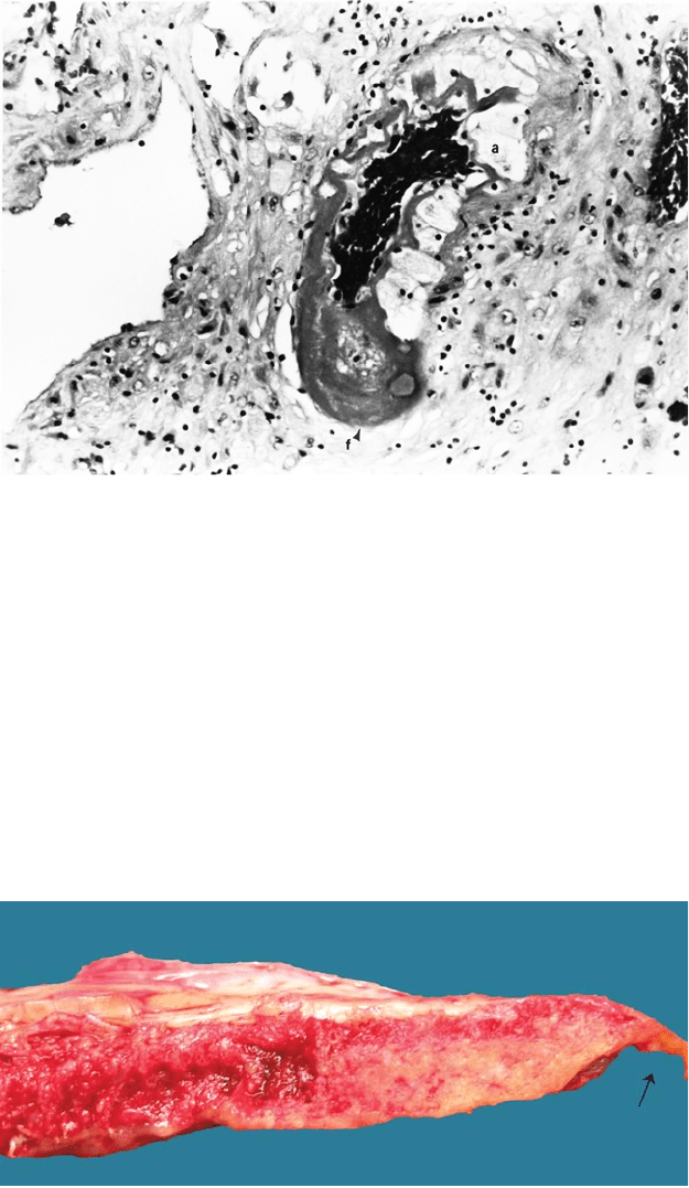
Figure 5.14. Histology of an old infarct reveals ghost outlines of villi enmeshed in
fibrin. Nuclear staining has been lost in all the trophoblast and most of the remain-
der of the villi. Viable small villi are present adjacent to the infarct on the right.
Figure 5.15. Villous structure is often altered in noninfarcted areas of placentas
associated with maternal vascular disease. The villi are smaller than expected for
gestation with dark smudgy syncytial knots, a change sometimes called “accel-
erated maturation.” Extremely small villi may be present (arrow).
of staining and fibrin is deposited (Figure 5.14). Associated ischemic
villous pathology is seen adjacent to infarcts and often diffusely in the
placenta (Figure 5.15). Maternal vascular lesions are sometimes found in
attached decidua (Figure 5.16). Vascular disease in the placenta associ-
ated with growth restriction and preterm delivery.
76 Chapter 5 Lesions of the Villous Tissue
Figure 5.16. Decidual vessels at the base or in the attached decidua may show
vascular lesions characteristic of maternal hypertensive disease. This vessel from
a severe preeclamptic shows atherosis, a vasculopathy characterized by dense
fibrin deposition and lipid-filled atherotic cells (a).
Figure 5.17. There is a yellow-white, somewhat triangular lesion at the margin
of this placenta. This is not a marginal infarct. It is shiny and does not have the
characteristic solid granular appearance. This is fibrin deposition and there is
decidual necrosis on the adjacent membranes (arrow). These processes are far
more common than true infarction at the placental margin.
Marginal infarction is overdiagnosed by many observers. Fibrin depo-
sition and necrotic decidua at the edge may be confusing (Figure 5.17).
Even occasional small (<0.5cm) gross infarcts are physiologically
insignificant in most cases and the extremely small ones (1 mm–2 mm)
do not warrant individual description.
Retroplacental Hemorrhage
Blood clots on the maternal surface of the placenta are caused by bleed-
ing from decidual vessels in areas of premature placental separation and
may relate to significant maternal or fetal disease. Trauma, hypertensive
disorders, chorioamnionitis, smoking, and possibly cocaine use have
been associated with retroplacental hemorrhage. It is preferable to use
descriptive terms for this process rather than “abruptio placenta,” a clin-
ical expression implying pain and bleeding. Although some retroplacen-
tal hemorrhages correspond to clinical abruptio placenta, many grossly
identified hematomas are unsuspected.
Very recent and at times massive placental separation often has little,
if any, gross or histologic change. The placenta may appear to be nor-
mally separated. Excessive blood clot received with a specimen, partic-
ularly if somewhat granular and formed, may be the first and sometimes
only clue to retroplacental hemorrhage. Most genuine fresh retropla-
cental hemorrhages are at least slightly adherent to the maternal surface,
as compared with gelatinous postpartum clot.
The gross morphology of retroplacental hemorrhage depends on the
duration and degree of blood trapping. When bleeding is contained
behind the placenta, the villous tissue becomes compressed by clot
(Figure 5.18). If the pregnancy continues, the separated area will infarct
because its blood supply has been lost. Lesions may be subtle on the
Retroplacental Hemorrhage 77
A
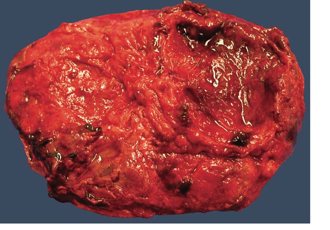
Figure 5.18. Inspection of the uncut maternal surface is important in recogniz-
ing retroplacental hemorrhages. In large lesions the formed clot may become
separated from the placenta.A depressed cavity remains on the maternal surface
into which the clot will often conform. (A) This placenta shows one obvious area
of separation on the right with some yellow coloration at the base indicating
infarction.
(Continued)