Kaplan Cynthia G., MD Color Atlas of Gross Placental Pathology
Подождите немного. Документ загружается.

98 Chapter 6 Multiple Gestations
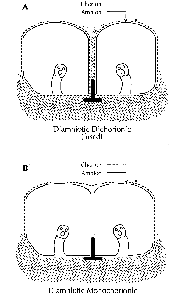
Figure 6.1. Diagrammatic views of the types of fused twin placentas with two
amniotic sacs. “T” sections are taken from the point where the dividing mem-
branes meet the fetal surface. (A) The dichorionic placenta has two sacs each
enclosed by amnion and chorion. There is chorionic tissue (stipples) in the divid-
ing membranes, forming a ridge on the surface. (B) The monochorionic placenta
shows no chorionic material in the dividing membranes and the chorion forms
a continuous plate on the surface of the placenta. The dividing membranes
consist of only two amnions.
Two-thirds of monozygotic twins are monochorionic, and the remainder
dichorionic.
Like-sexed monozygotic dichorionic twins cannot be differentiated
from like-sexed dizygotic dichorionic twins by placental exam. Only
genetic testing will definitively distinguish them. In the United States at
least 80% of like-sexed dichorionic twins are dizygotic, based on the inci-
dence of twin types. The incidence of monozygotic twins had been con-
stant throughout the world at about 1/300 births. Assisted reproductive
techniques have been found to double the rate of monozygotic twins.The
incidence of dizygotic twins is quite variable in different populations
around the world and this is the type of twinning that is familial.
Examination of Twin Placenta
Placentas received with totally separate disks are virtually always
dichorionic, even when their reconstructed morphology suggests they
were originally a single disc. These are examined as one would singleton
placentas, perhaps with the addition of some dividing membranes if
present. Minimally fused placentas are also usually dichorionic. The
dividing membranes are similar in both fused and separate dichorionic
placentas and gross determination of the chorionicity is quite simple. The
dividing membranes are evaluated for thickness and opacity. Dichorionic
membranes are relatively thick and opaque (Figure 6.2, Figure 6.3), and
there is a ridge where the dividing membranes meet the fetal surface
(Figure 6.4). If one tries to completely remove dichorionic dividing
Examination of Twin Placenta 99
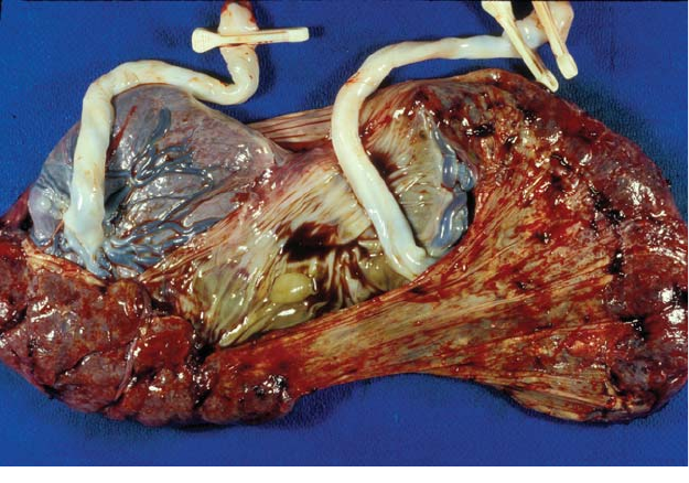
Figure 6.2. This near term dichorionic twin placenta has two separate disks con-
nected by membranes. A draped piece of thick, dividing membranes can be seen
between the cords. Note there is fresh meconium on the dividing membranes on
the side with two clamps.
100 Chapter 6 Multiple Gestations
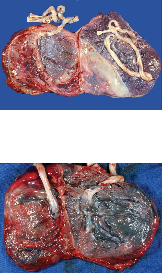
Figure 6.3. This placenta has two separate disks. There has been unequal chori-
onic “fusion” and the dividing membranes meet the fetal surface overlying the
placenta of A with 1 clamp. Such lines are common on the surface of dichorionic
placentas. These placentas can be manually separated with some superficial
disruption.
Figure 6.4. This dichorionic placenta has a “fused” disk. The dividing membranes
have been largely removed. Note the ridge of chorionic tissue between the cords
where the membranes had met the surface, diagnostic of a dichorionic placenta.
Separation of the two placentas occurs along this ridge, and can usually be done
with traction on each side. The vessels of these placentas do not connect. Note
the yolk sac remnant at 12 o’clock.
membranes by separating the layers, the surface will be disrupted
and the placentas will separate. In contrast, monochorionic placentas
have nearly transparent membranes and are easily removed leaving a
continuous monochorionic plate. No ridge is seen (Figure 6.5, Figure 6.6).
Chorionicity can be histologically confirmed in two ways. “T” sections
include dividing membranes at a point where they reach the placental
surface (Figure 6.1). Such sections are readily made on fixed dichorionic
placentas; however in monochorionic ones it is difficult to keep the
amnions intact. A roll of the dividing membranes can be made similar to
what is done with the peripheral membranes. The dividing membrane is
composed of 3 to 4 layers in dichorionic twins, and only two layers in
monochorionic placentas (Figure 6.7).
Monochorionic placentas virtually always show one or more vascular
anastomoses (Figure 6.8). These vascular anastomoses lead to the spe-
cific problems of monozygotic twins and it is important to document
them. Diagrams are useful in complicated cases. Arteries always pass
over veins. By visually following large superficial vessels one will iden-
tify many of the vascular connections between the two sides and deter-
mine sites of likely deep anastomoses. Once the placenta is fixed, this is
all that will be possible. In fresh placentas, a small syringe can be used
to inject a vessel by entering it proximal to the presumed point of
Examination of Twin Placenta 101
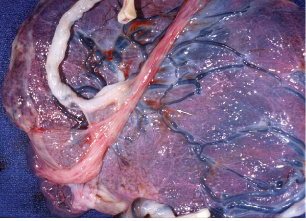
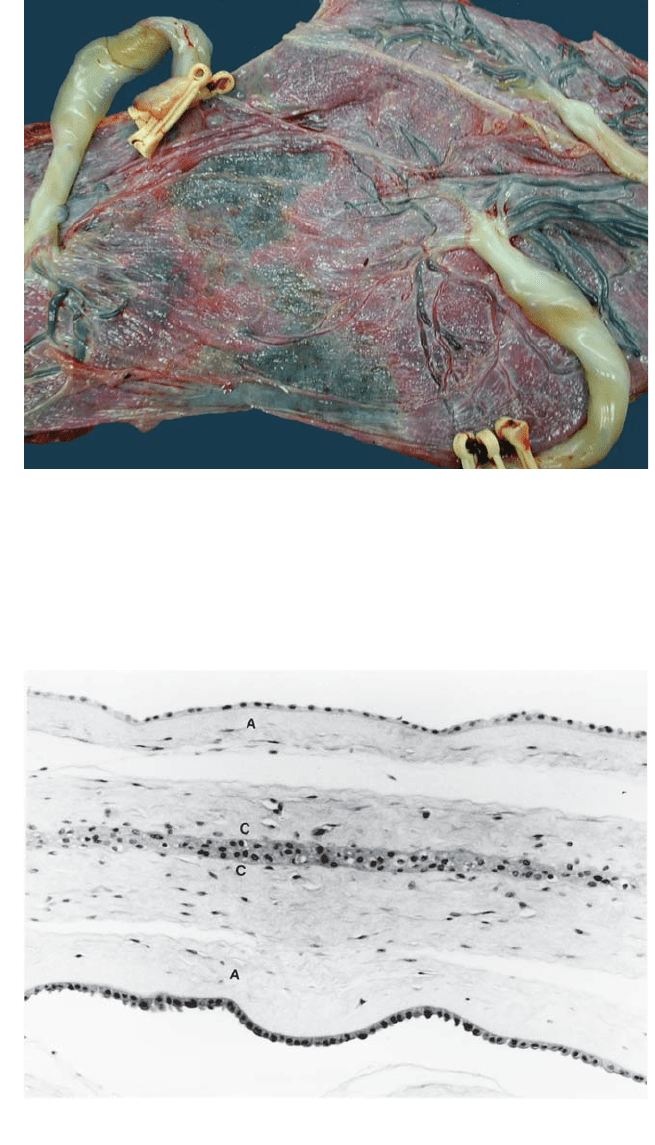
Figure 6.5. The extremely thin and delicate dividing membranes are folded on
the surface of this monochorionic placenta. Note how little substance they have
compared to those is Figure 6.2. Vessels dispersing from the cords can be seen
to have complicated connections (arrow). In humans, only monochorionic
placentas have vascular anastomoses. One cord shows a web to the dividing
membranes (arrowheads).
102 Chapter 6 Multiple Gestations
A
Figure 6.6. The dividing membranes of a monochorionic placenta can be readily
separated, leaving a smooth, continuous chorionic surface between the two cord
insertions. This triplet placenta had three amnions and 2 chorions. The amnions
have been removed leaving a continuous plate between the monochoronic set
and a ridge to the dichorionic triplet (upper right). In sectioning the dividing
membranes in higher multiples, there may be several rolls made. A consistent
means of submitting these should be adopted, such as A-B in A, B-C in B, and
C-A in C.
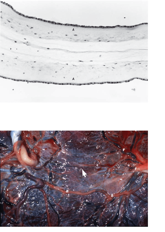
Figure 6.7. (A) Microscopic view of dividing membranes in a roll from a dichori-
onic twin gestation shows they are composed of amnion from each twin con-
taining epithelial cells and attached connective tissue (A) and chorion from each
in which the two chorionic layers may fuse (C).Any chorionic tissue in the divid-
ing membranes indicates a dichorionic placenta.
Examination of Twin Placenta 103
B
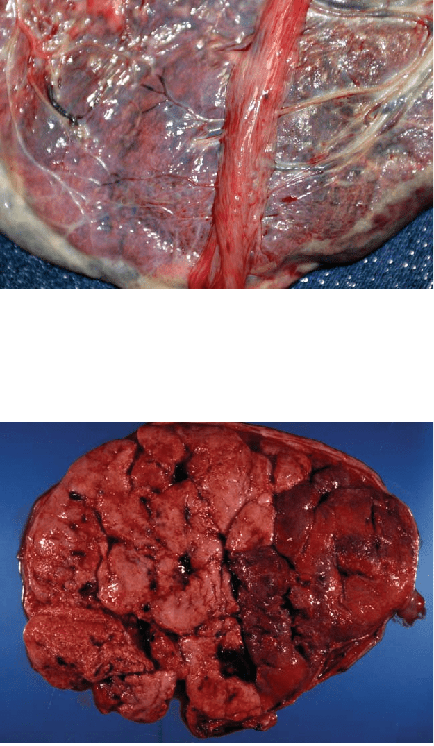
Figure 6.7. (B) Dividing membranes of a monochorionic gestation only show two
layers of amnion (A).
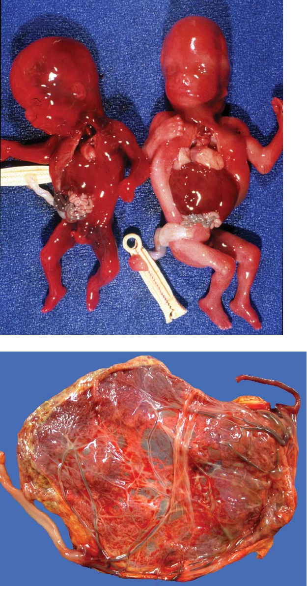
Figure 6.8. Vascular anastomoses in a monochorionic placenta are shown after
removal of the amnions. The lower anastomosis shows large arteries (“a”) from
each cord fusing in the center. The vessels, clear from injected water, are recog-
nized as arteries because they pass over other vessels. Injection is usually not
necessary to identify such large connections. Above this (arrow) is an area sug-
gestive of a deep artery to vein connection. Arteries and veins usually run
together as pairs. Here, an artery (a) from one cord ends without a parallel
returning vessel. A similar vein (v) ends just adjacent to it. While such anasto-
moses are important and most implicated in transfusion syndrome, they are dif-
ficult to inject because of pressure or disruption in the circuit. Recognition of
their usual gross morphology is thus important.
anastomosis and manually occluding backflow.The water, milk, air or dye
may be seen crossing to the other side. Small deep anastomoses are most
difficult to identify, and injection is frequently not be successful due to
disruption or incomplete filling.
What now remains to be done is the usual evaluation of the cord,
membranes, and villous tissue. A single placental disk is measured
overall. Fused dichorionic placentas can usually be separated manually
with traction starting at the edge where the dividing membranes
reach the surface. They are then examined as singletons. Monochorionic
placentas, however, cannot be separated by traction and require
cutting. This is done along the approximate line where each circulation
ends. The distribution of veins can be used as there are fewer venous
anastomoses than arterial ones. A visual assessment of the percentage
of the placentas belonging to each twin is also used. Division of the
disks is accurate in dichorionic placentas. The weight or size of
each monochorionic portion is at best an estimate since there is
considerable deep overlap. Differences between the sides such as
villous color should be noted. Tissue from each placental portion should
be placed in its own container. Hopefully the cords have been labeled.
If not, they should be arbitrarily designated and the materials kept
separate. The usual routine placental blocks are submitted along
with dividing membranes if present. In monochorionic placentas, blocks
should contain villous tissue clearly from the circulatory region of each
twin. Sections of the transitional zone may highlight differences in villous
structure.
Problems Unique to Monochorionic Twins
The vascular anastomoses virtually always present in monochorionic pla-
centas cause special problems. Unbalanced cross-circulation can lead to
the transfusion syndrome. In chronic cases the classic presentation is an
anemic, growth-retarded donor twin with oligohydramnios and a larger,
plethoric recipient with polyhydramnios (Figure 6.9). Hydrops may
occur in either infant.The donor usually has a pale placenta from anemia
while the recipient’s placenta is deep red and congested (Figure 6.10).
There may be microscopic differences in villous structure and matura-
tion. These are usually subtle, even in clear-cut chronic transfusions.
Acute transfusion syndromes also occur. One fetus can bleed through
the anastomoses into the placenta of the other when pressures drop after
the first is delivered or dies. At times this can reverse the gross appear-
ance of a chronic transfusion (Figure 6.11). Very premature delivery is
common in severe chronic transfusion syndromes, often occurring in the
second trimester. Death of both twins is common (Figure 6.12). If only
one twin dies, the chronic transfusion will stop, however there is about
a 20% risk of vascular disruptive anomalies (e.g. porencephalic cysts,
intestinal atresias) in the surviving infant (Figure 6.13). It is believed that
circulatory changes similar to those in seen acute transfusion syndrome
104 Chapter 6 Multiple Gestations
Problems Unique to Monochorionic Twins 105
Figure 6.9. This monochorionic placenta is from a set of twins with well-
developed chronic twin-twin transfusion syndrome. The placenta on the left is
quite shiny and its sac had polyhydramnios. The surface on the right is dull with
slight nodularity. This is amnion nodosum in the sac with minimal fluid.
Figure 6.10. The maternal surface of this monochorionic placenta was from
twins with severe chronic transfusion syndrome. There is a marked difference in
color between the two sides with the paler but larger portion associated with the
anemic donor twin. Such a striking difference is rare.
106 Chapter 6 Multiple Gestations
Figure 6.11. These two
17 week fetuses show
evidence of both acute
and chronic transfusion
syndrome. The larger fetus
had a hypertrophied heart,
evidence of recipient status
in chronic transfusion
syndrome. It is, however,
quite pale, having acutely
lost most of its blood
volume through an
anastomosis into the
smaller twin (chronic
donor, acute recipient)
who died first.
Figure 6.12.
Intrauterine death of
both twins occurred
in this preterm
pregnancy. The
placenta is
monochorionic and
both cords have
marginal to
velamentous
insertions. The fetus
on the right,
associated with the
hemolytic cord and
surface, died first.
Its placenta was
congested with
blood, lost by
the other twin
through vascular
anastomoses.
occur around the time of death and cause damage at that time from
anemia and hypovolemia. The incidence of disruptions in the survivor
does not seem to increase with long duration of the pregnancy after the
demise.
A small portion of monochorionic twin placentas entail relatively late
splits in the conceptus. Monoamniotic pregnancies are usually diagnosed
prenatally by ultrasound. Numerous large anastomoses typically occur
in such placentas, and chronic transfusion syndrome is rarely a problem.
Cord entanglement leads to very high morbidity and mortality. A
monoamniotic state should only be diagnosed if there is a layer of
amnion covering the fetal surface between the cords (Figure 6.14).
Most monochorionic twin placentas which apparently lack divid-
ing membranes are actually disrupted diamniotic monochorionic
placentas. A particular form of vascular anastomoses in a monochorionic
placenta permits the development of acardiac twins. Such fetuses are
passively perfused by their co-twin and lack cardiac development
from the circulatory reversal. External and internal development is
strikingly abnormal (Figure 6.15). Occasionally placentas are found
which show intermediate forms between the classic configurations
(Figure 6.16).
Problems Unique to Monochorionic Twins 107
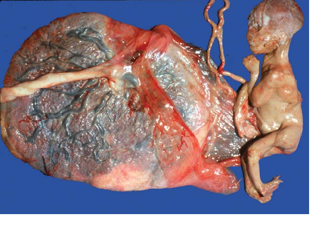
Figure 6.13. Death of one twin at 19 weeks led to the formation of a fetus
papyraceous in this term pregnancy. Morphology was adequately preserved to
permit the histologic confirmation of the dividing membranes as monochorionic.
While the etiology of the death of the infant is not fully determinable at this time,
transfusion syndrome is likely. The surviving twin was at increased risk for vas-
cular disruptive anomalies, but was uninvolved. The risk for disruption seems to
become greater as the gestational age at fetal death increases.
