Jan Lindhe. Clinical Periodontology
Подождите немного. Документ загружается.

IMPLANT PLACEMENT IN THE ESTHETIC ZONE • 927
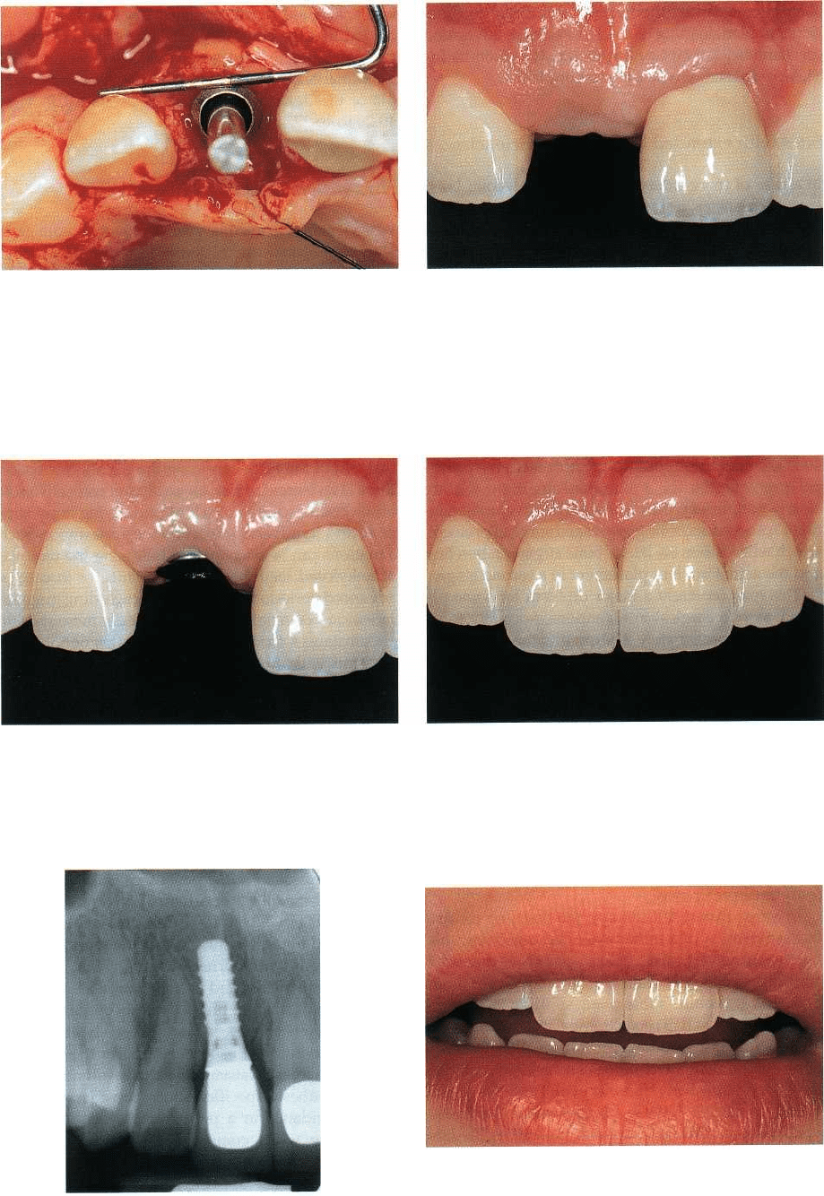
Fig. 40-32. During implant surgery one can note that all
Fig. 40-33. After three months of healing the labial view
key parameters characterizing an optimal implant posi-documents a slight excess of keratinised peri-implant
tion (shoulder sink depth, orofacial point of emer-
mucosa in a coronal direction, which is a prerequisite
gence, implant axis) could be satisfied. for the development of the final esthetic soft tissue con-
tours. The first step of the subsequent procedure will
consist of the insertion of a longer titanium healing
cap,
following a minor mucosaplasty.
Fig. 40-34. Two weeks after mucosaplasty and ex- Fig. 40-35. The two ceramo-metal crown restorations —
change of healing caps the initiation of a harmoniouslyone tooth borne (site 21) and one implant borne (site
scalloped labial soft tissue course is apparent. Further-
11) — display little difference in appearance since sym-
more, the access from the surface to the underlying im-
metry has been respected from the line of mucosal
plant shoulder has been established.
emergence to the incisal edge.
Fig. 40-36. The 1-year follow-up radiograph confirms
the stability of the osseointegrated 10-mm titanium
screw implant.
Fig. 40-37. An esthetically pleasing overall integration
of
the two maxillary anterior restorations is underlined
by a
close-up view of the patient
'
s unforced smile.
928 • CHAPTER 40
such circumstances the healing time prior to func-
tional implant loading remains the same as advocated
for standard implant protocols (i.e. two months for
SLA-coated screw-type titanium implants).
Sites with extended horizontal deficiencies
In a case of more extended horizontal alveolar bone
crest deficiencies, a simultaneous implant placement
and lateral bone augmentation procedure becomes
technically more difficult and less predictable, as the
ultimate goal remains an optimal "restoration-
driven"
implant positioning (Figs. 40-26, 40-27). The
described extended horizontal bone deficiency may
often, on the one hand, not permit an acceptable pri-
mary implant stability to be achieved, and on the other
hand may lead to a vestibular bone dehiscence that
does not have a distinct two-wall morphology. Fur-
thermore, the labial implant contour would be more
prominent than the respective surrounding bone (Fig.
40-28). Under these specific circumstances the princi
-
pal prerequisites for a simultaneous approach are
clearly not present, thus leading to the recommenda-
tion to proceed according to a staged surgical protocol,
which will address the lateral bone augmentation first
and the actual implant placement in a second stage.
This may represent a major problem for some pa-
tients, as two surgical interventions, normally sepa-
rated by approximately six months, are necessary,
leading to a total treatment time of eight months or
more. It is therefore indispensable to thoroughly in-
form the patient about both the reasons for the staged
approach associated to implant therapy, and the pos-
sible conventional prosthodontic alternatives (e.g. a
traditional tooth-borne FPD, eventually in combina-
tion with a connective tissue grafting procedure to
optimize the deficient edentulous ridge in view of an
optimal and esthetic pontic). The patient will then be
in a position to give his or her informed consent to
either of the two therapeutic modalities, according to
individual preference.
In a case of implant therapy, the first step consists
of the elevation of a rather extended mucoperiosteal
flap featuring vertical releasing incisions, as the added
site volume (due to the block graft and barrier mem-
brane) will require subsequent splitting of the pe-
riosteum prior to flap repositioning and suturing (Fig.
40-29). Numerous studies reporting results of various
bone augmentation techniques and related materials
have been published (Hiirzeler et al. 1994, Buser et al.
1996, Ellegaard et al. 199Th, Chiapasco et al. 1999, 2001,
von Arx et al. 2001a,b, Zitzmann et al. 2001). To date,
autogenous bone block grafts, mostly harvested from
the chin or the retromolar area, in combination with
e-PTFE barrier membranes, still have the best clinical
long-term documentation (Buser et al. 2002). These
authors presented prospectively documented 5-year
data of 40 consecutively treated patients, according to
a
staged protocol. On all laterally augmented sites
implants could be subsequently inserted. It was con-
cluded that the clinical results of implants placed in
regenerated bone were comparable to those reported
for implants in non-regenerated bone. A clinical exam
-
ple of the described approach is presented in Figs.
40-29 to 40-37.
Sites with major vertical tissue loss
When it comes to maxillary anterior single-tooth gaps
with significant vertical tissue loss, the predictable
achievement of an esthetically pleasing treatment out
-
come, ideally providing a so-called perfect illusion
with respect to its integration in the surrounding natu
-
ral dentition, gets difficult. As pointed out earlier in
this chapter, there exists a close relationship between
the interproximal bone height and the associated soft
tissue level (Figs. 40-7, 40-8). If the coronal border of
the alveolar bone is no longer within the physiological
distance of approximately 2 mm from the interproxi-
mal CEJ of the teeth confining the edentulous space,
there is an increased risk for an altered respective soft
tissue course (due to a lack of underlying bony sup-
port) and its adverse impact on the appearance. Such
situations can be encountered following the removal
of ankylosed teeth or failing implants, or in case of
advanced periodontal tissue loss — including gingival
recession — on neighboring teeth. Under these specific
circumstances, the final decision whether or not to use
implants will ultimately depend on the one hand on
the careful and comprehensive evaluation of all of the
therapeutic modalities available for anterior tooth re-
placement (Table 40-3), and on the other hand the
patient's individual smile line and expectations. This
process includes an objective analysis of the advan-
tages and eventual shortcomings associated with each
modality.
To illustrate these clinically relevant aspects, the
initial situation and the subsequent implant treatment
of a 35-year-old female patient consulting with an
ankylosed maxillary deciduous left canine, are pre-
sented in Figs. 40-38 to 40-46. The preoperative analy
-
sis had led to the conclusion that the fabrication of a
conventional tooth-borne three-unit FPD, using the
intact lateral incisor and first premolar as abutments
and featuring a canine pontic, was not opportune from
several points of view. Among these should be par-
ticularly mentioned aspects related to the question-
able mechanical resistance of the resulting conven-
tional prosthesis, specific occlusal considerations (e.g.
canine guidance in a pontic area), lack of esthetic
superiority when compared to a virtual implant-
borne
fixed restoration, and last but not least the con
flict
with the general principle of minimal invasive
ness (
maximum preservation of intact tooth structure).
Once the decision was made, both the implant sur-
gical and the restorative strategies focused on improv
-
ing or at least optimally exploiting the pre-existing
IMPLANT PLACEMENT IN THE ESTHETIC ZONE • 929
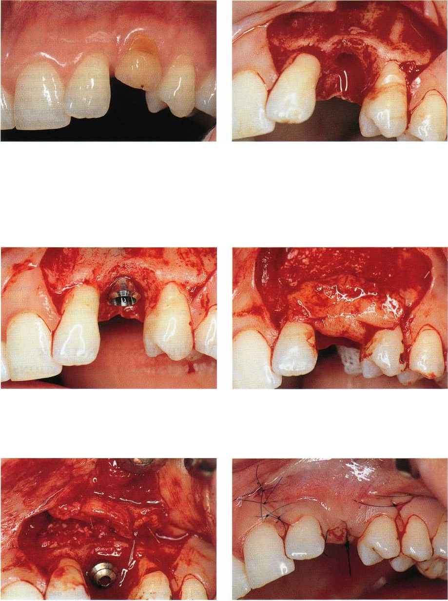
Fig. 40-38. Preoperative view of a 35-year-old female
Fig. 40-39. One month after removal of the deciduous
patient consulting with a persisting primary tooth in
canine, the root of which was severely resorbed, a mu-
the position of the maxillary left canine. Note the ir-
coperiosteal flap with vertical releasing incisions is ele-
regular course of the adjacent gingiva in general and
vated and the preparation of a calibrated implant bed
the loss in vertical tissue height in particular.
performed. One can note an increased distance be-
tween the cemento-enamel junction and the coronal
border of the alveolar bone and the left lateral incisor.
Fig. 40-40. Buccal view after insertion of the implant.
Fig. 40-41. In a case of rather thin mucosa, the utiliza-
tion of a connective tissue graft, harvested from the pal-
ate, may be indicated to create a sufficient thickness of
soft tissue at the implant site.
Fig. 40-42. Prior to flap closure, the connective tissue
Fig. 40-43. Coverage of most of the healing cap during
graft is secured to the flap with bioabsorbable sutures.suturing is recommended, leading to a submerged or
at least to a "semi-submerged" healing mode.
limited esthetic potential of the site. From the surgical localized lateral bone augmentation (simultaneous side, this
comprised a deeper than normal implant approach) procedure (Fig. 40-42) and a coronally re-
shoulder sink depth (
Fig. 40-40), the use of a connec- positioned flap (Fig. 40-43). The metal-ceramic im-
tive tissue graft on the vestibular aspect (Fig. 40-41), a
plant restoration featured a transverse screw-reten-
930 • CHAPTER 40
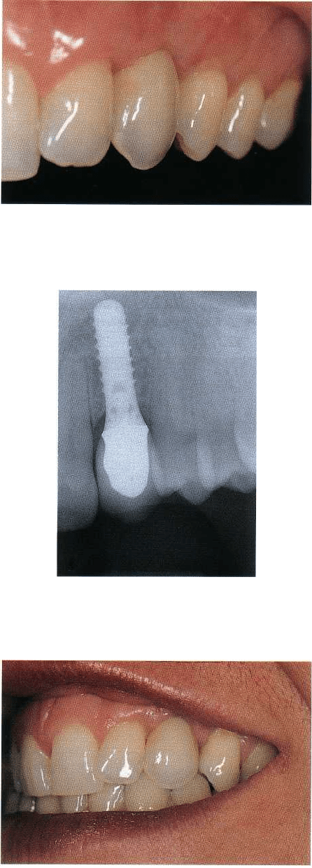
Fig. 40-44. The clinical aspect after insertion of the cera
-
mometal implant crown reveals stable and esthetic peri
-
implant soft tissue contours.
Fig. 40-45. The 2-year follow-up radiograph confirms
the stability of the osseointegrated 10 mm solid screw
titanium implant.
Fig. 40-46. On a left-lateral view, during the patien
t
'
s
forced smiling, one can note that the lack of vertical
soft tissue in the interproximal area has been compen
sated for with an apically extended interdental
contact
line.
tion to provide maximum space for esthetic porcelain
stratification and a long contact line on the mesial
aspect to compensate for the missing interdental soft
tissue height (Figs. 40-44 to 40-46).
A more severe preoperative situation of vertical
tissue deficiency, combined with a marked horizontal
bone defect, is presented in Figs. 40-47 and 40-49. This
19-year-old female patient lost her maxillary right
lateral incisor due to a localized periodontal problem.
Again, the comprehensive site analysis concluded
that
a single-tooth implant restoration was the best
compromise in view of major disadvantages associ-
ated with all of the conventional prosthodontic op-
tions. From a purely esthetic point of view, none of the
therapeutic modalities had the potential to predict-
ably lead to a perfect re-establishment of a symmetri-
cal, harmoniously scalloped soft tissue course at its
original physiological level. However, a rather low
lip-
line during the patient's normal communication
and
unforced smiling permitted the least invasive
approach to be chosen. Following a lateral connective
tissue and bone augmentation procedure (Fig. 40-50),
an implant could be inserted in an acceptable position
and subsequently restored with a screw-retained
crown. The final frontal view, allowing a direct com-
parison between the intact (Fig. 40-51) and the re-
stored side, clearly demonstrates the current esthetic
limitations associated with implant therapy in sites
with a marked vertical tissue deficiency (Fig. 40-52).
MULTIPLE-UNIT ANTERIOR
FIXED IMPLANT RESTORATIONS
The normal consequence following loss of two or
more
adjacent upper anterior teeth comprises a flattening
of the edentulous segment. In particular one
can
observe the disappearance, in an apical direction,
of
the crestal bone originally located between the in
cisor
teeth. This phenomenon is not, or only minimally,
present at the interproximal aspect of the re
maining
anterior teeth and thus explains the funda
mental
difference between a maxillary anterior single-
tooth
gap and a multi-unit edentulous segment.
If two standard screw-type titanium implants are
inserted to replace two missing maxillary central inci-
sors (Figs. 40-53, 40-54), an additional peri-implant
bone remodeling process will take place. In the frontal
plane, two different characteristic processes, one be-
tween the natural tooth and the implant and the other
between the two implants, can be distinguished. At
the site between tooth and implant, the tooth-sided
interproximal bone height should theoretically re-
main
at its original location, i.e. within 2 mm from the
CEJ,
from where the implant-sided interproximal
bone
height drops in an oblique manner towards the
first
implant-to-bone contact, normally located ap-
proximately 2 mm apically of the junction ("micro-
gap") between the implant shoulder and the abutment
or suprastructure. This phenomenon has been re-
ferred to in the literature as "saucerization" or establ-
IMPLANT PLACEMENT IN THE ESTHETIC ZONE • 931
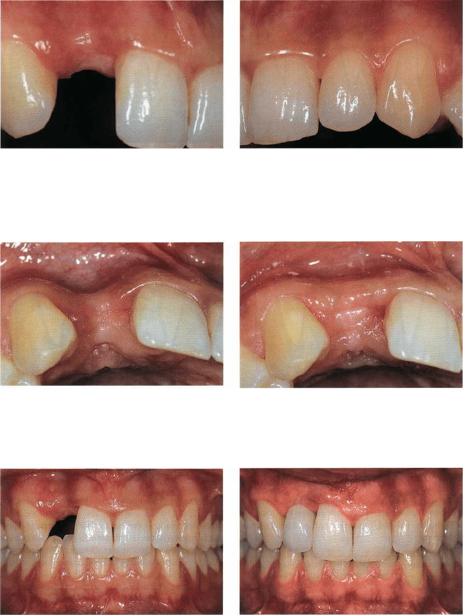
Fig. 40-47. Labial close-up view of the maxillary right
anterior region of a 19-year-old female patient. The in-
terdental soft tissue height distal to the central incisor
and the corresponding underlying alveolar bone height
are markedly reduced, leading to exposure of the
cemento-enamel junction.
Fig. 40-48. The contralateral side of the dental arch
shows perfectly intact and harmonious conditions with
respect to the course of the gingiva.
Fig. 40-49. On the occlusal view of the edentulous site a
significant lateral crest deficiency becomes apparent,
which calls for both a bone and soft tissue augmenta-
tion procedure, particularly if an implant solution is
planned.
Fig. 40-50. Six months after combined lateral bone and
soft tissue augmentation, the site appears to be compat-
ible with "restoration-driven" implant placement.
Fig. 40-51. The buccal view in centric occlusion posi
tion
before therapy summarizes the problems associ
ated
with localized vertical tissue deficiencies: lack of a
harmoniously scalloped soft tissue course in general
and missing interdental papillae in particular.
Fig. 40-52. The corresponding view after lateral bone
and
soft tissue augmentation on the one hand and in
sertion
of an implant borne single-tooth restoration on
the site of
the right lateral incisor on the other hand, un
derlines the
resulting shortcomings with respect to es
thetic
parameters. Vertical tissue deficiencies – which at
present cannot be predictably compensated for –
clearly
compromise the overall integration of an other-
wise
successful treatment.
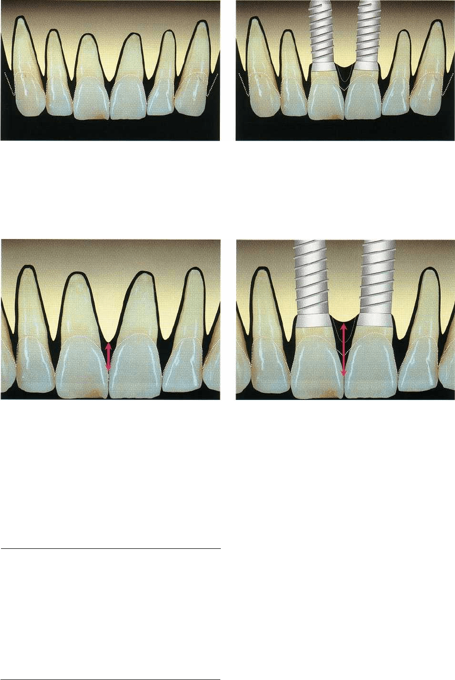
932 • CHAPTER
40
Fig. 40-53. Schematic representation of the six maxil-
lary anterior teeth, including their bony support and
the course of the marginal soft tissue, corresponding
ideally approximately to the cemento-enamel junction
(dotted line).
Fig. 40-54. Loss of the two central incisors and their
subsequent replacement by implant restorations nor-
mally leads to well-defined bone loss ("micro-gap", es-
tablishment of a "biologic width") around the implant
sites. The main consequence from an esthetic point of
view consists of vertical soft tissue deficiencies, namely
between adjacent implants (dotted lines).
Fig. 40-55. Schematic close-up view of the relationship
between cemento-enamel junction, alveolar bone and
course of the gingiva in the maxillary incisor area.
Table 40.8. Basic considerations related to anterior
fixed multiple-unit implant restorations in sites
with
horizontal and/or vertical soft and hard tissue
deficiencies
Achievements
Predictable and reproducible results
regarding lateral bone augmentation using
barrier membranes supported by autografts:
• allows implant placement in patients
with a low lip line
Limitations
Vertical bone augmentation is difficult to
achieve and related surgical techniques lack
prospective clinical long-term documentation
Interimplant papillae cannot predictably be
re-established as of yet
Fig. 40-56. Same area after implant therapy. The red ar-
row represents the distance between the interimplant
bone crest and the interdental contact point. The lack of
bony support for the interdental soft tissue often causes
the appearance of black triangles, compromis
ing the
esthetic treatment outcome.
ishment of a "biologic width" (Hermann et al. 1997,
2000, 2001a,b). In contrast, the interimplant bone
height normally decreases further in an apical direc-
tion, once the respective abutments or suprastructures
are connected to the implant shoulder. This process is
mostly accompanied by a loss of interimplant soft
tissue
height and hence may lead to unsightly, so-
called "black
interdental triangles". The schematic
close-up views
comparing the original dentate situ
ation with the status
after integration of two adjacent implant restorations,
clearly demonstrate the negative consequences on the
course of the marginal soft tissue
line in a case of
multiple adjacent maxillary anterior implants (Figs. 40-
55 and 40-56).
The basic considerations related to the current state
of achievements and limitations of maxillary anterior
fixed multiple-unit implant restorations in sites with
and
without horizontal and/or vertical soft and hard
tissue
deficiencies are summarized in Table 40-8.

IMPLANT PLACEMENT IN THE ESTHETIC ZONE • 933
Fig. 40-57. Clinical close-up view of the maxillary ante-
rior segment of a 32-year-old female patient following
placement of two 12 mm solid screw implants accord-
ing to a one-stage transmucosal surgical protocol.
Fig. 40-58. The conditioning of the peri-implant mu-
cosa in view of the future restorations has been per-
formed by means of auxiliary plastic components fea-
turing the possibility of individualizing the emergence
profile.
Fig. 40-59. The corresponding clinical close-up view,
Fig. 40-60. Clinically, a slight fill-in of interimplant mu
-
taken shortly after insertion of the two screw-retained
cosa and an overall stable soft tissue situation can be
ceramometal restorations, documents the effect of a
noted after 6 years of clinical service.
long interdental contact line, the presence of pro-
nounced mesial ridges and a slight increase of color
saturation in the cervico-interdental area. Such techni
-
cal measures contribute to the compensation of a flat
and more apically located labial mucosa line.
Fig. 40-61. In order to compensate for the reduced
height of the interimplant soft tissue, the ceramist has
used an apically prolonged interdental contact line in
the form of so-called "mini-wings". These interdental
ceramic extensions are made of a more saturated root-
like porcelain and are slightly displaced to the palatal
aspects of the crowns. This approach results in restora-
tions that integrate successfully, although being physi-
cally larger than the original anatomical crowns.
Fig. 40-62. Six years after placement of the 12 mm solid
screw titanium implants, the respective radiographs re-
veal stable conditions at the osseointegrated interface
and adequate marginal adaptation.
934 • CHAPTER 40
Sites without significant tissue deficiencies
Due to the previously described shortcomings inher-
ent in multiple adjacent implant restorations, the clini
-
cal decision-making process will thus address both
the height of the patient's smile-line (low, medium,
high) and the individual gingival phenotype ("thick
and low scalloped or thin and high scalloped"). In the
presence of a favorable gingival morphotype, some
restorative "tricks", including peri-implant soft tissue
conditioning and particular interproximal crown de-
sign, need to be implemented to predictably achieve
an acceptable esthetic compromise (Figs. 40-57 to 40
-
62). Peri-implant soft tissue conditioning is primarily
achieved by using either healing caps featuring an
appropriatelyshaped, continuously increasing (in a
coronal direction) axial emergence profile, or by
means of plastic components permitting the customi-
zation of the best suited axial contour in the region
from the implant shoulder or abutment to the mucosal
margin (Fig. 40-58). The particular suprastructure de
-
sign concerns the interimplant aspect, where instead
of an interdental contact point a long and slightly
palatal contact line is developed in the form of two
adjacent "wings", which are more color-saturated in
order to create a discrete shade transition ("blending-
in") at the mucosal margin. If the mesial oblique tri-
angular ridges of the two adjacent implant restora-
tions are located at their normal location, the ceramic
crowns will not — despite their increased vestibular
diameter — optically appear larger (Fig. 40-61). This
design reduced the interimplant cervical triangle to a
minimum at the moment of the crown insertion (Fig.
40-59), and favoured a coronal soft tissue increase,
clearly visible at the 6-year clinical follow-up (Fig.
40-
60).
parameters such as phonetics and minimal room re-
quired for the tongue. As this chapter focuses primar
-
ily on fixed maxillary anterior implant restorations,
we refer to the relevant respective literature.
Another approach consists of one of the various
lateral bone augmentation procedures reported in the
literature (Buser et al. 1996, 1999, Chiapasco et al. 1999,
von Arx et al. 2001a,b, Zitzmann et al. 2001, Buser et
al. 2002), which ultimately should lead to a more
"
restoration-driven" implant placement, ideally com-
patible with a straightforward fixed implant prosthe-
sis featuring a continuous, flat axial emergence profile.
To date a scalloped course of the peri-implant mucosa
cannot be predictably achieved around multiple adja-
cent maxillary anterior fixed implant restorations, and
as an increased clinical crown length is normally in-
herent in this approach as well, the preoperative as-
sessment of the patient's lip line or smile line (Jensen
et al. 1999) is of primary importance during the related
decision-making process.
Sites with major vertical tissue loss
The replacement of multiple missing adjacent maxil-
lary anterior teeth with a fixed implant prosthesis still
represents a major therapeutic challenge in the pres-
ence of combined major horizontal and vertical alveo-
lar ridge deficiencies. Vertical bone augmentation
techniques, as for example the distraction osteogene-
sis procedure (Chiapasco et al. 2001), hold promise for
the future but at present are lacking clinical long-term
documentation.
As a consequence, the treatment of choice consists
in most instances of an implant assisted (e.g. spherical
attachments, bar devices) removable overdenture.
Sites with extended horizontal deficiencies
If the absence of multiple adjacent teeth in the anterior
maxilla is accompanied by a marked, but primarily
horizontal, resorption of the edentulous alveolar bone
crest towards the palate, one can adopt two different
strategies. One consists of a so-called "bone-driven"
implant placement which will lead to a distinct palatal
implant position. In most instances this strategy calls
for an implant assisted overdenture-type prosthesis
which can more easily compensate for the discrepancy
between the required position of the teeth to be re-
placed and the actual implant location, when com-
pared to a fixed implant prosthesis. Furthermore, the
denture flange can solve quite efficiently shortcom-
ings related to esthetics, phonetics and/or insufficient
labial and facial tissue support. Normally, denture
stability and subjective comfort are excellent and —
owing to its removable nature — access for oral hygiene
is easy (Mericske-Stern 1998, Kiener et al. 2001). One
should be aware, however, that this approach also has
its inherent limits and has to take into account crucial
CONCLUSIONS AND
PERSPECTIVES
When it comes to implants to be inserted within the
esthetic zone in view of a fixed restoration, a deep
placement — close to or at the alveolar bone crest level
— of the shoulder of implants often specifically de-
signed for this indication, permits the suprastructure
margin below the mucosa to be hidden, and the devel
-
opment of a gradual harmonious emergence profile
from the implant shoulder to the surface, so that the
resulting clinical crown replicates the profile of the
natural control tooth despite a slightly more palatal
implant position. This in turn leads to a secondary
peri-implant bone loss or bone remodeling — particu-
larly in a case of multiple adjacent implants — due to
the reorganization of a biologic width (Hermann et al.
1997, 2000, 2001a,b). Under these particular circum
-
stances, screw-retained restorations, based on prefab
-
ricated, machined components, will assure a maxi-
mum marginal adaptation, favoring the maintenance
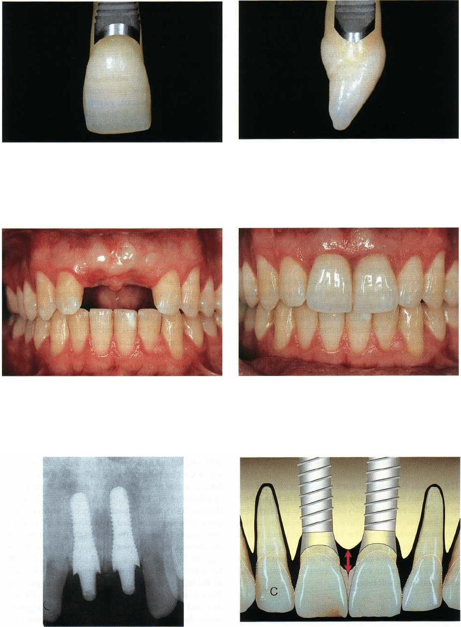
IMPLANT PLACEMENT IN THE ESTHETIC ZONE • 935
Fig. 40-63. Instead of the traditional implant design, fea-
turing a flat rotation symmetrical coronal aspect, a scal-
loped connection, inspired by the natural cemento-
enamel junction, may lead to a more superficial im
plant
insertion and by this to the preservation of more bone in
the interproximal area.
Fig. 40-64. Comparison in the sagital plane of a natural
maxillary central incisor and a titanium implant featur-
ing a scalloped design at its coronal end. The radius
corresponds to the amount of bone which might theo-
retically be preserved.
Fig. 40-65. Vestibular view in centric occlusion position
of a 24-year-old male patient. The two maxillary cen
tral
incisors have been lost due to a traumatic injury.
Fig. 40-66. After one year of clinical service, the pres-
ence of a harmoniously scalloped marginal soft tissue
course, including the most critical interimplant area,
can be noted.
Fig. 40-67. The 1-year follow-up radiograph shows pro-
totype of titanium implants featuring a scalloped de-
sign
at their coronal end. This design permits a more
superficial implant insertion, aiming at a better preser-
vation of interimplant alveolar bone.
Fig. 40-68. Schematic representation of the theoretical
advantages of a scalloped implant design: more super-
ficial implant placement, increased bone and soft tissue
preservation particularly in the interimplant area, and
improved esthetics (in combination with interdental
"
mini-wings").
93
6
• CHAPTER
40
of the long-term stability of the esthetic result (Belser
1999, Belser et al. 1998, 2000). The currently flat, "ro
-
tation-symmetrical" design of standard screw-type
titanium implants, leading to a marked submucosal
implant shoulder position at the interproximal aspect,
may not represent, however, the optimal design, in
particular in the context of multiple adjacent implants.
Scalloped implant design
As pointed out earlier in this chapter, the traditional
implant design may lead to esthetic shortcomings in
a
case of multiple adjacent maxillary anterior fixed
implant restorations. One could hypothesize in this
context whether a modified design at the coronal end
of the implant, in the sense of a scalloped, more "CEJ
-
like" configuration, might lead to an improved pres-
ervation of peri-implant bone at the interproximal
aspect in general, and between adjacent implants in
particular. One of the possible design solutions and its
anticipated theoretical impact on bone and esthetic
parameters are presented in Figs. 40-63, 40-64 and
40-68. More specifically, this approach ultimately aims
at creating an interimplant bone height and resulting
soft tissue level situation compatible with generally
accepted esthetic criteria. Among these one should
primarily mention the establishment and/or mainte-
nance of a harmoniously scalloped course of the mar-
ginal peri-implant mucosa. At present, the combina-
tion of the following three elements appears impor-
tant:
1.
screw-type titanium implant body, featuring opti-
mal surface characteristics
2.
tooth-colored transmucosal portion with adequate
axial emergence profile and scalloped coronal end
3.
mechanically sound suprastructure-connection,
permitting both screw-retention and cementation.
The clinical potential of such a novel, scalloped im-
plant design is anecdotally documented in Figs. 40-65
to 40-67, presenting a 24-year-old male patient who
had lost his two maxillary central incisors in the
course of an accident. The 1-year clinical and radio-
graphic follow-up appears to support — at least short
-
term — the hypothesis that such an approach may
preserve to a certain extent interimplant crestal bone
and overlaying soft tissue.
Segmented fixed implant restorations in the
edentulous maxilla
Another particular challenge from both a surgical and
a prosthodontic point of view represents the implant-
supported fixed prosthetic rehabilitation of the eden-
tulous maxilla. Undoubtedly esthetic considerations
and certain aspects associated with the patient's sub-
jective comfort — both during the actual treatment
phase and once the prosthesis is completed — also play
a major role in this context. We will limit our reflec-
tions to (1) specific aspects of pre-implant diagnosis,
(
2) the importance of implant number, alignment and
spatial distribution, and (3) conception of the supra-
structure.
These elements are addressed in the form of a re-
spective clinical case presentation, involving a 67-
year-old female patient, edentulous in the maxilla
(
Figs. 40-69 to 40-89). Besides the traditional clinical
and radiologic investigation, an in-mouth try-in of the
envisioned treatment objective in the form of a set-up
of teeth without vestibular denture-type flange is of
primary importance (Fig. 40-73). Among other as-
pects, this approach will allow the visualization of the
length of the clinical crowns of the future fixed im-
plant prosthesis, and the evaluation of whether a fixed
prosthesis will provide sufficient lip and facial sup-
port (Fig. 40-74). A surgical guide, derived from the
described tooth set-up, will guarantee that the future
implant positions are in accordance with the deter-
mined tooth positions. Whenever possible, parallel-
ism of implants is recommended, as it permits an
eventual early or immediate loading approach
(
Szmukler-Moncler et al. 2000, Cooper et al. 2001,
Andersen et al. 2002, Cochran et al. 2002), and facili-
tates the subsequent clinical and laboratory proce-
dures. Although little scientific evidence exists to in-
dicate how many implants of which dimension and in
what position are required for a predictable and long-
lasting fixed implant rehabilitation of an edentulous
maxilla, some clinical trends — mostly derived from
traditional prosthodontic experience — do exist. If one
plans to extend the prosthesis to the first molar area,
and if the anatomical conditions allow the use of
standard-size (length and diameter) implants, be-
tween six and eight implants seems reasonable. How-
ever, in order to increase the overall prosthetic versa-
tility and to be able to apply the principle of segment-
ing, which includes the ease of eventual reinterven-
tions in a case of localized complications (Priest 1996,
Goodacre et al. 1999, Lang et al. 2000, Johnson &
Persson 2001), eight implants may be considered ade
-
quate. The recommended respective positions are on
both sides of the jaw — the sites of the central incisors,
the canines, the first premolars and the first molars
(
Fig. 40-76). This approach will ultimately allow the
fabrication of four independent three-unit FPDs, with
all the related technical and clinical advantages (Figs.
40-78 to 40-89). Some of the scientific data available to
date and supporting the concept of smaller segments
rather than full-arch splinting will be presented and
discussed in Chapter 41.
In conclusion, the concepts and therapeutic modali
-
ties do exist nowadays to solve —by means of implants
— elegantly as well as predictably a majority of clinical
situations requiring the replacement of missing teeth
in the esthetic zone, and the most promising novel
approaches and perspectives can already be identified
on a not too distant horizon.
