Jan Lindhe. Clinical Periodontology
Подождите немного. Документ загружается.


IMPLANT PLACEMENT IN THE ESTHETIC ZONE • 9
1
7
Table 40.2. Patient expectations related to maxillary
anterior edentulous segments
Table 40.3. Therapeutic modalities for tooth replace-
ment in the esthetic zone
•
Long-lasting esthetic and functional result with a high degree
of predictability
•
Minimal invasiveness (preservation of tooth structure)
•
Maximum subjective comfort
•
Minimum risk for complications associated with surgery and
healing phase
•
Avoidance of removable prostheses
•
Optimum cost effectiveness
ble 40-1, Fig. 40-1). This list is completed by an addi-
tion of subjective criteria associated with esthetic in-
tegration, such as variations in the arrangement and
positioning of front teeth, relative crown length and
negative space.
Depending on the type of a given initial clinical
situation requiring the replacement of one or several
teeth, the patient's expectations may vary from the
achievement of an almost perfect illusion, i.e. that the
untrained eye cannot easily distinguish the restora-
tion from the surrounding natural dentition, to the
acceptance of various degrees of compromise from a
purely esthetic point of view. The latter case is not
infrequent after multiple anterior tooth loss in combi-
nation with significant hard and soft tissue deficien-
cies. In relation to maxillary anterior edentulous seg-
ments, patients expect in general a long-lasting func-
tional and esthetic result with a high level of predict-
ability (Table 40-2). To this primary objective are nor-
mally added a number of secondary goals which in-
clude parameters such as minimal invasiveness, low
risk associated to eventual surgery, overall simplicity
and cost effectiveness.
Prior to selecting an implant-based solution, one
should comprehensively review all of the possible
treatment modalities available (Table 40-3) which
have the potential to solve a given clinical problem,
and carefully ponder their respective advantages and
eventual shortcomings, and only then take the deci-
sion together with the adequately informed patient.
Currently, the restorative spectrum in the case of miss
ing maxillary anterior teeth comprises conventional
FPDs, resin-bonded bridges, removable partial den-
tures (RPDs), tooth-supported overdentures and im-
plant-supported fixed or removable prostheses. Fur-
thermore, one should not forget that occasionally or-
thodontic therapy, e.g. closure of limited edentulous
spaces, can represent an effective and elegant alterna
-
tive or adjunction to a prosthetic treatment. However,
the availability of scientific evidence — when possible
at its highest level — for the planned treatment modal
-
ity, should be the key parameter for the final choice.
In this clinical decision-making process certain cri-
teria, as for example the compromised structural, peri
-
odontal and/or endodontic status of potential natural
•
Conventional fixed partial dentures (FPDs), comprising
cantilever units
•
Resin-bonded ("adhesive") bridges
•
Conventional removable partial dentures (RPDs)
•
Tooth-supported overdentures
•
Orthodontic therapy (closure of edentulous spaces)
•
Implant-supported prostheses (fixed, retrievable or removable
suprastructures)
•
Combinations of the above
Table 40.4. Criteria favoring implant-borne restora-
tions
•
Normal wound healing capacity
•
Intact neighboring teeth
•
Unfavorable ("compromised")
potential abutment teeth
•
Extended edentulous segments
•
Missing strategic abutment teeth
•
Presence of diastemas
abutments, or the extended dimension of the edentu-
lous segment, are among the factors favoring an im-
plant-borne restoration rather than a tooth-supported
fixed prosthesis (Table 40-4).
Esthetic considerations related to maxillary
anterior implant restorations
h1
the context of the natural dentition, long clinical
crowns, the irregular contour of the gingival margin,
i.e. any abrupt change in vertical tissue height be-
tween neighboring teeth, and the loss of papillary
tissue often have an adverse influence on dental-facial
esthetics (Seibert & Lindhe 1989). Furthermore, the
same authors have underlined that in the case of a
high
scalloped gingival rnorphotype
(in contrast to a rather
low
scalloped gingival morphotype)
there is mostly an
unpre
dictable relationship between the underlying
bone
and the gingival contour, often leading to so
called "black hole cases" and presenting a high risk
for los
ing soft tissue (e.g. gingival or mucosal
recession at
the labial aspect of teeth or implants),
particularly in
relation to restorative procedures, as
for example in
sertion of retraction cords and
impression taking.
Another esthetically relevant concern lies in the fact
that under normal conditions a maxillary front tooth
extraction leads on average to approximately 2 mm
loss in vertical tissue height. The mean length of the
clinical crown of a maxillary central incisor is 10.2 mm,
the one of a lateral incisor 8.2 mm and that of a canine

918 • CHAPTER
40
Table 40.5. Evaluation of anterior tooth-bound eden-
tulous sites prior to implant therapy
•
Mesio-distal dimension of the edentulous segment, including
its comparison with existing contralateral control teeth
•
Three-dimensional analysis of the edentulous segment
regarding soft tissue configuration and underlying alveolar
bone crest (ref. "bone-mapping")
•
Neighboring teeth:
•
volume (relative tooth dimensions), basic features of tooth
form and three-dimensional position and orientation of
the
clinical crowns
•
structural integrity and condition
•
surrounding gingival tissues (course/scalloping of the
gingival line)
•
periodontal and endodontic status/conditions
•
crown-to-root ratio
•
length of roots and respective inclinations in the frontal
plane
•
eventual presence of diastemata
•
Interarch relationships:
•
vertical dimension of occlusion
•
anterior guidance
•
interocclusal space
•
Esthetic parameters:
•
height of upper smile line ("high lip" versus "low lip")
•
lower lip line
•
course of the gingival-mucosa line
•
orientation of the occlusal plane
•
dental versus facial symmetry
•
lip support
10.4 mm. Consequently, any kind of maxillary anterior
restoration should aim at staying within reasonable
limits of these average morphological dimensions, if
a
harmonious and esthetically pleasing result is to be
achieved. Ultimately, an anterior implant restoration
should correspond closely to an ovate pontic of a
conventional FPD with respect to the relevant soft
tissue parameters (Kois 1996).
Numerous publications, mostly in the form of text-
books, book chapters, reviews, case reports and de-
scriptions of clinical and laboratory procedures and
techniques, have addressed various aspects specifi-
cally related to esthetics and osseointegration (Parel &
Sullivan 1989, Gelb & Lazzara 1993, Jaggers et al. 1993,
Vlassis et al. 1993, Bichacho & Landsberg 1994, Ghalili
1994, Landsberg & Bichacho 1994, Neale & Chee 1994,
Studer et al. 1994, Carrick 1995, Corrente et al. 1995,
De Lange 1995, Garber 1995, Garber & Belser 1995,
Jansen & Weisgold 1995, Khayat et al. 1995, Touati
1995, Brugnolo et al. 1996, Davidoff 1996, Grunder et
al. 1996, Hess et al. 1996, Marchack 1996, Mecall &
Rosenfeld 1996, Bain & Weisgold 1997, Bichacho &
Landsberg 1997, Chee et al. 1997, Garg et al. 1997,
Spear et al. 1997, Salinas & Sadan 1998, Jemt 1999,
Table 40.6. Optimal three-dimensional implant posi
-
tioning ("restoration-driven implant placement") in
anterior maxillary sites.
Implant = apical extension of
the ideal future restoration
•
Correct vertical position of implant shoulder (sink depth)
using the cemento-enamel junction of adjacent teeth as
reference:
•
no visible metal
•
gradually developed, flat axial profile
•
Correct oro-facial position of point of emergence for future
suprastructure from the mucosa:
•
similar to adjacent teeth
•
flat emergence profile
•
Implant axis compatible with available prosthetic treatment
options (ideally: implant axis identical with "prosthetic axis")
Price & Price 1999, Belser et al. 2000, Tarnow et al.
2000).
In view of maxillary anterior implant restorations,
the systematic and comprehensive evaluation of eden
tulous sites, including the surrounding natural denti
-
tion, is of paramount importance (Table 40-5). Key
parameters comprise the mesio-distal dimension of
the edentulous segment, the three-dimensional analy
sis of the underlying alveolar bone crest, the status of
the neighboring teeth, and interarch relationships as
well as specific esthetic parameters.
As one should consider the implant as the apical
extension of the ideal future restoration and not the
opposite, a respective optimal three-dimensional
("
restoration-driven") implant position is mandatory
(
Table 40-6). Consequently, parameters addressing
vertical (sink-depth) and oro-facial implant shoulder
location, have been defined, as well as guidelines
related to the long axis of the implant, as the latter has
a significant impact on the subsequent technical pro
-
cedures during suprastructure conception and fabri-
cation.
Recently, the ITI Consensus Conference has ap-
proved the distinctly submucosal implant shoulder
location in the maxillary anterior segment in order to
respond to natural esthetic demands (Buser & von Arx
2000). As the current implant design — in contrast to
the scalloped cemento-enamel junction — features a
straight horizontal, "rotation-symmetrical" restora-
tive interface, interproximal implant crown margins
are often located several millimeters submucosally,
and thus difficult to reach by the patien
t
'
s routine oral
hygiene efforts (Belser et al. 1998). Mainly for this
reason a screw-retained implant suprastructure (Sut
-
ter et al. 1993, Hebel & Gajjar 1997, Keller et al. 1998)
is preferred to a cemented one, as it benefits from the
surface quality and marginal fidelity of prefabricated,
machined components, and avoids potential prob-
lems associated with cement excess that may be diffi
-
cult to reach and thoroughly eliminate.

IMPLANT PLACEMENT IN THE ESTHETIC ZONE •
919
ANTERIOR SINGLE-TOOTH
REPLACEMENT
Favorable 5-year multicenter results for 71 single-
tooth replacements in the anterior maxilla (implant
success rate of 96.6%) were reported by Henry et al.
(
1996); however, this group mentioned an associated
10% esthetic failure rate. In a retrospective study on
236 patients treated with single-tooth implant restora
-
tions in the anterior maxilla (Walther et al. 1996), a
Kaplan-Meier survival rate of 89% was found for an
observation period of 10 years. The failure rate for
lateral incisor replacement was lower than the one for
central incisors. Furthermore, 5% of the related pros-
thetic suprastructures had to be replaced during the
10 years of observation. Kemppainen et al. (1997)
prospectively documented 102 implants (ASTRA/
ITI)
for single-tooth replacement in the anterior max
illa of
82 patients and found survival rates of 97.8%
and
100%, respectively, after 1 year. Still related to
single-
tooth maxillary anterior implants, a prospective
study on 15 patients revealed a 100% implant
survival rate after two years of function (Palmer et al.
1997). At crown insertion (6 months after implant
placement) the mean bone level was located 0.47 mm
apically to the top of the implants. No significant
additional changes in crestal bone level occurred dur-
ing the remainder of the study.
Today, it is generally accepted that the final implant
shoulder sink depth for esthetic fixed single-tooth
restorations can be determined primarily by the loca-
tion of the cemento-enamel junction (CEJ) of the
neighboring teeth and by the level of the free gingival
margin at the vestibular aspect of these same teeth.
This means that the implant shoulder is positioned 1-2
mm more apically to the labial CEJ of the adjacent
teeth (Belser et al. 1998, 2000). However, the noticeable
esthetic progress made in this kind of implant resto-
ration is the result of recent developments in the ab-
sence of extensive long-term documentation. Because
the exclusive use of clinical signs for establishing peri
-
implant health or disease may not be sufficient, the
evaluation of additional objective parameters is
needed. A number of diagnostic tests have been util-
ized by clinicians to supplement clinical signs with
objective methods. These tests include microbiologic
monitoring, proteolytic bacterial enzyme markers,
markers of tissue destruction, and finally, markers of
tissue repair and regeneration. In this context peri-im
-
plant crevicular fluid (PICF) analysis has become the
focus of intense investigation. It has been observed
that the volume of crevicular fluid did not differ be-
tween implant sites and natural teeth and that the
features of inflammation seem to be the same around
teeth and implants. In addition, the histologic ar-
rangement of peri-implant soft tissues resembles ba-
sically that observed around natural teeth, although
featuring also some aspects of scar tissue (Abraham-
Table 40.7 Basic considerations related to anterior
single-tooth replacement
Achievements
Predictable and reproducible results regarding
both esthetic parameters and longevity in sites
without significant vertical tissue deficienies
Well defined and well established surgical
protocols:
•
restoration-driven
implant placement
Adequate and versatile restorative protocols
and prosthetic components:
•
occclusal/transverse screw-retention
• angulated abutments
•
high-strength ceramic components
Limitations
Combined vertical bone and soft tissue deficienies:
•
following removal of ankylosed teeth or
failing implants
•
advanced loss of periodontal tissues,
including gingival recession, on
neighboring teeth
•
limited scientific documentation related to
vertical
bone augmentation
and
distraction
osteogenesis
son et al. 1996, Berglundh & Lindhe 1996, Abraham
-
son et al. 1997, Lindhe & Berglundh 1998).
Giannopoulou et al. (2002) investigated the effect of
intracrevicular restoration margins on peri-implant
health of 61 maxillary anterior implants — mainly
single-tooth replacements — in 45 patients up to 9
years. Results revealed that the only statistically sig
-
nificant differences between baseline and follow-up
examination concerned pocket probing depth (PPD)
and the distance between the implant shoulder and
the mucosal margin (DIM measurements), which
slightly increased over time. The remainder of the
clinical measurements and almost all of the microbi
-
ologic and biochemical parameters analysed did not
significantly change. Probably the most critical pa-
rameter from a purely esthetic point of view is the DIM
value, particularly on the labial aspect of the maxillary
anterior implants investigated in this study. A mean
value of -1.5 ± 1.1 mm was found at baseline exami
-
nation, and a slight increase (—1.7 ± 1.1 mm) at the
follow-up. This indicates that the risk for exposure of
the implant-to-crown interface or margin can be con
-
sidered low. These findings corroborate recently pub-
•
technique offers efficacy and predictability
•
simultaneous
or
staged approach
depending on defect extension and defect
morphology
Lateral bone augmentation by means of
alveolar
bone
crest
splitting
and/or various
osteotome
techniques:
•
limited clinical long-term documentation
Sites with
Lateral bone augmentation using autografts
buccal bone
and
barrier
membranes:
deficienies

920 • CHAPTER
40
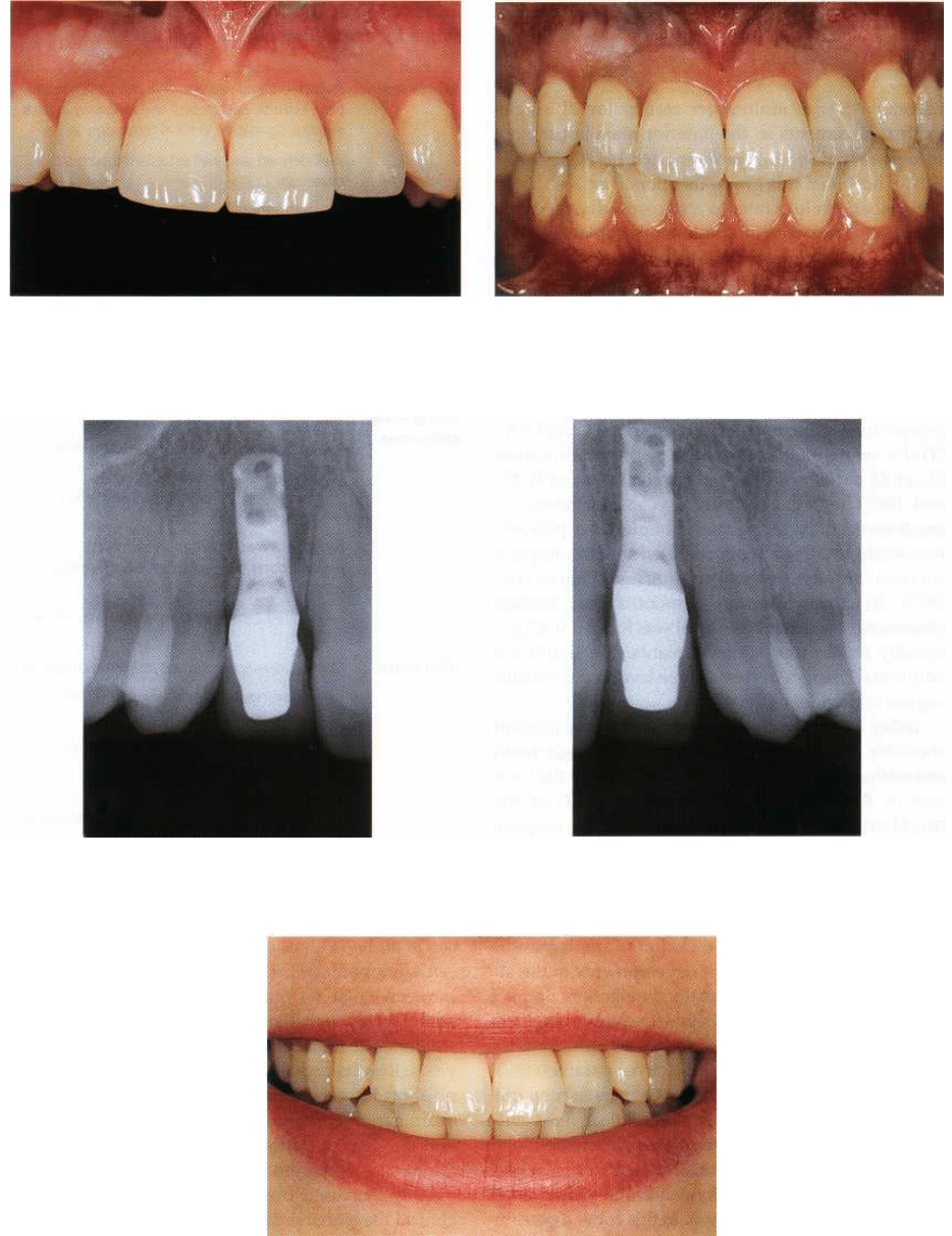
Fig. 40-2. 10-year follow-up of a 28-year-old female pa-
Fig. 40-3. The frontal view in centric occlusal position
tient. Both congenitally missing lateral incisors were re-
documents the harmonious integration of the two im-
placed by implants, restored with screw-retained porce-plant restorations after 10 years of clinical service.
lain-fused-to-metal crowns.
Fig. 40-4. 10-year postoperative radiograph of the max-Fig. 40-5. 10-year postoperative radiograph of the max-
illary right lateral single-tooth implant restoration.
illary left lateral single-tooth implant restoration.
lished data addressing similar parameters (Grunder were observed between the above results and the 2000).
The consistently negative Periotest scores con- number of years that the implants had been in func-
firmed
the stability and osseointegrated status of the tion. Based on these clinical, microbiologic and bio-
implants
examined. Furthermore, no associations chemical data, and on an observation period of 4-9
Fig. 40-6. During unforced smiling an adequate balance
between implant-crowns and natural dentition can be
noticed.

IMPLANT PLACEMENT IN THE ESTHETIC ZONE • 921
Fig. 40-7. Schematic representation of an intact maxil-
lary right anterior segment. The alveolar bone follows
the scalloped course of the cemento-enamel junction
for a distance of approximately 2 mm (white dotted line)
, whereas, accordingly, the gingival tissue occupies
completely the interdental area.
Fig. 40-8. Schematic representation of the same seg-
ment after loss of the lateral incisor. While the inter-
proximal bone height has basically been maintained,
the corresponding gingival tissue is flattened due to a
lack of support originally provided by the now missing
tooth.
Fig. 40-9. The treatment objective in the case of an ante
-
rior single-tooth replacement is an implant restoration
with a gradually developed, flat emergence profile
from
the implant shoulder to the peri-implant mucosal
surface. Ideally, the clinical crown of the implant resto-
ration should aim at replicating the clinical crown of
the
corresponding contralateral tooth.
Fig. 40-10. Schematic comparison in the sagital plane
between a natural maxillary incisor and a respective
implant borne single-tooth restoration. The decrease of
alveolar bone height on the labial and palatal aspect fol-
lowing tooth loss leads to a more palatal implant posi-
tion when compared to the original root position,
which
in turn influences the axial profile of the restora
tion.
years (mean: 6.8 years), it was concluded that in pa-
tients with appropriate oral hygiene, implant-sup
ported
maxillary anterior crowns with distinctly in
tracrevicular
margins did not predispose to unfavor
able peri-implant
host and microbial responses. In
particular, overall
healthy and stable peri-implant tis
sue conditions – a
paramount criterion when it comes
to esthetic implant
crowns – were consistently en-
countered and
maintained longitudinally. One of the
patients
participating in this study and who recently
passed the
10-year clinical and radiographic follow-
up control, is
presented in Figs. 40-2 to 40-6. An ade
quate esthetic
integration of the two single-tooth restorations,
replacing the congenitally missing lateral incisors, could
be achieved and maintained over time.
In a simplistic way, the morphologic and esthetic
consequences in the frontal plane of the loss of a single
maxillary incisor, when compared to the original in-
tact
situation, can be summarized as follows: mainte
nance
of the tooth-sided interproximal bone height at
the
neighboring teeth, and vertical loss ("flattening")
of the
corresponding gingival tissue due to a lack of support
originally provided by the now missing tooth
(Figs. 40-7
and 40-8). In case of an anterior single-tooth
replacement, the related implant restoration should
aim
at replicating the clinical crown of the contralat
eral
control tooth from the line of soft tissue emergence
to
the incisal border. Additionally, a gradually developed,
flat emergence profile from the implant shoul
der to the
peri-implant mucosal margin is mandatory
(Figs. 40-9
and 40-10).
The basic considerations related to maxillary ante-

922 • CHAPTER
40
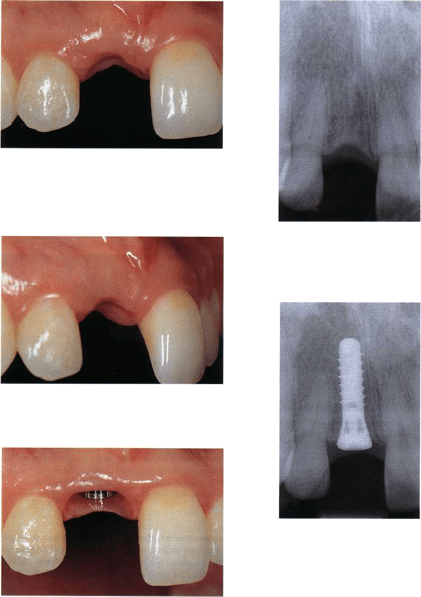
Fig. 40-11. Preoperative close-up view of the upper
right
anterior region of a 22-year-old female patient
with a
missing right central incisor. The scalloped course of
the gingiva is maintained, featuring inter-
proximal soft
tissue at the level of the cemento-enamel
junction.
Fig. 40-13. The oblique close-up view confirms optimal
conditions for the insertion of an implant, namely inter-
proximal soft tissue height and no significant loss of
the
buccal bone plate.
Fig. 40-14. Clinical view of the maxillary anterior im-
plant site 8 weeks after insertion of a solid screw im-
plant according to a one-stage transmucosal surgical
protocol. A harmonious peri-implant soft tissue profile
has been established by means of a titanium healing
cap featuring a respective emergence profile and thus
offering adequate interproximal soft tissue support.
rior single-tooth replacement, including the respec
tive
general achievements and limitations, and ad-
Fig. 40-12. The corresponding radiograph displays fa-
vorable bony conditions in view of implant therapy.
Note in particular the interproximal bone height, fol-
lowing the cemento-enamel junction for a distance of
less than 2 mm.
Fig. 40-15. The corresponding radiograph displays a
continuous close contact between bone and implant
and confirms that the vertical interproximal bone level
has been maintained.
dressing edentulous segments with different types of
labial bone deficiencies, are presented in Table 40-7.
Sites without significant tissue deficiencies
An increasing body of evidence indicates that the most
determinant parameter for achieving an esthetic sin-
gle-tooth restoration is the interproximal bone height at
the level of the teeth confining the edentulous gap.
The
related bone should be within a physiologic dis-

IMPLANT PLACEMENT IN THE ESTHETIC ZONE • 923
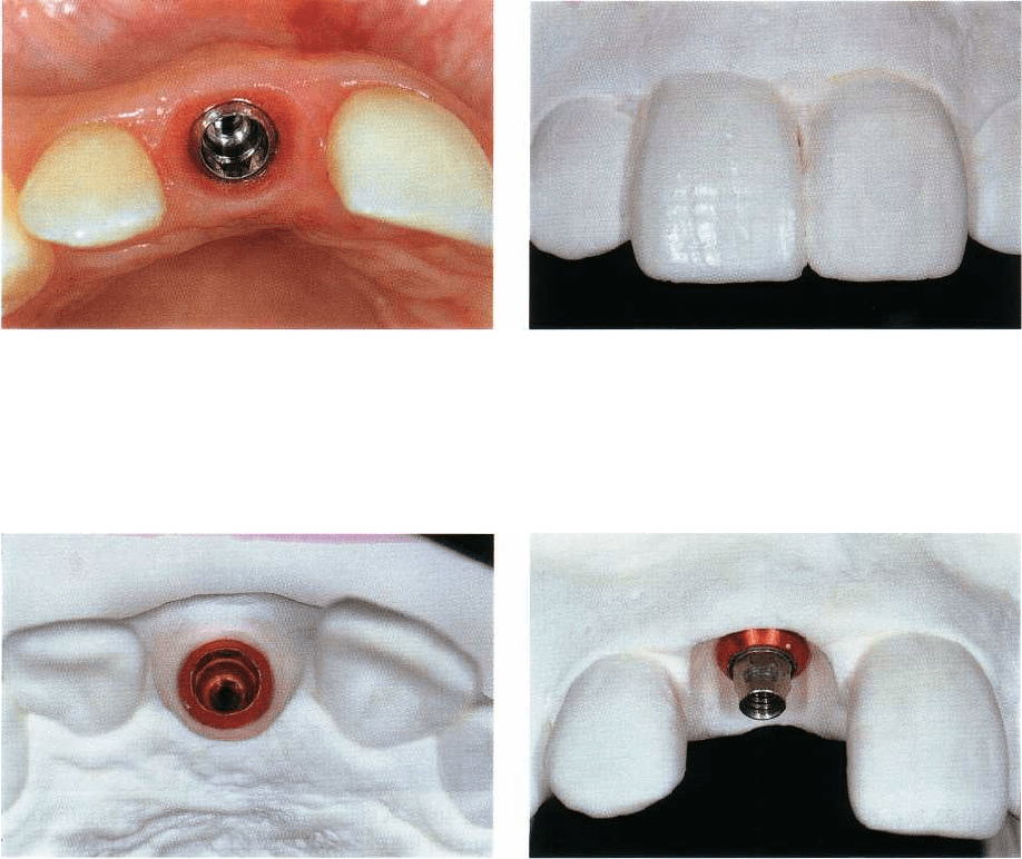
Fig. 40-16. The occlusal view reveals an implant posi-
tion in the orofacial plane that is in accordance with the
adjacent natural roots and thus permits development
of a flat emergence profile.
Fig. 40-17. On a stone model derived from the clinical
situation, the laboratory technician defines the treat-
ment objective in wax. At this stage priority is given to
esthetic principles and maintenance of symmetry
rather than to the actual position of the underlying im-
plant.
Fig. 40-18. The configuration of the peri-implant soft
tissue is subsequently adapted on the stone model ac-
cording to the diagnostic wax-up. Ultimately, it will be
the restoration itself that completes the last phase of
soft tissue conditioning by subtle respective physical
displacement.
tance, i.e. approximately 2 mm, of the cemento-enamel
junction (CEJ) and thus be providing the essential
support for the overlaying soft tissue compartments.
Consequently, preoperative diagnosis will include in-
terproximal radiographic bone height assessment and
periodontal probing of the soft tissue attachment
level.
If the comprehensive presurgical analysis of a given
maxillary anterior single-tooth gap has confirmed on
the
one hand a favorable vertical level of both soft
tissue and
underlying alveolar bone at the interproximal aspect of
the two adjacent teeth (Figs. 40-11, 40-12,
40-13), and no
major vestibular bone deficiencies on
the other hand,
the site can be considered compatible
with a
straightforward implant surgical protocol. In
order to
ensure the best probability of a successful and long-
lasting esthetic treatment outcome, the actual
implant
placement has to be carried out meticulously
according
to the surgical guidelines defined in Table
40-6. These
guidelines include key-parameters such as
Fig. 40-19. An appropriate secondary titanium compo
-
nent (abutment) is selected as support for the planned
screw-retained implant restoration.
low-trauma surgical principles in general and precise
three-dimensional ("restoration-driven") implant po-
sitioning in particular. In the case of standard single-
tooth sites, most surgeons do not advocate the use of
a
surgical guide or stent, as the adjacent teeth and
associated anatomical structures normally offer suffi-
cient morphologic landmarks to safely reach the
therapeutic objective. As far as the detailed surgical
protocol is concerned, readers are referred to Chapter
37, "The surgical site". Buser and von Arx (2000) have
published the surgical step-by-step procedure related to
maxillary anterior single-tooth implants, and insisted
on a slightly palatal incision technique to pre-
serve a
maximum of keratinized mucosa on the labial
aspect of
the future implant restoration. Another crucial
parameter is the maintenance of at least 1 mm of bone
plate on the vestibular aspect of the implant in
order to
minimize the risk for peri-implant soft tissue
recessions,
a factor parameter when it comes to esthetics. Under
such conditions one may consistently

924 • CHAPTER 40
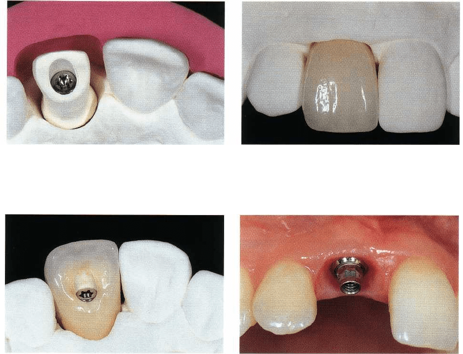
Fig. 40-20. Using a silicon template as guide, a prefabri
cated ceramic blank is inserted and subsequently
re
duced to provide adequate space for the external
layers
of cosmetic porcelain.
Fig. 40-21. Labial view of the completed ceramo-ce
-
ramic restoration on the master cast.
Fig. 40-22. In particular, the completed screw-retained
all-ceramic restoration displays a high degree of trans
-
lucency on its incisal third.
achieve postsurgical treatment outcomes featuring
unaltered vertical soft tissue and underlying bone
levels at the interproximal aspect of the adjacent natu
-
ral teeth (Figs. 40-14, 40-15, 40-16).
Once osseointegration is confirmed radiologically
and clinically, the clinical situation is transferred to the
master model by means of an impression, normally
assisted by auxiliary components in the form of pre-
fabricated impression copings. On the master model,
which in turn contains a replica (analogue) of the
implant, the laboratory technician defines the final
configuration of the single-tooth implant restoration
by means of a diagnostic wax-up (Fig. 40-17). Under
normal circumstances, i.e. when the natural contralat
-
eral control tooth corresponds mostly to the esthetic
and functional requirements of an appropriate "target
model", the technician basically copies the clinical
crown of this control tooth in wax, regardless of the
actual underlying implant position. At this stage a
close-to-ideal restoration is planned, while its connec
-
tion to the underlying implant will be addressed later.
This approach comprises the minute shaping of the
peri-implant soft tissue configuration (on the master
model in the form of stone), in view of an identical
emergence from the labial and interproximal soft tis-
sue margin, like the one observed on the natural tooth
Fig. 40-23. A titanium abutment will serve as infrastruc
-
ture for the transocclusally screw-retained high-
strength all-ceramic restoration.
site (Fig. 40-18). Only after having completed this
preparatory step, will the ceramist select the most
adequate secondary component (i.e. abutment), de-
pending on the three following cardinal criteria (Fig.
40-19):
1.
implant shoulder depth in relation to the labial
mucosal margin
2.
oro-facial implant shoulder position with respect to
the future line of emergence of the suprastructure
3.
long axis of the implant.
In most instances, preference will be given to a screw
-
retained implant suprastructure, unless a combina-
tion of mesiostructure and cemented restoration is
chosen. Screw-retention is primarily preferred due to
a marked submucosally located implant shoulder, in
particular at the interproximal aspect, which may ren
-
der the removal of excess cement difficult, and which
is mostly not within reach of the patient's routine oral
hygiene measures. In addition, screw-retained su-
prastructures benefit from the close-to-perfect surface
quality characteristics and the marginal precision of
machined, prefabricated components. Nowadays sev-
eral of the leading implant systems also offer high-
strength ceramic tertiary components which may

IMPLANT PLACEMENT IN THE ESTHETIC ZONE • 925
Fig. 40-24. The one-year postoperative radiograph con
-
firms favorable conditions at the bone-to-implant inter
-
face. Note a high degree of radio-opacity of the all-ce
-
ramic substrate, permitting the evaluation of the fidel
-
ity of the marginal adaptation.
positively contribute to the esthetic treatment out-
come, particularly in the case of a rather thin labial
peri-implant mucosa (Fig. 40-20). Another parameter
which is of primary importance when it comes to
esthetic considerations relates to maxillary anterior
implant restorations and is associated with the supras
tructure design itself at the interproximal aspect. In
order to provide optimal conditions for the related soft
tissue, a long interdental contact line is established,
located slightly more towards the palatal aspect of the
restoration (Figs. 40-21, 40-22). This design offers op
-
timal support for the interproximal soft tissue and
thereby reduces the potential hazard of a so-called
"
black triangle" (Figs. 40-23, 40-24, 40-25). In this con
-
text some studies have indicated that there exists a
predictable relationship between the location of the
interdental contact point and the associated alveolar
bone crest when it comes to presence or absence of
interdental papillae fully occupying the interdental
space of maxillary anterior teeth (Tarnow et al. 1992,
Tarnow & Eskow 1995).
Sites with localized horizontal deficiencies
In a case of a localized (minor) horizontal deficiency,
i.
e. a confined vestibular alveolar bone crest defect at
the vestibular aspect of a maxillary anterior single-
tooth gap, one prefers to place the implant and simul
-
taneously undertake a lateral bone augmentation pro
-
cedure, on condition that several well-defined prereq-
uisites are fulfilled. These include an implant place-
ment in accordance with the guidelines presented in
Table 40-6 ("restoration-driven" implant placement),
the achievement of an adequate primary stability and
a
resulting cervical dehiscence-type bony defect
Fig. 40-25. An acceptable overall integration of the met
-
al-free implant-borne restoration on site 11 can be
noted.
which is compatible with a predictable bone augmen-
tation procedure. More specifically, the dehiscence
should have the form of a two-wall bony defect,
whereas the labial aspect of the inserted implant
should not exceed the surrounding bone contours.
Under such conditions, the treatment of choice con-
sists of the application of autogenous bone chips,
harvested at the site of the implant surgical interven-
tion. The bone chips which can be combined with one
of the numerous available bone substitutes (e.g.
BioOss @) if necessary, will provide adequate support
for a subsequently adapted barrier membrane. The
described grafting material is finally complemented
with "bone slurry", constantly collected during the
entire procedure. Subsequently, a bioabsorbable mem
brane is applied prior to repositioning and tension-
free suturing of the mucoperiosteal flap. This impli-
cates a rather extended flap design, comprising verti-
cal releasing incisions.
In conclusion, a simultaneous lateral augmentation
procedure is recommended if the three following con-
ditions are present:
1.
ideal three-dimensional ("restoration-driven") im-
plant position
2.
adequate primary implant stability
3.
localized two-wall bony defect, exceeding the la
bial
contour of the implant and hereby assuring an
appropriate bone regeneration potential and pro-
viding the necessary stability to the applied bone
graft.
Under these specific conditions, the implant can be
functionally loaded after 2-4 months, depending on
size and configuration of the respective bone defect.
It is not infrequent in the anterior maxilla, due to its
specific alveolar bone crest morphology, that "restora
-
tion-driven" rather than "bone-driven" implant posi-
tioning leads to a fenestration-type defect in the apical
area of the implant. If adequate primary implant sta-
bility can be obtained, a similar simultaneous lateral
bone augmentation procedure, as described for local-
ized dehiscence-type defects, appears feasible. Under

926 • CHAPTER 40
Fig. 40-26. Schematic representation of a horizontal sec-
tion at the cemento-enamel junction level of the maxil-
lary right anterior segment.
Fig. 40-27.
"
Restoration-driven" implant placement in
the
horizontal plane at the site of the maxillary right lat
eral
incisor. In order to maintain at least 1 mm of alveo
lar
bone also on the labial aspect, the implant has to be
inserted approximately 1-2 mm more to the palate
when
compared to the adjacent roots.
Fig. 40-28. In case of an extended lateral bone defi-
ciency, where an adequately placed implant would
largely exceed the vestibular border of the alveolar
bone crest, a lateral bone augmentation procedure
(
staged approach) is indicated
Fig. 40-29. After elevation of a mucoperiosteal flap a se-
vere extended resorption – namely on the vestibular as-
pect – of the edentulous alveolar ridge becomes appar-
ent. Such a morphology is hardly compatible with "res-
toration-driven" implant placement.
Fig. 40-30. An autogenous bone graft, harvested from
Fig. 40-31. Six months after the lateral ridge augmenta-
the patient's chin region, has been secured with a fixa-
tion procedure the clinical occlusal view documents
tion screw and its periphery filled in with additional
that uneventful healing has occurred and that the oro-
bone chips prior to membrane placement.
facial ridge profile has been improved.
