Jan Lindhe. Clinical Periodontology
Подождите немного. Документ загружается.


ALVEOLAR BONE FORMATION • 867
Fig. 38-1. Histological section illus
trating a bone multicellular unit
(
BMU). Note the presence of a re-
sorption front with osteoclast
(
OC) and a deposition front that
contains osteoblasts (OB), and
osteoid (OS). Vascular structures
(V) occupy the central area of the
BMU. RL = reserval line; LB =
lamellar bone.
progenitor cells are present in the bone marrow, in the
endosteum and in the periosteum that cover the sur-
faces of bone. Such cells possess an intrinsic capacity
to proliferate and differentiate into osteoblasts. Induc
-
ible osteogenic precursor cells, on the other hand,
represent mesenchymal cells present in other organs
and tissues (e.g. muscles) that may become bone-
forming cells when exposed to specific stimuli. As
osteogenesis is always closely related to the ingrowth
of vascular tissue, the stellate-shaped perivascular cell
(the
pericyte) is
considered to be the main osteopro-
genitor cell. The differentiation and development of
osteoblasts from osteoprogenitor cells are dependent
on the release of bone morphogenetic proteins (BMP)
and other growth factors such as insulin-growth factor
(IGF), platelet-derived growth factor (PDGF) and fi-
broblast growth factor (FGF).
The bone formation activity is consistently coupled
to bone resorption that is initiated and maintained by
osteoclasts.
Osteoclasts are multinucleated cells that
originate from hemopoietic precursor cells.
Modeling and remodeling
Once bone has formed, the new mineralized tissue
starts to be reshaped and renewed by processes of
resorption and apposition, i.e. through
modeling
and
remodeling.
Modeling represents a process that allows
a change in the initial bone architecture. It has been
suggested that external demands (such as load) on
bone tissue may initiate modeling. Remodeling, on
the
other hand, represents a change that occurs within
the
mineralized bone without a concomitant alteration of
the architecture of the tissue. The process of
remodeling is important (1) during bone formation,
and (2) when old bone is replaced with new bone.
During bone formation remodeling enables the sub-
stitution of the primary bone (woven bone), which has
low load bearing capacity, with lamellar bone which
is
more resistant to load.
The bone remodeling that occurs in order to allow
replacement of old bone with new bone involves two
processes: bone resorption and bone apposition (for-
mation). These processes are coupled in time and are
characterized by the presence of so called
bone mul-
ticellular units
(BMUs). A BMU (Fig. 38-1) is comprised
of (1) a front osteoclast residing on a surface of newly
resorbed bone – the resorption front, (2) a compart-
ment containing vessels and pericytes, and (3) a layer
of osteoblasts present on a newly formed organic
matrix – the deposition front. Local stimuli and release
of hormones, such as parathyroid hormone, growth
hormone, leptin and calcitonin, are involved in the
control of bone remodeling. Modeling and remodel
ing
occur throughout life to allow bone to adapt to
external and internal demands.
BONE HEALING — GENERAL
ASPECTS
Healing of an injured tissue usually leads to the for-
mation of a tissue that differs in morphology or func-
tion from the original tissue. This type of healing is
called
repair.
Tissue
regeneration,
on the other hand, is
a
term used to describe a healing that leads to com-
plete restoration of morphology and function.
The healing of bone tissue includes both regenera-
tion and repair phenomena depending on the charac-
ter of the injury. For example, a properly stabililized,
narrow bone fracture (e.g. green stick fracture) will
heal by regeneration, while a larger defect in the bone
will often heal with repair. There are certain factors
that may interfere with the bone tissue formation
following injury, such as:
1.
failure of vessels to proliferate into the wound
2.
improper stabilization of the coagulum and granu-
lation tissue in the defect
3.
ingrowth of "non-osseous" tissues with a high pro-
liferative activity
4.
bacterial contamination.
868 • CHAPTER 38
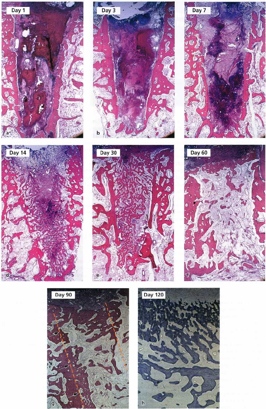
Fig. 38-2. Overall pattern of bone formation in an extraction socket. For details see text.
ALVEOLAR BONE FORMATION •
869
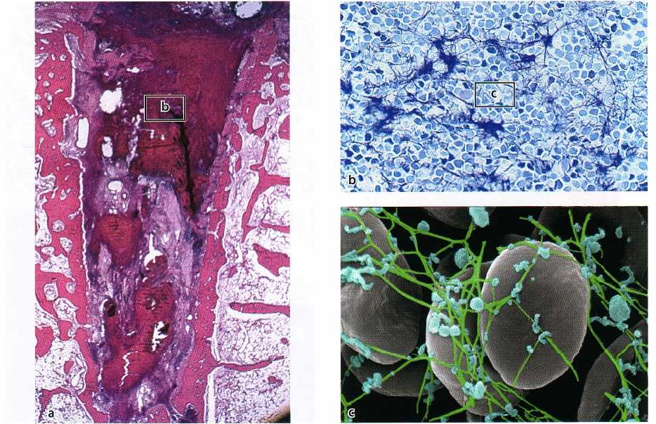
Fig. 38-3. Histologic section representing 1 day of healing (a). The socket is occupied with a blood clot that
contains
large numbers of erythrocytes (b) entrapped in a fibrin network, as well as platelets (blue cells in c).
The healing of a wound includes four phases:
1.
blood clotting
2.
wound cleansing
3.
tissue formation
4.
tissue modeling and remodeling.
These phases occur in an orderly sequence but, in a
given site, may overlap in such a way that in some
areas of the wound, tissue formation may be in pro-
gress while in other areas tissue modeling is the domi
-
nating event.
Model of bone tissue formation
The closure of an extraction socket may serve as a
model to describe tissue events that lead to bone
formation in a defect in the alveolar process. The
healing of such an extraction socket is described in Fig.
38-2. The mandibular premolars in a group of dogs
were extracted, and the healing of the extraction sites
was monitored in biopsies obtained at various time
intervals following tooth removal.
Overall pattern
of bone formation
The empty socket was first filled with blood and a
coagulum (clot) was formed (Fig. 38-2a). Inflamma-
tory cells migrated into the coagulum and the process
of wound cleansing was initiated (Fig. 38-2b). Vascu-
lar tissue and mesenchymal cells entered into the
coagulum and a granulation tissue was produced
(
Fig. 38-2c). This granulation tissue was gradually
replaced by a provisional connective tissue (Fig. 38-
2d) and the formation of new bone (woven bone)
started (Fig. 38-2d,e). The socket (identified by dotted
lines) was gradually filled with this woven bone (Fig.
38-2e) that later on was modeled and remodeled into
lamellar bone and marrow (Figs. 38-2f,g,h). Note the
dotted lines and the arrows which indicate the border
between the old bone and the newly formed bone
Important events in bone formation
Blood clotting:
Immediately after tooth extraction,
blood from the severed blood vessels will fill the
cavity. Proteins derived from vessels and damaged
cells initiate a series of events that lead to the forma-
tion of a fibrin network.
Platelets
form aggregates
(
platelet thrombi) and interact with the fibrin network
to
produce
a
blood clot
(a coagulum) that effectively
plugs
the severed vessels and stops the bleeding (Fig.
38-3).
The blood clot acts as a physical matrix that
directs
cellular movements and contains substances
that are
of importance for the continuation of the
healing
process. Thus, the clot contains substances
that (1)
influence mesenchymal cells (i.e.
growth fac
tors),
and (
2) affect inflammatory cells. These sub-
stances will
induce and amplify the migration of various types of
cells, as well as their proliferation, differ
entiation and
synthetic activity within the coagulum.
Although the blood clot is crucial in the initial
phase of wound healing, its removal is mandatory to
870 • CHAPTER 38
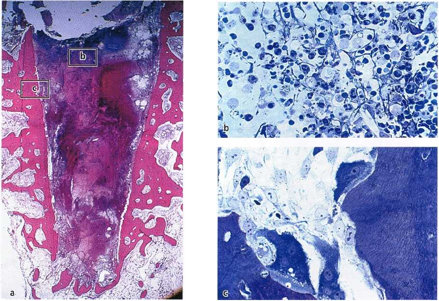
Fig. 38-4. Histologic section representing 3 days of healing (a). Note the presence of neutrophils and macrophages
that engaged in wound cleansing and the breakdown of the blood clot (b). Osteoclastic activity on the surface of
the walls of the old bone that lined the socket (c).
enable the formation of new tissue. Thus, within a few
days after the tooth extraction, the blood clot will start
to break down, i.e. "fibrinolysis" starts (Fig. 38-4).
Wound cleansing:
Neutrophils and macrophages mi-
grate into the wound, engulf bacteria and damaged
tissue (Fig. 38-4) and clean the site before tissue for-
mation starts. The neutrophils enter the wound early
while macrophages come into the scene somewhat
later. The macrophages are not only involved in the
cleaning of the wound but they also release several
growth factors and cytokines that further promote the
migration, proliferation and differentiation of me-
senchymal cells. Once the debris has been removed
and the wound has become "sterilized", the neutro-
phils undergo a programmed cell death (i.e.
apoptosis)
and are removed from the site through the action of
macrophages. The macrophages subsequently with-
draw from the wound.
In the extraction socket, a portion of the trauma-
tized bone facing the wound will undergo necrosis
and
will be removed by osteoclastic activity. Thus,
osteoclasts also may participate in the wound cleans-
ing phase of the bone healing.
Tissue formation:
mesenchymal, fibroblast-like cells
which migrate into the wound from, for example, the
bone marrow, start to proliferate and deposit matrix
components in an extracellular location (Fig. 38-5). In
this manner a new tissue, i.e.
granulation tissue,
will
gradually replace the blood clot. From a didactic point
of view the granulation tissue may be divided into two
portions: (1) early granulation tissue, and (2) late
granulation tissue. A large number of macrophages, a
few mesenchymal cells, small amounts of collagen
fibers and sprouts of vessels make up the early granu
-
lation tissue. The late granulation tissue contains few
macrophages, but a large number of fibroblast-like
cells and newly formed blood vessels present in a
connective matrix. The fibroblast-like cells continue
(
1) to release growth factors, (2) to proliferate, and (3)
to deposit a new extracellular matrix that guides the
ingrowth of new cells and the further differentiation
of
the tissue. The newly formed vessels provide the
oxygen and nutrients that are needed for the increas-
ing number of cells in the new tissue. The intense
synthesis of matrix components exhibited by these
mesenchymal cells is called
fibroplasia
while the for-
mation of new vessels is called
angiogenesis.
Through
the combined fibroplasia and angiogenesis
a provi-
sional connective tissue is
established (Fig. 38-6).
The transition of the provisional connective tissue
into bone tissue occurs along the vascular structures.
Thus, osteoprogenitor cells (e.g. pericytes) migrate
and
gather in the vicinity of the vessel. They differen
tiate
into osteoblasts that produce a matrix of collagen
fibers which takes on a woven pattern. The osteoid is
hereby formed and the process of mineralization is
initiated in its central portions. The osteoblasts con-
tinue to lay down osteoid and occasionally cells are

ALVEOLAR BONE FORMATION • 871
Fig. 38-5. Histologic section representing 7 days of healing (a). Note in the upper portion in the socket a richly vas-
cularized early granulation tissue with large numbers of inflammatory cells can be seen (b), while in more apical ar-
eas a tissue including large numbers of fibroblast-like cells is present, i.e. late granulation tissue (c).
Fig. 38-6. Histologic section representing 14 days of healing (a). In the marginal portion in the wound a provisional
connective tissue rich in fibroblast-like cells is formed (b). The formation of woven bone has at this time interval al-
ready begun in more apical regions of the bone defect (c).
872 • CHAPTER 38
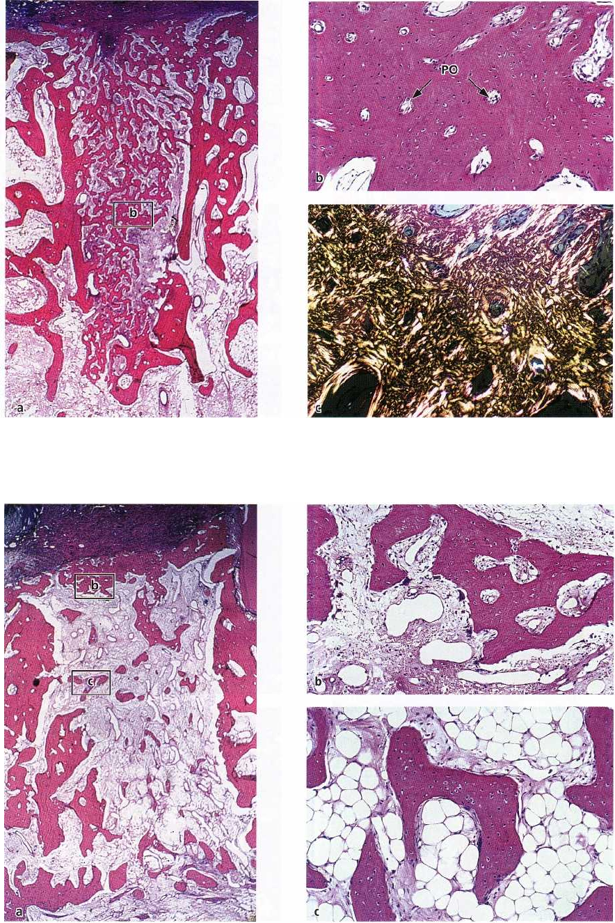
Fig. 38-7. Histologic section representing 30 days of healing (a). The socket is filled with woven bone. This woven
bone contains a large number of cells and primary osteons (PO;b). The woven pattern of the collagen fibers of the
woven bone is illustrated in (c) (polarized light).
Fig. 38-8. Histologic section representing 60 days of healing (a). A large portion of the woven bone has been re-
placed with bone marrow through osteoclastic activity and subsequent bone marrow formation, i.e. modeling (b).
Note in (c) the large number of adipocytes that reside in a tissue that still contains portions of woven bone.
ALVEOLAR BONE FORMATION • 873
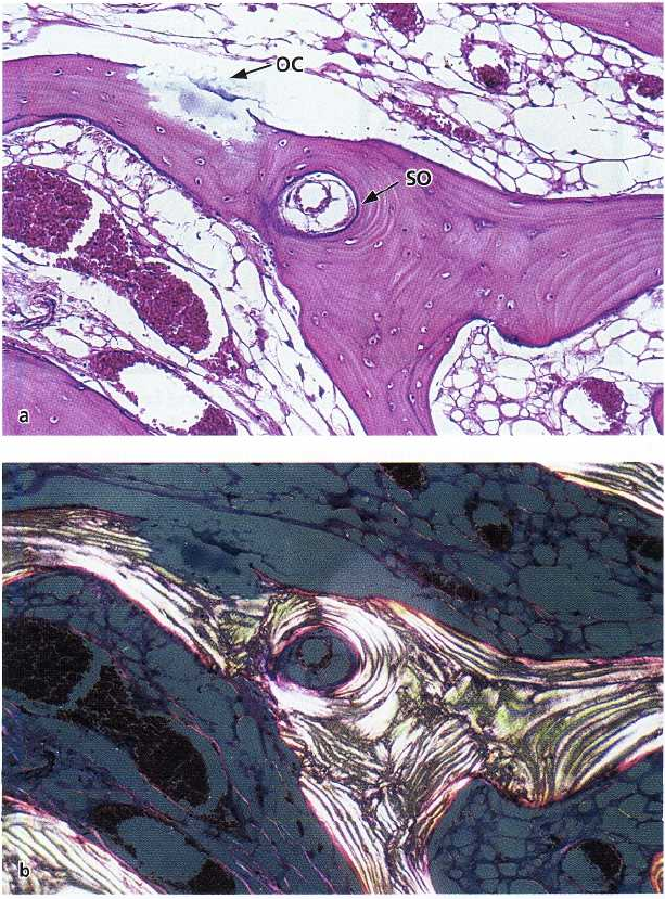
Fig. 38-9. Histologic section repre
-
senting a bone tissue after 60 days
of healing. Note in (a) the pres-
ence of osteoclasts (OC) on the
surface of a secondary osteon (SO)
within the bone trabeculae. (b)
The concentric pattern of the colla
gen fibers of the lamellar bone
within the secondary osteon (po-
larized light).
trapped in the matrix and become osteocytes. The
newly formed bone is called
woven
bone
(Fig. 38-7).
The woven bone is the first type of bone to be
formed and is characterized by (1) its rapid deposition
along the route of vessels, (2) the poorly organized
collagen matrix, (3) the large number of osteoblasts
that are trapped in its mineralized matrix, and (4) its
low load-bearing capacity. The woven bone forms as
finger-like projections along the newly formed ves-
sels. Trabeculae of woven bone are shaped and encir-
cle the vessel. The trabeculae become thicker through
the deposition of further woven bone, cells (osteocyts)
are entrapped and the first set of osteons, the
primary
osteons
are organized. The woven bone is occasionally
reinforced by the deposition of so called
parallel-fibered
bone
that has its collagen fibers organized not in a
woven but in a concentric pattern.
Tissue modeling and remodeling:
The initial bone forma-
tion is a fast process. Within a few weeks, the entire
extraction socket will be occupied with woven bone
or
as this tissue is also called,
primary bone spongiosa.
The
woven bone offers (1) a stable scaffold, (2) a solid
surface, (3) a source of osteoprogenitor cells, and (4)
ample blood supply for cell function and matrix min-
eralization.
The woven bone with its primary osteons is gradu-
ally replaced by lamellar bone and bone marrow (Figs.
38-8, 38-9) through the processes of modeling and
remodeling as described earlier (Fig. 38-10). In the
remodeling process the primary osteons are replaced
with secondary osteons. The woven bone is first
through osteoclastic activity resorbed to a certain
level. This level of the resorption front will establish
the so-called reversal line, which is also the starting
point for the new bone formation building up a sec-
ondary osteon. Although the modeling and remodel-
ing may start early it will take several months until all
woven bone in the extraction socket is replaced by
bone marrow and lamellar bone (Fig. 38-11).
In summary:
Following tooth extraction, the first 24
hours are characterized by the formation of a
blood clot
and the starting of
hemolysis
(Fig. 38-12a). Within 2-3
days the blood clot is contracting and is replaced by
the
formation of a granulation tissue
with the
blood vessels
and collagen fibers (Fig. 38-12b). After 3 days, an
increased density of fibroblasts is visible in the clot
and the
proliferation of epithelium
from the wound mar-
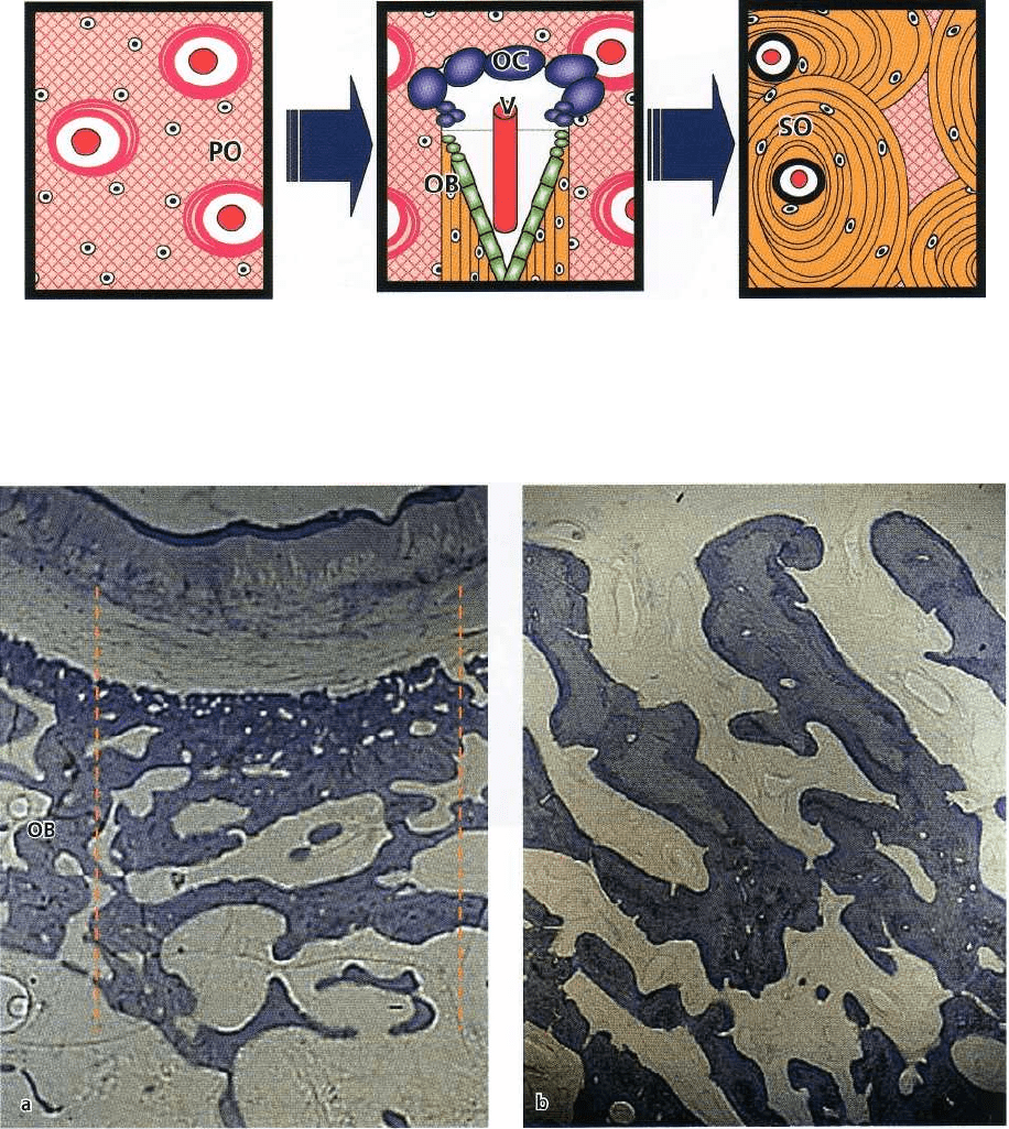
Fig. 38-10. Schematic drawing describing the transition between woven bone and lamellar bone, i.e. remodeling.
Woven bone with primary osteons (PO) is transformed into lamellar bone in a process that involves the presence of
BMUs. The BMU contains osteoclasts (OC), as well as vascular structures (V) and osteoblasts (OB). Thus, the
osteoblasts in the BMU produce bone tissue that has a concentric orientation around the vessel, and secondary
osteons (SO) within lamellar bone are hereby formed.
Fig. 38-11. After 6 months of healing the extraction site (within the area delineated by the vertical, dotted lines (a))
is
more or less fully healed. The site contains lamellar bone and marrow (b) and some remaining woven bone.
Through a process of modeling and remodeling the newly formed bone is now continuous with the "old bone"
(
OB (a)) of the neighboring areas.
gins is apparent. Remodeling of the sockets begins
with
the
presence of osteoclasts
inducing bone resorp
tion (
Fig. 38-12c). One week after extraction, the socket
is
characterized by granulation tissue consisting of a
vascular network,
young
connective tissue, osteoid forma
tion in the apical portion and epithelial coverage over
the
wound (Fig. 38-12d). One month following extrac
tion,
the socket is characterized by a
dense connective
tissue
overlying the residual sockets, which are now
filled with
granulation tissue.
A trabecular pattern of
bone starts emerging. Wound
coverage by epithelium is
complete (Fig. 38-12e). Two months following extrac-
tion, bone formation in the socket is complete. The
bony
height of the original sockets has not yet been
reached
and the trabecular pattern is still undergoing
remodeling (Fig. 38-12f).
874 • CHAPTER 38
Woven bone
BMU
Lamellar bone

ALVEOLAR BONE FORMATION • 875
Tooth extraction
48-72 h after extraction
96 h after extraction
Hemorrhagia,
Bleeding,
Blood clot
Blood clot,
Beginning of
granulation tissue formation
Residual blood clot,
Granulation tissue,
Epithelial proliferation
7 days after extraction
Young connective tissue,
Primary osteoid formation,
Epithelial proliferation
21 days after extraction
Connective tissue,
Osteoid start of mineralization,
Reepithelialization
6 weeks after extraction
Connective tissue,
Woven bone, trabeculae,
Reepithelialization
Fig. 38-12. Healing of extraction sockets (Amler 1969). (a) Bleeding and formation of a blood clot immediately after
tooth extraction. Blood vessels are closed by thrombi and a fibrin network is formed. (b) Already during the first 48
hours, neutrophil granulocytes, monocytes and fibroblasts begin to migrate along the fibrin network. (c) The blood
clot is slowly replaced by granulation tissue. (d) Granulation tissue forms predominantly in the apical third of the
alveolus. Increased density of fibroblasts. After 4 days, contraction of the clot and beginning proliferation of the
oral
epithelium. Osteoclasts are visible at the margins of the alveolus. Osteoblasts and osteoids seem to appear in
the
bottom of the alveolus. (e) Reorganization of the granulation tissue through formation of osteoid trabeculae.
Epithelial proliferation from the wound margins on top of the young connective tissue. Again, the formation of
osteoid trabeculae is evident from the wall of the alveolus in a coronal direction. After 3 weeks some of the trabecu-
lae start to mineralize. (f) Radiographically, bone formation may be visible. The soft tissue wound is closed and epi-
thelialized after 6 weeks. However, bone fill in the alveolus takes up to 4 months and does not seem to reach the
level of the neighboring teeth.
The placement of bone-grafting materials to favor
876 • CHAPTER 38
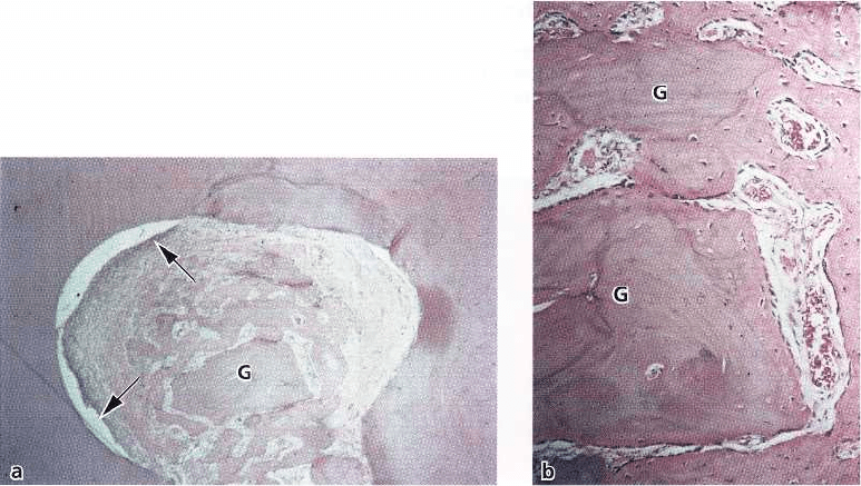
Fig. 38-13. Microphotograph (a) demonstrating bifurcation defect 3 weeks after grafting with autogenous cancel
lous jaw bone (G). New bone has invaded the defect, and the bone grafts have exerted an osteoconductive
function.
Epithelium (arrows) has migrated into one side of the defect. (b) Higher magnification of (a) showing
that new
bone has formed around the bone grafts (G), which have lost their vitality, indicated by the empty
osteocyte lacu
nae.
Bone grafting
Although bone tissue exhibits a large regeneration
potential and may restore its original structure and
function completely, bony defects may often fail to
heal with bone tissue. In order to facilitate and/or
promote healing, bone grafting materials have been
placed into bony defects. It is generally accepted that
the biologic mechanisms forming the basis for bone
grafting include three basic processes:
osteogenesis,
osteoconduction
and
osteoinduction.
Osteogenesis
occurs when viable osteoblasts and pre-
cursor osteoblasts are transplanted with the grafting
material into the defects, where they may establish
centers of bone formation. Autogenous iliac bone and
marrow grafts are examples of transplants with osteo
-
genic properties (see Chapter 33).
Osteoconduction
occurs when non-vital implant mate-
rial serves as a scaffold for the ingrowth of precursor
osteoblasts into the defect. This process is usually
followed by a gradual resorption of the implant ma-
terial. Autogenous cortical bone or banked bone allo-
grafts may be examples of grafting materials with
osteoconductive properties (Fig. 38-13). Such grafting
materials, as well as bone-derived or synthetic bone
substitutes, have similar osteoconductive properties.
However, degradation and substitution by viable bone
is often poor. If the implanted material is not
resorbable, which is the case for most porous hydroxy
-
lapatite implants, the incorporation is restricted to
bone apposition to the material surface, but no substi
-
tution occurs during the remodeling phase.
Osteoinduction
involves new bone formation by the
differentiation of local uncommitted connective tissue
cells into bone-forming cells under the influence of
one or more inducing agents.
Demineralized bone ma-
trix
(DMB) or
bone morphogenetic proteins
(BMP) are
examples of such grafting materials (Bowers et al.
1989a,b, Sigurdsson et al. 1994).
It often occurs that all three basic bone-forming
mechanisms are involved in bone regeneration. In
fact, osteogenesis without osteoconduction and
osteoinduction is unlikely to occur, since almost none
of the transmitted cells of autogenous cancellous bone
grafts survive the transplantation. Thus, the grafting
material predominantly functions as a scaffold for
invading cells of the host. In addition, the osteoblasts
and osteocytes of the surrounding bone lack the abil-
ity to migrate and divide which, in turn, means that
the transplant is invaded by uncommitted mesenchy-
mal cells that later differentiate into osteoblasts.
On that basis it is appropriate to define three basic
conditions as prerequisites for bone regeneration:
1.
the
supply of bone forming cells
or cells with the
capacity to differentiate into bone forming cells
2.
the presence of
osteoinductive stimuli
to initiate the
differentiation of mesenchymal cells into osteo-
blasts
3.
the presence of an
osteoconductive environment
form
ing a scaffold upon which invading tissue can pro-
liferate and in which the stimulated osteoprogeni-
tor cells can differentiate into osteoblasts and form
bone.
