Jan Lindhe. Clinical Periodontology
Подождите немного. Документ загружается.

MUCOGINGIVAL THERAPY — PERIODONTAL PLASTIC SURGERY • 605
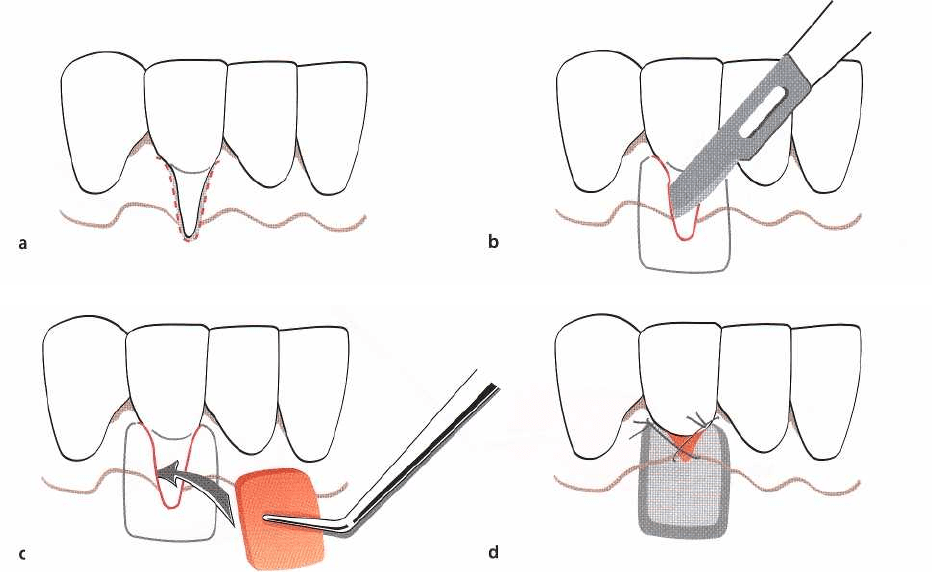
a
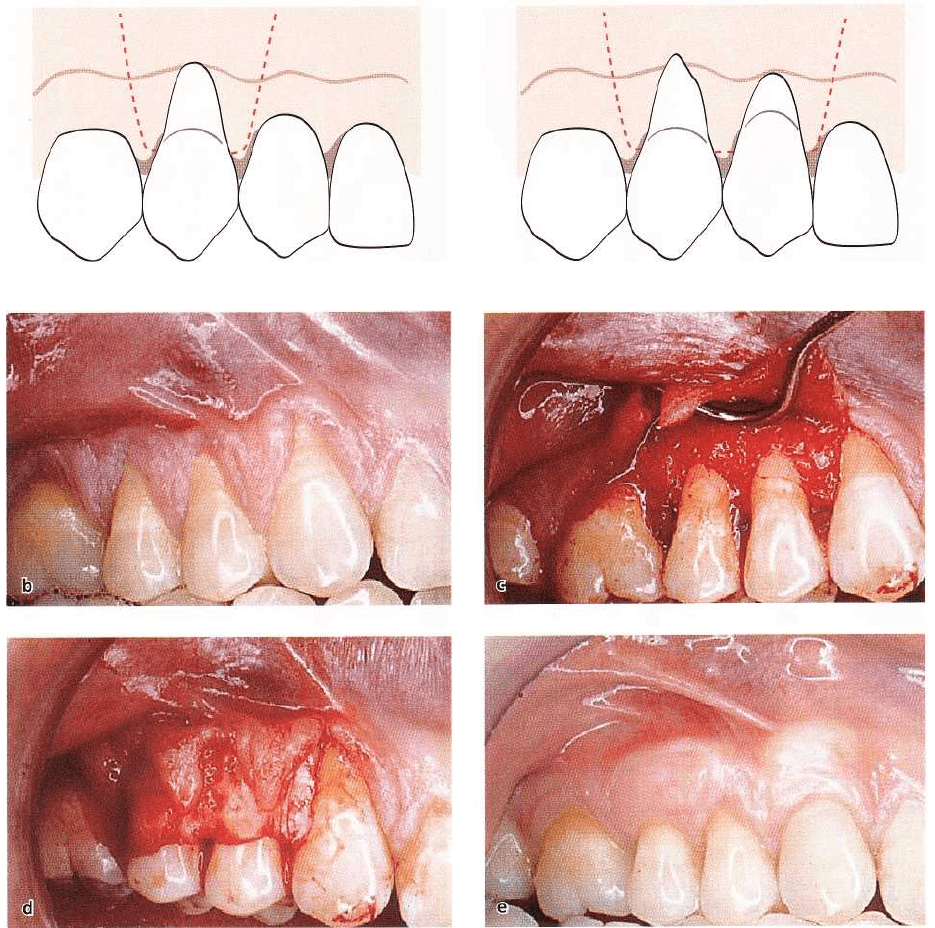
Fig. 27-41. (a-d)
Free connective tissue graft combined with a coronally advanced flap procedure
(see text for explanation).
(
e) The 1-year post-treatment result.
Free soft tissue graft procedures
A free soft tissue graft of masticatory mucosa is usu-
ally selected when there is no acceptable donor tissue
present in the area adjacent to the recession defect or
when a thicker marginal tissue is desirable. The pro-
cedure can be used for the treatment of a single tooth
as well as for groups of teeth. The graft used may
either be (1) an epithelialized graft or (2) a subepi-
thelial connective tissue graft of palatal masticatory
mucosa.
Epithelialized soft tissue graft
The epithelialized free soft tissue graft procedure can
be performed either as a two-step surgical technique,
where an epithelialized free soft tissue graft is placed
apical to the recession and following healing is
coronally positioned over the denuded root (Fig. 27-
39) (Bernimoulin et al. 1975, Guinard & Caffesse 1978),
or as a one-step technique, in which the graft is placed
directly over the root surface (Fig. 27-40) (Sullivan &
Atkins 1968a,b, Miller 1982). The latter technique has
been the one most commonly used.
Technique
The principles of utilizing free mucosal grafts were
outlined by Sullivan & Atkins (1968a,b) and later
modified by Miller (1982).
•
Before any incisions the exposed root surface is
carefully scaled and root planed (Fig. 27-40a). The
convexity of the root may be reduced to minimize
the mesiodistal avascular recipient bed.
•
As in the treatment with pedicle grafts, the prepa-
ration of
the recipient bed is
crucial for the success of
free graft procedure. A 3-4-mm-wide recipient con-
nective tissue bed apical as well as lateral of the
606 • CHAPTER
27
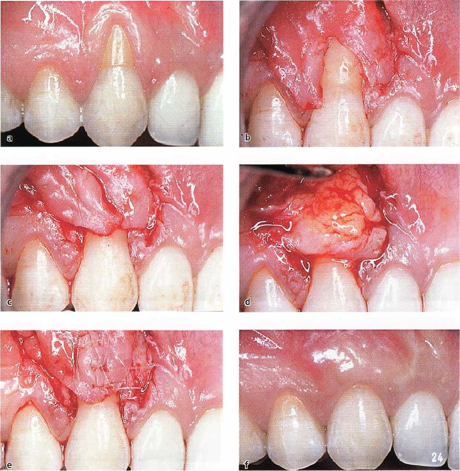
Fig. 27-42. (a-e)
Free connective tissue graft combined with a double papilla flap procedure.
(f) The 1-year post-
treatment
result.
recession defect should be prepared (Fig. 27-40b).
The area is demarcated by first placing a horizontal
incision, at the level of the cemento-enamel junc-
tion, in the interdental tissue on each side of the
tooth to be treated. Subsequently, two vertical inci-
sions, extending from the incision line placed in the
interdental tissue to a level approximately 4-5 mm
apical of the recession, are placed. A horizontal
incision is then made, connecting the two vertical
incisions at their apical termination. Starting from
an intracrevicular incision, a split incision is made
to sharply dissect the epithelium and the outer por
-
tion of the connective tissue within the demarcated
area.
•
To ensure that a graft of sufficient size and proper
contour is removed from the donor area, a foil tem-
plate of the recipient site is prepared. This template
is transferred to the donor site, the palatal mucosa
in the region of the premolars, and the required size
of the graft is outlined by a shallow incision. A graft
with a thickness of 2-3 mm is then dissected from
the donor area (Fig. 27-20c-d). It is advocated to
place sutures in the graft before it is cut completely
free from the donor area since this may facilitate its
transfer to the recipient site. Following the removal
of the graft, pressure is applied to the wound area
for control of bleeding.
•
The graft is immediately placed on the prepared
recipient bed. In order to immobilize the graft at the
recipient site the sutures must be anchored in the
periosteum or in the adjacent attached gingiva.
Adequate numbers of sutures are placed to secure
a
close adaptation of the graft to the underlying
connective tissue bed and root surface (Fig. 27-40c).
MUCOGINGIVAL THERAPY — PERIODONTAL PLASTIC SURGERY •
607
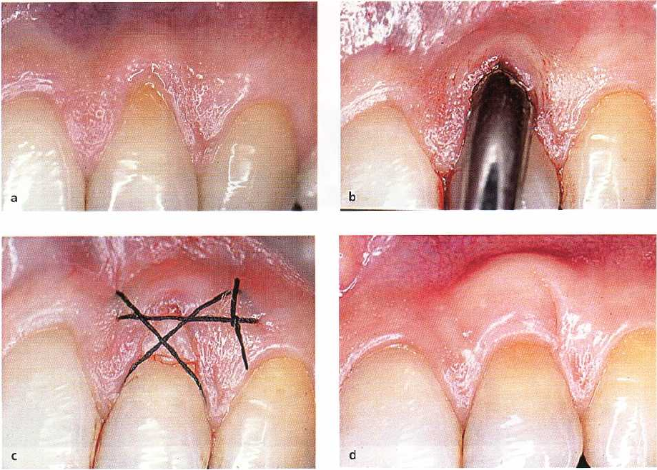
Fig. 27-43. (a-c)
Free connective tissue graft procedure — the "envelope technique
"
(see text for explanation) (Courtesy of
Dr M. Cattabriga). (d) The 1-year post-treatment result (Courtesy of Dr P. Cortellini).
Before the placement of a periodontal dressing,
pressure is exerted against the graft for some min-
utes in order to eliminate blood between the graft
and the recipient bed. The wound in the donor area
in the palate is, following the control of the bleeding,
covered by a periodontal dressing. To maintain the
dressing in place during the healing phase, the use
of an acrylic plate may often be required.
• The sutures and periodontal dressing are usually
maintained for 2 weeks. The appearance of a grafted
area after 3-month healing is shown in Fig. 27-40d.
A gingivoplasty may sometimes be indicated to
achieve a satisfactory esthetic appearance of the
grafted area (Fig. 27-40e-f).
Connective tissue graft
The technique utilizing a subepithelial soft tissue
graft, i.e. the connective tissue, involves the placement
of the graft directly over the exposed root and the
mobilization of a mucosal flap to be coronally (Fig.
27-
41) or laterally (Fig. 27-42) moved for coverage of
the
graft (Langer & Langer 1985, Nelson 1987, Harris
1992, Bruno 1994). An alternative technique is to place
the base of the connective tissue graft within an "en-
velope" prepared by an undermining partial thick-
ness incision from the soft tissue margin, i.e. part of
the graft will rest on the root surface coronal to the soft
tissue margin (Fig. 27-43) (Raetzke 1985, Allen 1994).
For the treatment of multiple adjacent recessions, a
multi-envelope recipient bed ("tunnel") may be pre-
pared (Zabalegui et al. 1999). The subepithelial con-
nective graft is harvested from the palate or the retro-
molar pad by the use of a "trap door" approach (Fig.
27-44d-f). Compared to the epithelialized graft, the
connective tissue graft is preferable due to less inva-
sive palatal wound and improved esthetic result.
6o8 •
CHAPTER
27
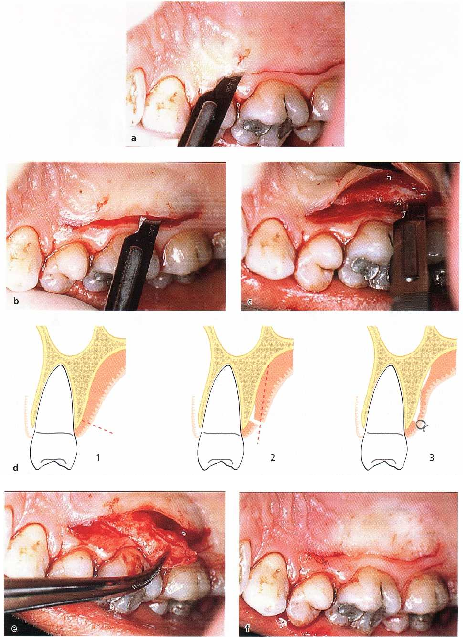
Fig. 27-44a-f.
"Trap-door" technique for harvest of a free connective tissue graft
(see text for explanation).
Technique
face of the interdental tissue on each side of the teeth
Connective tissue graft covered by a coronally advanced flap
to be treated (Fig. 27-41a). The incision should be
(Fig. 27-41)
placed just coronal to the intended level of root
• A horizontal incision is first made in the facial sur-
coverage. Care should be taken not to decrease the
MUCOGINGIVAL THERAPY — PERIODONTAL PLASTIC SURGERY • 609
Fig. 27-45a-c.
Free connective tissue graft procedure — the "envelope technique".
Schematic drawings illustrating the sur
-
gical technique (see text for explanation).
height of the papillae. Subsequently, starting from
the line of incision in the interdental area at the
mesial and distal termination of the surgical area,
two divergent, vertical incisions are placed and ex-
tended well beyond the mucogingival line.
•
A split thickness flap is then prepared by sharp
dissection and elevated to such an extent that it can
be coronally repositioned at the level of the ce-
mento-enamel junction without tension (Fig. 27-
41c).
•
A subepithelial connective tissue graft of mastica-
tory mucosa is harvested on the palatal aspect of the
maxillary premolars (or from the retromolar pad)
by the use of a "trap door" approach (Fig. 27-44).
Before incisions are placed, the available thickness
of the mucosa is estimated by the use of the tip of
the syringe. A horizontal incision, perpendicular to
the underlying bone surface, is made approxi-
mately 3 mm apical to the soft tissue margin (Fig.
27-44a). The mesiodistal extension of the incision is
determined by the graft size required. To facilitate
the removal of the graft, a vertical releasing incision
can be made at the mesial termination of the pri-
mary incision. An incision is then placed from the
line of the first incision and directed apically to
perform a split incision of the palatal mucosa (Fig.
27-44b-f). A small periosteal elevator or the scalpel
is used to release the connective tissue graft from
the bone. Sutures may be placed in the graft before
it is released completely free from the donor area to
facilitate its placement at the recipient site.
•
The graft is immediately placed in the recipient site
(Fig. 27-41d) and secured in position with inter-
rupted sutures or a sling suture. The mucosal flap
is then sutured to cover the connective tissue graft.
Interrupted sutures are placed in the papilla regions
as well as along the wound of the vertical incisions.
A surgical dressing may be applied for protection of
the area during the first week of healing.
The "envelope" technique (Fig. 27-45)
•
With the use of the "envelope" technique the recipi-
ent site is prepared by first eliminating the sulcular
epithelium by an internal beveled incision (Fig. 27-
45a). Secondly, an "envelope" is prepared apically
and laterally of the recession by split incisions (Fig.
27-45b). The depth of the preparation should be 3-5
mm in all directions. In apical direction, the prepa-
ration of the site should extend beyond the mu-
cogingival junction to facilitate the placement of the
connective tissue graft and to allow for coronal
advancement of the mucosal flap at time of sutur-
ing.
•
A foil template may be used for the harvest of an
appropriately sized connective tissue graft. The
graft, which is obtained by the "trap door" ap-
proach described above (Fig. 27-44), is inserted into
the prepared "envelope" and positioned to cover the
exposed root surface (Fig. 27-45c-d).
•
Sutures are placed to secure graft position (Fig. 27-
45d). A crossed sling suture may be placed to
coronally advance the mucosal flap. Pressure is ap-
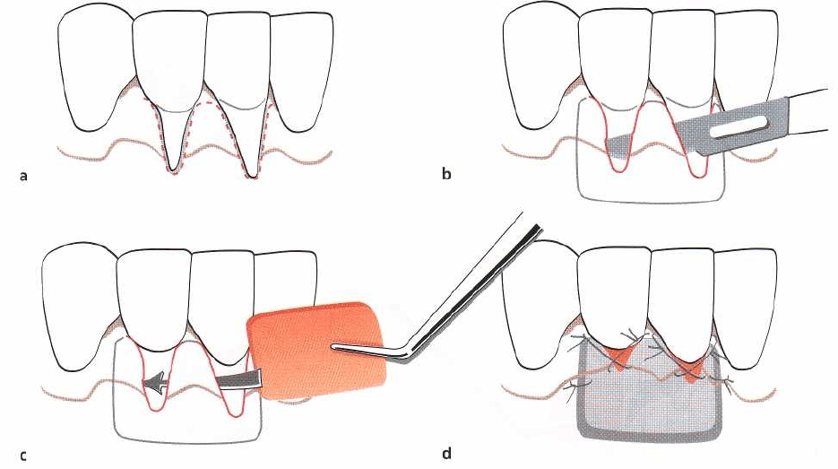
610 • CHAPTER
27
Fig. 27-46a-d.
Free connective tissue graft procedure — the
"
tunnel technique
"
.
Schematic drawings illustrating the surgi-
cal technique (see text for explanation).
plied for 5 min to closely adapt the graft to the root
surface and covering soft tissue. Application of a
periodontal dressing is usually not required.
Fig. 27-43 shows the treatment of a recession defect
with the "envelope" technique.
The "tunnel" technique (Fig. 27-46)
•
In case multiple adjacent recessions are to be
treated, "envelopes" are prepared for each tooth as
described above. However, the lateral split incisions
are extended so that the multi-envelopes are con-
nected mesially and distally to form a mucosal tun
-
nel. Care should be taken to avoid detachment of
the papillae.
•
The graft is gently positioned inside the tunnel and
its mesial and distal extremities are fixed with two
interrupted sutures. Sling sutures may be placed to
coronally advance the mucosal flap over the ex-
posed portions of the connective tissue graft. Pres-
sure is applied for 5 min to closely adapt the graft
to the root surface and covering soft tissue. Appli-
cation of a periodontal dressing is often not re-
quired.
Clinical outcome of root coverage procedures
Independent of the modality of surgical procedure
used to obtain soft tissue root coverage, shallow resid
-
ual probing depths, gain in clinical attachment and an
increase in gingival height are the common charac-
teristics of treatment outcome. Although the major
indication for performing root coverage procedures is
esthetic demands by the patient, almost no studies
have included assessments of esthetics as an endpoint
of success. Instead, the common outcome variable
used is the amount of root coverage achieved, ex-
pressed as a percentage of the initial depth of the
recession. In some studies also the proportion of
treated sites showing complete root coverage is re-
ported.
Root coverage
An overall comparison of the treatment outcome of
root coverage procedures is hampered by the fact that
comparatively few studies have presented well-docu-
mented clinical results. A summary of published stud
-
ies providing data for calculation of the average
amount of the initial recession defect that has been
successfully covered following treatment (Table 27-1)
shows that an average of 63-86% root coverage may
be expected, depending on the treatment procedure
used. However, the variation (range) in treatment out
-
come for the various procedures, both within and
between studies, is large. This indicates that the pro-
cedures are operator sensitive and/or that factors in-
fluencing the treatment outcome have not been ade-
quately considered. Complete coverage of the reces-
sion defect is the ultimate goal of the therapy. Table
27-2 summarizes available data on the predictability
of complete root coverage with the use of the various

MUCOGINGIVAL THERAPY — PERIODONTAL PLASTIC SURGERY • 611
Table 27-1. Summary of the data available in the literature on the amount of root coverage obtainable with
various procedures
Root coverage procedure
Root coverage
No. of studies
No.
of patients/
teeth
Mean
%
of initial
recession
Range
Rotational flaps
10
222/235
68% 41-74
Coronally advanced flap
12
216/416
80% 55-99
Guided tissue regeneration
34
576/682
75% 48-94
Free connective tissue graft 30
589/796
86% 53-98
Epithelialized free soft tissue graft
16
335/491
63% 11-87
Table 27-2. Summary of the data available in the literature on the predictability of complete root coverage fol
-
lowing the use of various procedures
Root coverage procedure
Complete root coverage
No. of studies
No.
of patients/
teeth
Mean
%
of teeth
Range
Rotational flaps
1
30/30 43%
-
Coronally advanced flap
10
188/388
50% 9-95
Guided tissue regeneration
23
344/440
36% 0-75
Free connective tissue graft 25
532/715
61% 0-93
Epithelialized free soft tissue graft
10
253/380
28% 0-90
procedures. The average percentage of complete root
coverage following pedicle or free graft procedures
varies between 28% and 61%; the lowest figure for the
epithelialized soft tissue graft and the highest for the
connective tissue graft procedure. The lower predict-
ability of complete root coverage with the GTR proce-
dure, compared to the coronally advanced flap proce-
dure, has been associated with the problem of mem-
brane exposure during healing (Trombelli et al. 1995),
but whether a bioabsorbable or a non-biodegradable
barrier membrane is used does not seem to affect the
treatment outcome (Roccuzzo et al. 1996).
Short-term clinical trials comparing the treatment
outcome of the two modalities of free soft tissue grafts
(
Sbordone et al. 1988, Daniel & Cheru 1990, Jahnke et
al. 1993) have shown that the connective tissue graft
results in superior root coverage compared to the
epithelialized free soft tissue graft. The color match of
the connective tissue grafted area to the adjacent
gingiva is esthetically also more favorable with the
connective tissue graft than that of an epithelialized
free graft.
Factors influencing the degree of root coverage
Patient-related factors
As with other surgical periodontal treatment proce-
dures, poor oral hygiene following the procedure will
negatively influence the success of root coverage pro-
cedures (Caffesse et al. 1987). Also, the predominant
causative factor in the development of gingival reces-
sion is toothbrushing trauma, and hence this factor
has to be corrected to secure an optimal outcome of
any root coverage procedure. Whether smoking may
negatively influence the outcome of root coverage
procedures is still a controversial issue. Some studies
have reported less favorable outcome in terms of root
coverage following free graft and GTR procedures in
smokers (Miller 1987, Trombelli & Scabbia 1997,
Muller et al. 1998, Zucchelli et al. 2000), while other
studies have shown no differences between smokers
and non-smokers (Tolmie et al. 1991, Harris 1994).
Site-related factors
Among site specific factors, the level of interdental
periodontal support may be of greatest significance
for the outcome of root coverage procedures. From a
biological point of view complete root coverage is
achievable in Class I and II type recession defects (Fig.
612 •
CHAPTER
27
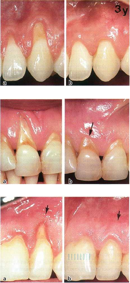
Fig. 27-47a-b. Buccal recession defects but no loss of
periodontal support at proximal surfaces (a). Complete
root coverage can be achieved. (b) 3-year follow-up.
Fig. 27-48. A deep buccal recession at tooth 11 (a). The
tooth has loss of support at proximal sites (Miller Class
III) and complete root coverage is not achievable. Also
neighboring teeth show recessions at all tooth surfaces.
(b) 2-year healing result following attempted root cov-
erage at the facial aspect of tooth 11. The coronal posi
-
tion of the soft tissue margin is defined by the exten-
sion of proximal loss of periodontal support.
Fig. 27-49. Increased dimension of keratinized tissue 1
year following root coverage with a coronally ad-
vanced flap procedure. Before (a) and 1-year postopera
-
tively (b). Arrows indicate the position of the mu-
cogingival line.
27-47), while when loss of connective tissue attach-
ment also involves proximal tooth sites (Class III-IV
recession defects), only partial facial root coverage is
obtainable (Miller 1985b) (Fig. 27-48).
An additional factor shown to influence the degree
of attainable root coverage is the dimensions of the
recession defect. Less favorable treatment outcome
has
been reported at sites with wide (> 3 mm) and
deep (>_
5 mm) recessions (Holbrook & Ochsenbein
1983, Pini
Prato et al. 1992, Trombelli et al. 1995).
Wennstrom &
Zucchelli (1996) reported in a study comparing the
treatment effect of coronally advanced
flap and free
connective tissue graft procedures that
complete root
coverage was observed in only 50% of
the defects with
an initial depth of 5 mm compared
to 96% in shallower
defects. In a controlled clinical
trial Pini Prato et al. (
1992) treated 50 teeth with a
coronally advanced flap
procedure, either with or
without the use of a non-degradable membrane bar-
rier. The mean percentage root coverage at the 18-
month follow-up examination was 73% with and 71%
without the use of a barrier, and a subsequent 4-year
follow-up report revealed long-term stability of
achieved root coverage for both procedures (Pini
Prato
et al. 1996). The authors suggested that a more
favorable result with respect to root coverage might
be
obtained with the GTR procedure in sites with deep
(>_
5 mm) recession defects as compared to the
coronally
advanced flap. At the 18-month examination the
coverage was 77% with and 66% without the
inclusion
of a membrane barrier in the treatment pro
cedure.
However, the data presented in Table 27-2,
showing
that the predictability of complete root cov
erage is
markedly reduced with the use of barrier
membranes,
limit the justification to utilize the GTR
procedure in
the treatment of recession defects.
MUCOGINGIVAL THERAPY — PERIODONTAL PLASTIC SURGERY • 613
The pretreatment gingival height apical to the re-
cession defect is not correlated to the amount of root
coverage obtained (Romanos et al. 1993, Harris 1994).
Technique-related factors
Several technique-related factors may influence the
treatment outcome of a pedicle graft procedure. A
positive association between recession reduction and
the thickness of the flap was shown by Baldi et al.
(
1999). Complete root coverage at sites with Miller
Class I-II recessions was obtained only when the flap
thickness was 0.8 mm. However, whether a full or
split thickness pedicle graft is used for root coverage
was not found to influence the treatment outcome
(
Espinel & Caffesse 1981).
Flap tension has been reported to be an important
factor for the outcome of the coronally advanced flap
procedure. The best clinical result is achieved if the
flap is passively adapted to the root surface (Allen &
Miller 1989, Pini Prato et al. 2000a). In the study by
Pini Prato et al. (2000a) the tension in coronally ad-
vanced flaps was measured to compare the amount of
recession reduction in sites with and without residual
flap tension. At the test sites, which had an average
residual tension of 6.5 g, the root coverage amounted
to 78% 3 months post-surgically and 18% of the treated
sites showed complete root coverage, whereas the
control sites without or with only minimal remaining
tension demonstrated a mean root coverage of 87%
and complete root coverage in 45% of the cases. Fur-
thermore, in the test group a statistically significant
negative association was shown between the magni-
tude of residual tension in the flap and the amount of
recession reduction.
Although the connective tissue areas lateral to the
recession defect may be considered important for the
retention of the advanced flap when positioned over
the root surface, the dimension of the interdental pa-
pilla is not a prognostic factor for the clinical outcome
of the root coverage procedure (Saletta et al. 2001).
With regard to free graft procedures, the thickness
of the graft is a factor influencing the success of treat
-
ment procedure (Borghetti & Gardella 1990). A thick-
ness of the free graft of about 2 mm is recommended.
Increased
gingival
height
An increased apicocoronal height of gingiva is found
following all procedures in which pedicle grafts of
adjacent gingiva or free grafts from the palate have
been placed over the recession defect. It is interesting
to note, however, that an increased gingival height is
also a common finding following a coronally ad-
vanced flap procedure only involving the existing
gingiva apical to the recession (Fig. 27-49). This find-
ing may be explained by several events taking place
during the healing and maturation of the marginal
tissue. Granulation tissue formation derived from the
periodontal ligament tissue will form a connective
tissue similar to the one of gingiva and with the po-
tential to induce keratinization of the covering epithe-
lium (Karring et al. 1971, Lundberg & Wennstrom
1988). A second factor to consider is the tendency of
the mucogingival line to regain its "genetically" de-
fined position following its coronal "dislocation" with
the coronally advanced flap procedure used to
achieve root coverage. Support for the concept that the
mucogingival line over time will regain its original
position is generated from a study by Ainamo et al.
(
1992). The authors performed an apically reposi-
tioned flap procedure in the mandibular anterior
tooth region, which resulted in a 3-mm apical dis-
placement of the mucogingival line. The re-examina-
tion after 18 years showed no differences in position
of the mucogingival line between sites treated with the
apically repositioned flap and contralateral control
sites treated with a procedure not interfering with the
mucogingival line, indicating that the mucogingival
line had regained its original position.
Soft tissue healing against the covered root
surface
Although successful treatment outcome of gingival
recessions by pedicle grafts or free grafts has been
reported in a number of publications (for review see
Wennstrom 1996), it is debated to which extent this
type of treatment results in new connective tissue
attachment or epithelial attachment. However, inde-
pendent of the quality of attachment formed, the root
coverage procedures evidently rarely result in the
formation of a deep periodontal pocket.
Healing of pedicle soft tissue grafts
In the areas surrounding the recession defect, i.e.
where the recipient bed consists of bone covered by
connective tissue, the pattern of healing is similar to
that observed following a traditional flap operation.
Cells and blood vessels from the recipient bed as well
as from the tissue graft invade the fibrin layer, which
gradually becomes replaced by connective tissue. As
early as 1 week later a fibrous reunion is established
between the graft and the underlying tissue.
Healing in the area where the pedicle graft is in
contact with the denuded root surface was studied by
Wilderman & Wentz (1965) in dogs. According to these
authors the healing process can be divided into four
different stages (Fig. 27-50).
The adaptation stage (from 0 to 4 days)
The laterally repositioned flap is separated from the
exposed root surface by a thin fibrin layer. The epithe
lium covering the transplanted tissue flap starts to
proliferate and reaches contact with the tooth surface
at the coronal edge of the flap after a few days.
The proliferation stage (from 4 to 21 days)
In the early phase of this stage the fibrin layer between
the root surface and the flap is invaded by connective
tissue proliferating from the subsurface of the flap. In
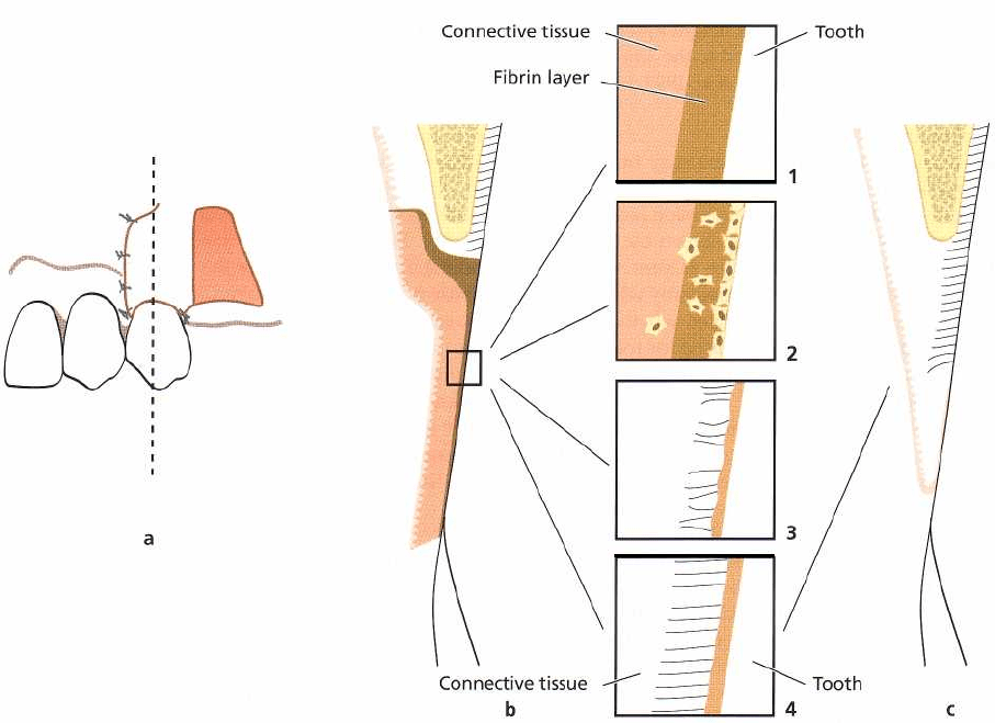
614 • CHAPTER
27
Fig. 27-50. Schematic drawing illustrating healing following treatment of a localized soft tissue recession with a
pedicle graft (a). (b) Cross-section through the area immediately after operation. The framed areas (1-4) illustrate the
four stages into which the healing process can be divided. (c) Area after healing. Approximately 50% of the suc-
cessfully covered defect may show new connective tissue attachment.
contrast to areas where healing occurs between two
connective tissue surfaces, growth of connective tissue
into the fibrin layer can only take place from one
surface. After 6-10 days a layer of fibroblasts is seen in
apposition to the root surface. These cells are believed
to differentiate into cementoblasts at a later stage of
healing. At the end of the proliferation stage, thin
collagen fibers are formed adjacent to the root surface,
but a fibrous union between the connective tissue and
the root has not been observed. From the coronal edge
of the wound, epithelium is proliferating apically
along the root surface. According to Wilderman &
Wentz (1965), the apical proliferation of epithelium
may stop within the coronal half of the defect, al-
though further downgrowth of epithelium was also
frequently observed.
The attachment stage (from 27 to 28 days)
During this stage of healing thin collagen fibers be-
come inserted in a layer of new cementum formed at
the root surface in the apical portion of the recession.
The maturation state
This last stage of healing is characterized by continu-
ous formation of collagen fibers. After 2-3 months,
bundles of collagen fibers are inserting into the cemen
-
tum layer on the curetted root surface in the apical
portion of the recession.
Results of experimental studies in monkeys and
dogs on the healing characteristics of the periodontal
wound have been interpreted to indicate that gingival
connective tissue lacks the ability to form a new con-
nective tissue attachment, but may induce root resorp
-
tion (see Chapter 28). This finding is of particular
interest when considering the rationale for the treat-
ment of recession defects by free or pedicle soft tissue
grafts. Since in these surgical procedures gingival con
-
nective tissue is placed in contact with a denuded root
surface, root resorption should be expected to occur.
The reason why it is not a common complication
following this type of treatment can be explained by
two possible events. Either cells from the periodontal
ligament form a fibrous attachment to the root surface
or epithelial cells proliferate apically, forming a root
protective barrier (long junctional epithelium) to-
wards the gingival connective tissue.
Histologic studies on whether it is the one or the
other type of attachment that results following treat-
ment of recessions with pedicle grafts indicate that
new connective tissue attachment with cementum for
-
mation may be formed in part of the defect. In the
study by Wilderman & Wentz (1965) a new connective
tissue attachment of around 2 mm and an epithelial