Hatano Y., Katsumura Y., Mozumder A. (Eds.) Charged Particle and Photon Interactions with Matter - Recent Advances, Applications, and Interfaces
Подождите немного. Документ загружается.

120 Charged Particle and Photon Interactions with Matter
decreases from 240 to 280 eV as the photon energy increases. Two maxima can be seen at about 290
and 305eV, accompanied by a minimum at about 295eV. From 310 to 400eV, the W-value remains
essentially constant.
An electron energy loss spectrum and a photoabsorption spectrum of methane show the four
peaks at 287, 288, 289.4, and 289.8eV (Brown et al., 1978; Wight and Brion, 1974). A strong reso-
nance is located at 288eV, and the three other peaks are weak in intensity. The transitions of an
electron from the inner C K-shell to Rydberg orbitals give rise to these peaks. The K-shell ioniza-
tion energy is 290.8eV (Pireaux et al., 1976). The oscillatory structure shown by the circles in
Figure6.11
is presumed to connect with the structure caused by transitions of the K-shell electron.
A
model, which is the same as that for ethylene, seems applicable for interpreting the structure
in the energy dependence of the W-value (Saito and Suzuki, 1986; Suzuki and Saito, 1987). Since
methane has no transition into the π* orbital, the oscillatory structure is simpler than that for
ethylene. Then, the energy region of methane classied by the photoabsorption process is 5, which
is smaller than that for ethylene. The W-value calculated is illustrated with a solid curve in Figure 6.11.
The calculation has reproduced the measured data essentially well, although it overestimates the
effect on PCI just above the C K-edge.
Propane has K-shell ionization energy of 290.6eV (Pireaux et al., 1976). Transitions of the 1s
electrons to unoccupied orbitals are supposed to take place slightly below the ionization threshold.
These types of excitation and ionization probably cause molecules to change into different types
of states of the molecular ion. The W-value is expected to be affected by these ion states near the
K-edge. The relative W-values derived from the measurements are shown with open triangles for
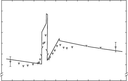
photon energies from 260 to 360eV in Figure 6.12 (Suzuki and Saito, 1985b). The solid curve indicates
a calculated prole based on the model mentioned below. The measured W-value slightly decreases
with increasing energy from 260 to 280 eV. The W-value shows a sharp rise at 286eV, goes to
a maximum at 291 eV, and then decreases to a minimum at 294eV. A linear dependence of the
W-value data on the photon energy is seen from 294 to 304 eV. The W-value is essentially constant
above
304
eV.
In order to clarify the origin of the variation in the W-value shown in Figure 6.12, Suzuki and
Saito (1985b) observed the photoabsorption spectrum of propane around the C K-edge. Although
the inner-shell excited states are not separated from one another owing to the low resolving power of
the monochromator used, the following items are possibly discernible. The absorption efciency is
260
1.00
1.04
1.08
C
3
H
8
Relative W-value
280 300
Photon energy (eV)
320 340 360
Figure 6.12 Photon W-value for C
3
H
8
as a function of soft x-ray energy. Open triangles indicate mea-
sured data. The solid curve denotes the calculated prole based on the model explained in Section 6.4.5.
(Reproduced
from Suzuki, I.H. and Saito, N., Bull. Chem. Soc. Jpn., 58, 3210, 1985b.)
New Directions in W-Value Studies 121
low below 285eV, becomes high at about 287eV, and reaches a maximum at around 291eV. Above
291eV, this efciency gradually decreases with increasing photon energy. Judging from the inner-
shell excitation spectra of methane and ethane (Eberhardt et al., 1976), the high efciency at 287eV
can probably be ascribed to a transition of C-atomic orbitals, 1s → 3s. In the cases of methane and
ethane, the transition (1s → 3s) is low in intensity. This type of transition is supposed to occur with
a higher probability in propane because of the poorer symmetry than those for methane and ethane.
This transition is followed by transitions to higher excited states (e.g., 1s → 3p) and to ionized states.
Bearing in mind the photoabsorption spectrum, a model similar to that for methane is proposed for
the interpretation of the energy dependence of the W-value (Suzuki and Saito, 1985b). The calculated
curve
has well reproduced the measured data and the model is essentially correct.
6.5 osCillatory variation in energy dependenCe oF photon
W-values
F
or
r
are
g
as
a
toms
and a
tomiC shell
eFF
eCts
6.5.1 introduction
Based on the progress in synchrotron radiation researches, many photoabsorption spectra have
been measured in a high resolution of photon energy, and electron emission spectra have also been
observed in a high resolution of electron energy (Svensson et al., 1988; Chen et al., 1989; Okada
et al., 2005; Kato et al., 2007). Further, several ions with various charges, produced by the photo-
ionization of atoms and molecules, have been observed by means of mass spectrometry (Hayaishi
etal., 1984; Saito and Suzuki, 1992, 1994, 1997; Tamenori et al., 2004; Suzuki et al., 2006). In some
of these studies, average charge states have been obtained for ions produced from rare gas atoms
irradiated with soft x-rays (Saito and Suzuki, 1992, 1994; Tamenori et al., 2004). Some measure-
ments were attempted for the determination of the absolute intensity of soft x-rays using a multi-
electrode ion chamber and other techniques as per the requirements in photon science and photon
engineering (Rabus et al., 1997; Saito and Suzuki, 1998, 1999). Photoionization cross sections of
rare gas atoms were obtained on an absolute scale in the soft x-ray region using a sophisticated ion
chamber technique (Suzuki and Saito, 2002, 2005). These developments have enabled us to deter-
mine absolute photon W-values of some gases in the sub-keV energy regions, even though it is very
difcult
for the proportional counter technique to provide accurate W-values on the absolute scale.
In
this section, photon W-values for rare gas atoms are presented, which were obtained using
monochromatic synchrotron radiation combined with the multielectrode ion chamber (Saito and
Suzuki, 2001a,b; Suzuki and Saito, 2001). In deriving these quantities from observed photoion cur-
rents, average charge states, which are called γ-values here, have been utilized. Branching ratios of
produced ion charges in individual photon energies are used in the calculation of the W-value on the
basis of a precise model, which takes atomic shell effects into consideration. First, a measurement
method and an analysis technique are given, and the result for Kr is discussed in detail in compari-
son with a calculated prole (Saito and Suzuki, 2001b). Then, results for Ar and Xe are presented
(Saito
and Suzuki, 2001a; Suzuki and Saito, 2001).
6.5.2 MeaSureMentS
Synchrotron radiation at the AIST (National Institute of Advanced Industrial Science and
Technology) was dispersed by a Grasshopper grating monochromator (Saito and Suzuki, 2001a,b;
Suzuki and Saito, 2001). The monochromatic photons entered the multielectrode ion chamber,
whose shape is cylindrical, 65mm in diameter and 1300mm in length. In order to reduce higher-
order radiation and scattered lights, the electron energy of the storage ring was often lowered from
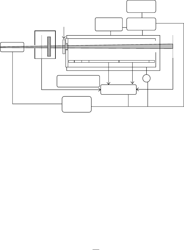
750MeV to 250–600MeV, and some thin lms were inserted into the soft x-ray beam. Figure 6.13 shows
the schematic diagram of the measurement system for W-values (Saito and Suzubi, 2001b). The
window of the ion chamber consists of a thin VYNS foil and a circular aperture of 2mm diameter.
122 Charged Particle and Photon Interactions with Matter
The chamber contains a set of six electrodes for detection of photoion currents and for suppression
of end distortion in electric elds. The sample gases of Kr, Ar, and Xe (research grade purity) were
supplied at about 10
−2
–10
3
Pa, the densities of which were monitored with a capacitance manometer
and controlled with an automatic valve system. A positive potential was applied to the outer cylin-
drical electrode. The photoion current at each inner electrode was transferred to a pico-ammeter and
then to a personal computer. The incident intensity of the soft x-rays was continuously monitored
with
the Au-plated Ni-mesh, illustrated in Figure 6.13.
6.5.3 analySiS
Let us consider that a total of N electrons are produced upon absorption of an incident soft x-ray of
energy E
p
in a sufciently high density gas (Saito and Suzubi, 2001b). Then, the photon W-value,
W
p
, is expressed by
W
E
N
p
p
= . (6.9)
The number of electrons, n, produced in the ion chamber with a certain gas density, p, is the sum
of contributions from two factors. The rst factor is the initial ionization through atomic multiple
photoionization effects, and the second is the secondary ionization effects originating from colli-
sions
of emitted electrons with ambient gases in the chamber. Thus,
n p p( ) ( ),= +γ δ (6.10)
where
γ
denotes the number of electrons ejected from the atom having absorbed a soft x-ray quantum
δ is the number of electrons secondarily produced
The
value of γ was determined previously for several rare gas atoms in the soft x-ray region using
a time-of-ight mass spectrometer technique (Saito and Suzuki, 1992; Suzuki and Saito, 1992).
Monochromator
Differential
pumping
system
Filter
Photon
monitor
Aperture
window
Capacitance
manometer
Soft x-ray beam
Ion chamber
Pico-ammeter
+V
Personal
computer
Turbo-molecular
pump
Gas supply
Pressure
controller
Photon
monitor
Figure 6.13 Schematic of an experimental system for measurements of the photon W-value using a multi-
electrode ion chamber. The personal computer controls the monochromator, the voltage supply (+V), and the
pressure
controller. (Reproduced from Saito, N. and Suzuki, I.H., Radiat. Res., 156, 317, 2001b.)
New Directions in W-Value Studies 123
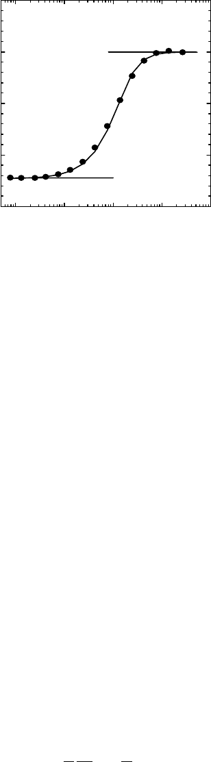
Asthe gas density decreases, the parameter value δ approaches zero and the n value becomes equal
to the γ value. On the other hand, the δ value approaches a constant value and the n value reaches a
plateau value of N, as the gas density increases. An example of results measured at a photon energy
of 400 eV is shown in Figure 6.14, where the number of electrons produced is represented as a func-
tion of the Kr density (Saito and Suzubi, 2001b). From the curve in Figure 6.14, constant values at
low- and high-density limits are easily estimated with a high precision. When the purity of the soft
x-ray beam was not very high, the plateau value did not show up clearly. This situation has been
overcome
by the improvement of the photon purity, as indicated above.
As
described previously on the absolute measurement of soft x-rays (Saito and Suzuki, 1998,
1999),
the photoion current, i, in the ion chamber under a certain gas density is given as
i enI l p L p= − − −
{ }
exp( ) exp( ) .σ σ1 (6.11)
Here, e, I, σ, L, and l denote the elementary charge, the photon absolute intensity, the photoabsorp-
tion cross section, the length of the electrode, and the length of the insensitive region at the front
end,
respectively. Then, the photoion current at a sufciently low gas density is
i e I l p L p= − − −
{ }
γ σ σexp( ) exp( ) .1 (6.12)
Similarly,
the photoion current at a sufciently high density is
i eNI l p L p= − − −
{ }
exp( ) exp( ) .σ σ1 (6.13)
The photoabsorption cross section has been obtained with ion currents from the two successive
electrodes,
i
1
and i
2
, under appropriate gas densities as
σ =
1
1
2
L
d
dp
i
i
ln . (6.14)
By using Equations 6.12 and 6.14, we have obtained the absolute intensity of the incident soft x-ray.
Then, the value of N, that is, the total number of electrons produced, has been derived from the
0.1 1 10
γ
100 1000
0
5
10
15
N
20
Number of electrons produced
Kr density (Pa)
Figure 6.14 Number of electrons produced in the ion chamber as a function of Kr gas density. Data are
plotted at a photon energy of 400.3eV. See Section 6.5.3 for N and γ. (Reproduced from Saito, N. and Suzuki,
I.H.,
Radiat. Res., 156, 317, 2001b.)
124 Charged Particle and Photon Interactions with Matter
obtained photon intensity using Equation 6.13. Finally, the photon W-value has been calculated from
the
derived N value, according to Equation 6.9.
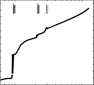
Figure
6.15 shows the value of γ for Kr in the region of 60–1100eV (Saito and Suzuki, 2001b).
These values have been derived from branching ratios into several charge states in multiple pho-
toionization (Suzuki and Saito, 1992). It is found that the γ-value shows a steep jump at the 3d
electron ionization threshold (93.8eV). This nding can be interpreted in terms of the formation of
doubly charged Kr ions through the 3d electron ionization. Even below the 3d threshold, the γ-value
is slightly higher than unity, showing that two valence electrons are appreciably ejected through
absorption of one photon. The value of γ increases gradually above the 3d threshold, and then
exhibits small upward steps at the 3p (214.4eV) and 3s (292.8eV) electron ionization thresholds.
Above the 3s threshold, the γ-value acquires an additional feature. This feature probably comes
from a shake-off process induced by the inner-shell ionization (Saito and Suzuki, 1994, 1997;
Suzuki et al.,
2006).
6.5.4 reSult of kr
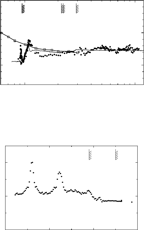
The photon W-value of Kr is shown on an absolute scale as a function of photon energy from 85 to
1000eV in Figure 6.16 (Saito and Suzuki, 2001b). The present data are denoted with solid circles.
The bars with hatching indicate the ionization thresholds of 3d, 3p, and 3s electrons. The open
square data points give the W-value for electrons, W
e
, which were reported earlier and are shown
in Figure 6.1 (Combecher, 1980). The solid curve represents the result calculated from the model
described below. The measured W
p
exhibit considerably lower values than those for electrons
below 91eV. The sharp peak structure appears just below the 3d threshold, which is shown in
Figure 6.17.
The value of W
p
steeply increases at the 3d ionization threshold and decreases slightly between
120 and 210eV. This W
p
shows a small oscillation near the 3p and 3s ionization thresholds, and
then essentially reaches a constant value above 400eV. The W
e
for electrons is close to the present
measured value of W
p
in energies between 200 and 300eV. The variation in W
p
near the ionization
thresholds is supposed to originate from inner-shell ionization effects, which is similar to those for
hydrocarbon molecules near the C 1s ionization thresholds, as described in Section 6.4.4 (Suzuki
and Saito, 1985a,b, 1987; Saito and Suzuki, 1986). Then, the mechanism underlying these phenom-
ena
is assumed to have a close relation with the model proposed in regard to these molecules.
100 1000
1
2
3
3s
3p
3d
γ-value
Photon energy (eV)
Figure 6.15 γ-Value (average charge state) of Kr as a function of photon energy. Bars with hatching denote
the ionization thresholds of 3d, 3p, and 3s electrons. (Reproduced from Saito, N. and Suzuki, I.H., Radiat.
Res.,
156, 317, 2001b.)
New Directions in W-Value Studies 125
6.5.5 preciSe Model of the atoMic Shell effect on variation in the W-value of kr
The model for explaining the oscillatory variation in the measured W
p
value is described as follows
(Saito and Suzuki, 2001b): The number of secondary electrons produced by the electrons emitted
from
a Kr atom is calculated using the experimental data of Combecher (1980).
(i) Below the 3d threshold: Single, double, and triple valence photoionizations take place.
In the case of single ionization, a valence electron of 4p or 4s with an energy of E
4p
or
E
4s
, respectively, is ejected (Svensson et al., 1988; Schmidt, 1997). The numbers of elec-
trons produced from collisions of the 4p and 4s photoelectrons with ambient atoms can
be calculated to be E
4p
/W
e
(E
4p
) and E
4s
/W
e
(E
4s
), respectively. The branching ratio of the
4p and 4s ionizations is simply assumed to be the multiplicity of the two orbitals, that is,
3:1. Then the total number of electrons produced by single valence ionization, N
s
, is given
90 92 94 96
24
26
28
3d
3/2
3d
5/2
W-value
Photon energy (eV)
Figure 6.17 Photon W-value for Kr in the region of 3d thresholds as a function of photon energy. Solid
circles show measured data. The bars with hatching denote ionization thresholds for 3d
3/2
and 3d
5/2
electrons.
(Reproduced
from Saito, N. and Suzuki, I.H., Radiat. Res., 156, 317, 2001b.)
100 1000
25
30
35
3s
3p
3d
W-value
Photon energy (eV)
Figure 6.16 Photon W-value for Kr as a function of photon energy. Solid circles show data by Saito and
Suzuki, and open squares are data for electrons by Combecher (1980). The solid curve represents the photon
W-values calculated using the model explained in Section 6.5.5. The bars with hatching indicate the ionization
thresholds of 3d, 3p, and 3s electrons. (Reproduced from Saito, N. and Suzuki, I.H., Radiat. Res., 156, 317,
2001b.)

126 Charged Particle and Photon Interactions with Matter
by 1 +0.75E
4p
/W
e
(E
4p
) + 0.25E
4s
/W
e
(E
4s
). In the double photoionization case, two electrons
with energies E
1
and E
2
are emitted through photoabsorption. The excess energy (E
1
+E
2
)
is calculated by assuming that two 4p electrons are ejected in double photoionization. The
distribution of excess energy into the two electrons is simply assumed so that one elec-
tron has all excess energy and the other has no energy (E
2
= 0). Then the total number of
electrons produced via double ionization, N
d
, is expressed by 2 + E
1
/W
e
(E
1
). In the triple
photoionization case, three electrons, E
1
, E
2
, and E
3
, are ejected through photoabsorption,
and E
2
and E
3
are assumed to be zero. The total number of electrons, N
t
, is expressed as
3 + E
1
/W
e
(E
1
). The branching ratios of Kr
+
, Kr
2+
, and Kr
3+
below the 3d threshold, that is,
R
1
,R
2
, and R
3
, respectively, which can be cited from the literature (Saito and Suzuki, 1992,
1994), correspond to the branching ratios of single, double, and triple photoionizations,
respectively.
The photon W-value is derived as
W
E
R N R N R N
p
p
s d t
=
+ +
1 2 3
. (6.15)
Table
6.3 lists the term symbols used in the present model, along with their explanation.
(ii) Above the 3d threshold: Photoionization of a 3d electron mainly takes place (Henke etal.,
1993). The formed 3d hole is dominantly lled through the normal Auger transition, and
a double Auger decay slightly contributes to multiple ionization. The total number of elec-
trons in the normal Auger decay, N
nA
, is given by 2 + E
3d
/W
e
(E
3d
) + E
nA
/W
e
(E
nA
), where
E
nA
is the kinetic energy of Auger electrons, which is approximately calculated to be the
table 6.3
list
of t
erm
s
ymbols
u
sed
in the m
odel
p
roposed
for
p
hoton
W-
value
symbol Content
N
s
Total number of electrons through single valence ionization
N
d
Total number of electrons through double valence ionization
N
t
Total number of electrons through triple valence ionization
N
nA
Total number of electrons through the normal Auger decay
N
dA
Total number of electrons through the double Auger decay
N
sh
Total number of electrons through the photoionization shake-off followed
by
the normal
Auger
decay
N
shdA
Total number of electrons through the photoionization shake-off followed
by
the double
Auger
decay
R
1
Branching ratio of single ionization
R
2
Branching ratio of double ionization
R
3
Branching ratio of triple ionization
R
4
Branching ratio of quadruple ionization
R
iv
Fraction of valence i-fold ionization among the total valence ionization
R
nd
Ratio of the double Auger decay to the normal Auger decay
E
i
Energy of the ith photoelectron
E
nA
Energy of the normal Auger electron
E
dAi
Energy of the ith electron formed through the double Auger decay
E
sh
Energy of the ejected electron through photoionization shake-off
E
shA
Energy of the normal Auger electron after the photoionization shake-off
E
shdA
Energy of the double Auger electron after the photoionization shake-off

New Directions in W-Value Studies 127
energy difference between the ground state of 3d hole states and that of doubly charged
states (4p
−2
) (Radzig and Smirnov, 1985; Schmidt, 1997). The total number of electrons
in the double Auger decay, N
dA
, is given by 3 + E
3d
/W
e
(E
3d
) + E
dA1
/W
e
(E
dA1
), where E
dA1
denotes the kinetic energy for one of the double Auger electrons, and the energy of another
Auger electron, E
dA2
, is assumed to be zero. The energy E
dA1
is assumed to be the energy
difference between the ground state of 3d hole states and that of triply charged states. Since
normal Auger and double Auger processes produce Kr
2+
and Kr
3+
, the branching ratios of
these ions can be utilized for the ratios of normal to double Auger processes, which was
observed using a coincidence technique (Saito and Suzuki, 1997). We cannot neglect the
contribution of valence photoionization. The ratio of the single valence ionization corre-
sponds to the branching ratio of Kr
+
. We assume that the ratios of single, double, and triple
valence ionization above the 3d threshold are equal to those just below the 3d threshold,
say R
1v
, R
2v
, and R
3v
, respectively. Then the ratios of the double and triple valence ioniza-
tions are calculated to be R
1
R
2v
/R
1v
and R
1
R
3v
/R
1v
, respectively (Saito and Suzuki, 1992,
1994).
W
p
is expressed as follows:
W
E
R N R
R
R
N R
R
R
N R R
R
R
N R R
p
p
s
v
v
d
3v
1v
t
2v
1v
=
+ + + −
+ −
1 1
2
1
1 2 1 3 1nA
RR
R
N
3v
1v
dA
. (6.16)
(iii) At energies considerably higher than the 3d ionization threshold and lower than the 3p
threshold: A photoionization shake-off process occurs, in which a 3d electron and a
valence electron are ejected at the initial photoabsorption step. The atom having two
holes turns into a triply charged ion in most cases via Auger decay and slightly into a qua-
druply charged ion via double Auger decay. The kinetic energies for the initially ejected
electrons and the Auger electron are assumed to be E
sh
, and zero and E
shA
, respectively.
The total number of electrons via the photoionization shake-off and Auger decay (N
sh
) is
given by 3 + E
sh
/W
e
(E
sh
) + E
shA
/W
e
(E
shA
). In the case of the double Auger decay, the kinetic
energies of the two Auger electrons are assumed to be E
shdA
and zero. Then the total
number of electrons via the photoionization shake-off and double Auger decay (N
shdA
) is
given by 4 + E
sh
/W
e
(E
sh
) + E
shdA
/W
e
(E
shdA
). In these cases, the energies of E
sh
, E
shA
, and
E
shdA
are assumed to be the differences between the corresponding ground states among
the related hole states (Radzig and Smirnov, 1985; Schmidt, 1997). The branching ratios
of normal Auger, double Auger, shake-off + Auger, and shake-off + double Auger can
be calculated using the branching ratios of Kr ions and the ratio of the double to normal
Auger processes. The ratio of the double to normal Auger transitions (R
nd
) is cited from
the literature (Saito and Suzuki, 1997). Then W
p
is represented as
W
E
R N R
R
R
N R
R
R
N R R
R
R
N R R
p
p
s
2v
1v
3v
1v
t
2v
1v
nA
=
+ + + −
+ −
1 1 1 2 1 2 1d
RR
R
R N
R R
R
R
R R
R
R
R
2v
1v
nd dA
3v
1v
2v
1v
nd
+ − − −
3 1 2 1
NN R N
sh shdA
+
4
(6.17)
In this equation, R
4
indicates the branching ratio of the double Auger decay after the
photoionization shake-off process.
128 Charged Particle and Photon Interactions with Matter
(iv) Above the 3p ionization threshold and the 3s threshold: Similar assumptions have been
made for the calculation of W
p
. Partial photoabsorption cross sections of 3p and 3s elec-
trons are small, and the details of the assumptions have not considerably affected the cal-
culated
values of W
p
.
Let us compare the measured data with the result calculated by this model. The curve for W
p
calcu-
lated in this model has been plotted as the solid line in Figure 6.16. This curve has well reproduced
the energy dependence of the experimental photon W-value. This nding suggests that the present
model seems quantitatively valid. However, a slight difference between experimental and calcu-
lated W-values is seen in the region of 125–160 eV. A possible reason may be the effect of impurity
contained in the sample gas. If this is the case, the Penning ionization may create additional ions.
This effect will lead to a lower W-value than real. However, this kind of effect is supposed to induce
lower W-values at other photon energies. Figure 6.16 essentially indicates the agreement between
the experiment and the calculation in other energy regions. Therefore, the impurity effect does
not contribute much to the discrepancy near 140 eV. Another reason may be the mixing of higher-
order radiation in the incident soft x-ray. The second-order radiation can create ions in the chamber
about two times of the rst because of similar values of W
p
near 140 and 280 eV. The energy of the
electron beam in the storage ring was lowered to 350MeV in the measurement of W
p
in this region,
and the percentage of the second-order light was supposed to be extremely low. This supposition
was conrmed from the saturation curve with an increase in the gas density in the ion chamber, as
shown in Figure 6.14. Another possibility is related to the assumption of the ratio of photoionization
shake-off in the proposed model. This shake-off process forms many electrons simultaneously, and
thus makes secondary ionizations less efcient. A smaller number of the total electrons is given by
a
larger ratio of this shake-off process, that is, a larger W
p
is derived through this assumption.
When multiply charged Kr ions collide with neutral atoms, their charge states change due to the
collision:
Kr Kr Kr Kr KE
+
1
+ +n
n
+ → + +
−
( )
(6.18)
→ + + −
′
−
Kr Kr e KE
( 1)+ 2+n
(6.19)
The terms KE and KE′ denote energies of exothermic and endothermic reactions. In most cases,
the total charge of the two species is taken to be the same, as given in Equation 4.18, before and
after these collisions, because the ion does not have a high kinetic energy. The collision process
expressed by Equation 6.19 takes place only with an extremely low probability. This process con-
tributes to the increase in the total charge. Therefore, this process has a slight possibility of lower-
ing the measured W
p
near 140 eV. This possibility is probably supported by the fact that the ground
state of a quadruply charged Kr ion is positioned at 130eV. However, the photon energy for peak-
ing the Kr
4+
fraction in the initial photoionization step is about 290 eV and that for Kr
5+
is about
400eV, according to the data from the literature (Saito and Suzuki, 1992). The process expressed
by Equation 6.19 should play a more signicant role at energies of about 280eV than about 140 eV.
Finally, the discrepancy near 140eV cannot be claried at present, and several factors possibly con-
tribute
to this discrepancy.
The
W
p
obtained at 1 keV in the present study, which is 27.2 ± 1.0eV, is slightly higher than
24.0 ± 0.7eV at 5.9keV that was obtained previously using the proportional counter (Borges and
Conde, 1996). Since the curve of W
p
in Figure 6.16 shows a tendency of decreasing with increas-
ing photon energy, this trend seems to take the present W
p
value close to the previous one at that
energy. However, Kr has the L-shell absorption edges around 1.7 keV, and these edges probably
yield upward jumps in the W
p
. If the jump is not large, energy dependence will explain the differ-
ence
of W
p
at the two photon energies.
New Directions in W-Value Studies 129
Figure 6.17 shows some sharp peaks of the experimental W
p
in the energy region for excitation of
the 3d electron into Rydberg orbitals. According to the photoabsorption spectrum (Hayaishi et al.,
1984; Saito and Suzuki, 1992), each peak corresponds to a transition of the 3d electron into a Rydberg
orbital, that is, the peak at 91.2eV comes from the transition to the 3d
5/2
−1
5p state and the peak at
92.5eV originates from transitions to the 3d
3/2
−1
5p state and to the 3d
5/2
−1
6p state. These peaks are
connected to the energy of resonant Auger electrons. Based on the studies on resonant Auger transi-
tions, the decay processes are approximately classied into two types (Carlson et al., 1989; Okada
et al., 2005). The rst type is a spectator decay, in which the excited electron remains in the orbital just
occupied, and the second is a participator. Usually the spectator decay takes place at a high probability
when the relevant orbital is a Rydberg. Then the energy of the electron formed through the spectator
is considerably lower than the valence photoelectron at the same photon energy. This lower-energy
electron induces a lower ionization yield in the ion chamber than the photoelectron. This consideration
assumes that a higher W
p
is obtained at the Rydberg excitation than the valence orbital ionization. The
peak width for this excitation is very narrow, and the width measured has been governed by the mono-
chromator resolution. Therefore, the maximum value of W
p
at the Rydberg excitation is not conclusive,
owing to the moderate resolution of the present soft x-ray monochromator. Slightly lower values for W
p
are derived just above the ionization thresholds. This nding is presumed to come from the emission
of 3d electrons, which have very low energies at these energies. The low energy emission serves to
produce efcient yields of ionization in the gas system.
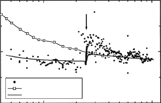
6.5.6 ar and xe
The photon W-value of Ar is shown in the absolute scale as a function of the photon energy from
50 to 1000eV in Figure 6.18 (Saito and Suzuki, 2001a). The present data are exhibited with solid circles.
The arrow denotes the 2p electron ionization threshold. The open squares indicate the W-value for
electrons, W
e
, which was reported previously (Combecher, 1980). The measured W
p
exhibits lower
values than those for electrons below 250eV. The W
p
increases sharply near the 2p threshold, which
shows slightly higher or almost the same values as W
e
. The absolute values for W
p
measured here
are presumed to be correct in consideration of the agreement with the data for electrons, W
e
, in the
100
Photon W-value
Electron W-value
Calculated photon W-value
20
30
W-value (eV)
40
Photon energy (eV)
2p
1000
Figure 6.18 Photon W-value for Ar as a function of photon energy. Solid circles show data by Saito and
Suzuki, and open squares are data for electrons by Combecher (1980). The solid curve represents the photon
W-values calculated using the model explained in Section 6.5.6. The arrow indicates the ionization threshold
of 2p electrons.
(Reproduced from Saito, N. and Suzuki, I.H., Radiat. Phys. Chem., 60, 291, 2001a.)