Elsevier Encyclopedia of Geology - vol I A-E
Подождите немного. Документ загружается.

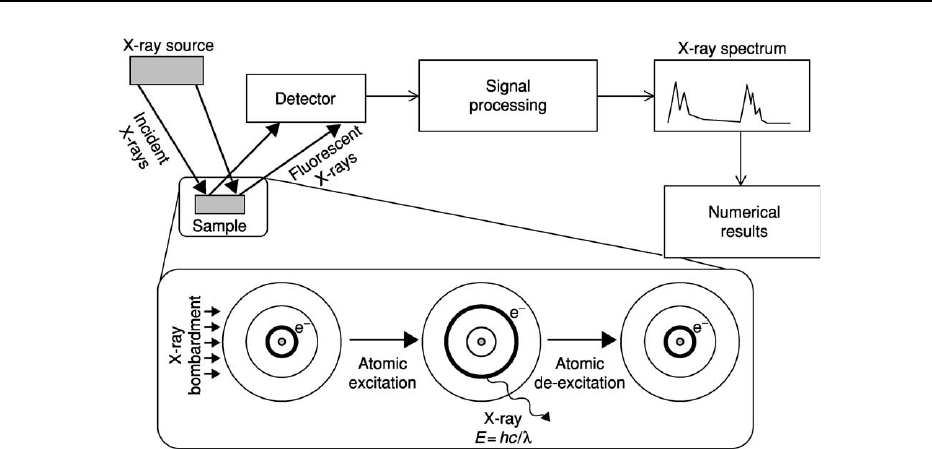
tube or even a radioactive source) strikes a sample,
the X-ray can be either absorbed by the atom or
scattered through the material. Some incident
X-rays are absorbed by the atom, and electrons are
ejected from the inner shells to outer shells, creating
vacancies. As the atom returns to its stable condition,
electrons from the outer shells are transferred to the
inner shells and in the process give off a characteristic
X-ray with an energy equal to the difference between
the two binding energies of the corresponding shells.
XRF is widely used to measure the elemental com-
position of natural materials, since it is fast and non-
destructive to the sample. It can be used to measure
many elements simultaneously. XRF can be used dir-
ectly on rock surfaces, although there is a danger of
natural heterogeneity resulting in variable results.
Rock, soil, and sediment samples are typically crushed
and made into pellets by compressing them or by
melting the whole sample and then quenching to
make a glass disk. XRF is useful for the geochemical
analysis of a wide range of metals and refractory and
amphoteric compounds (such as SiO
2
and Al
2
O
3
) and
even some non-metals (chloride and bromide). XRF
is also routinely used to measure the natural metal
content of liquid petroleum samples. The quality of
XRF data is a function of the selection of appropriate
standards. It is considered to be best practice to
use standards that are similar to the samples in ques-
tion to minimize matrix effects. XRF can measure
down to parts-per-million concentrations and lower,
depending on the element and the material.
X-ray Diffraction
X-rays have a similar wavelength to the lattice spacing
of common rock-forming minerals and have been used
to characterize the crystal structure and mineralogy
of Earth materials by using X-ray diffraction (XRD)
analysis. This is most commonly used to define the
presence of minerals, mineral proportions, mineral
composition (in favourable circumstances), and other
subtle mineralogical features of rocks, sediments, and
soils.
X-rays, even from a pure elemental source bom-
barded with electrons, have a collection of peaks –
X-rays characteristic of the quantized electron energy
levels – and bremsstrahlung. X-ray beams of a tightly
defined energy (and thus wavelength) have been used
to investigate and characterize the minerals present
in rocks, sediments, and soils by removing all but one
X-ray peak from the spectrum of wavelengths gener-
ated by a source element. X-rays are useful in investi-
gating mineral structure since they can be selected to
have a wavelength that is only just smaller than the
interlattice spacing (d-spacing) of common rock-
forming minerals. A number of X-ray sources have
historically been selected, but copper is the most
commonly employed, and the copper Ka peak is the
one that is directed onto samples. This has a charac-
teristic wavelength of 1.5418 A
˚
(0.15418 nm). This is
ideal for many minerals since they have high-order
(dominant, most obvious) lattice spacings of this size
or up to 10–15 times greater than this wavelength.
Figure 8 General set of processes involved in the generation of fluorescent X-rays by X-ray bombardment of atoms. The small grey
filled circle represents the nucleus; the outer rings represent electron orbitals. The thicker black circle represents the location of a
given electron (e
). With X-ray bombardment, the highlighted electron jumps to a higher orbital. The energized electron quickly falls
back to its original state, releasing a secondary X-ray. The range of elements in a sample leads to a range of characteristic fluorescent
X-rays with peak heights that are functions of the amounts of the elements in the sample.
ANALYTICAL METHODS/Geochemical Analysis (Including X-ray) 61
The melee of X-radiation from copper can be reduced
to the Ka peak and then directed by a series of
diffraction gratings, collimators, and slits.
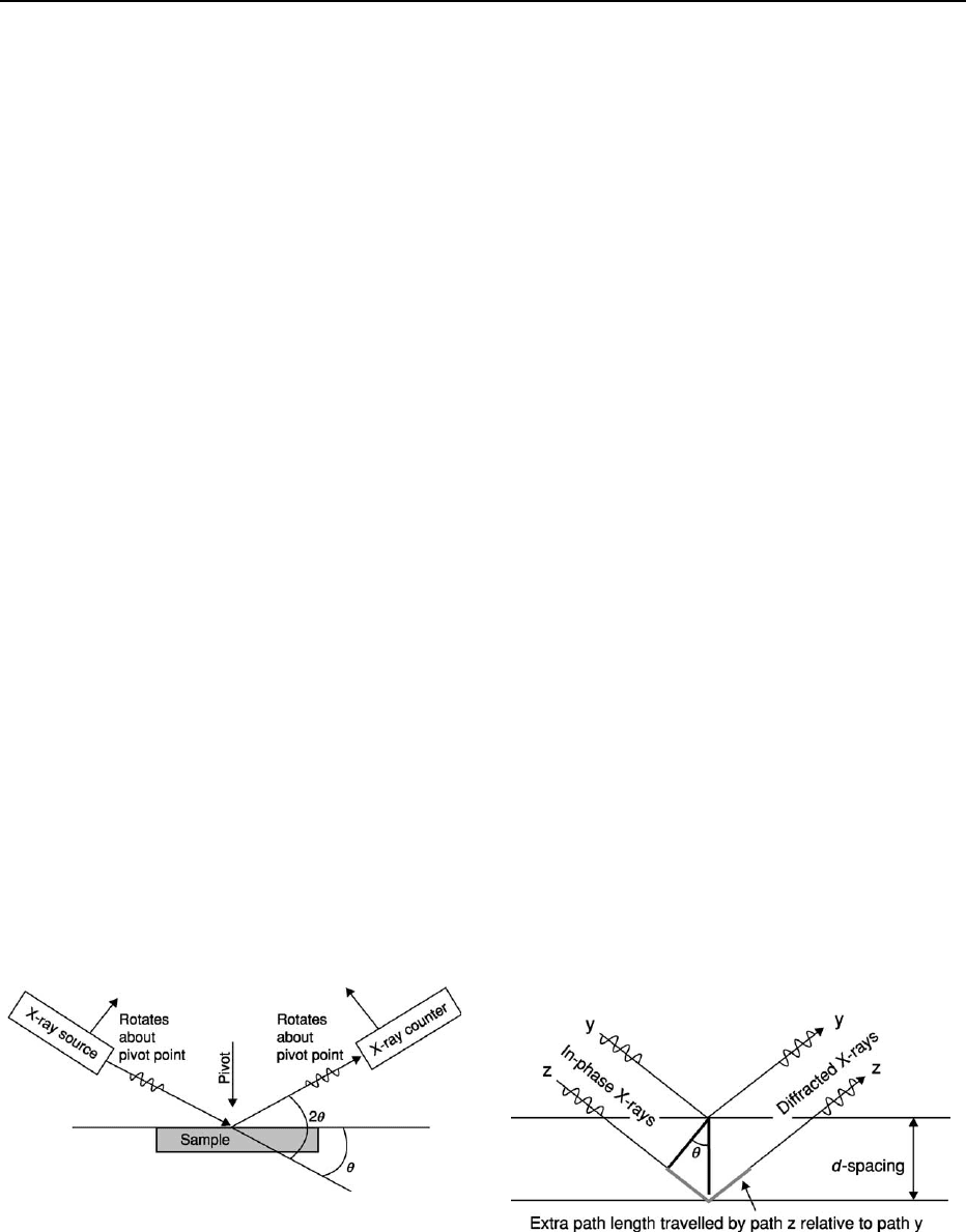
The main features of an XRD analyser include an
X-ray source with collimators, slits, etc, a sample,
and an X-ray detector (Figure 9). The source and
detector are both rotated about the sample, an arbi-
trarily fixed point, and define the same angle (y)
relative to the sample. The angle between the source
and the detector is thus 2y relative to the sample.
Diffraction occurs when X-rays, light, or any other
type of radiation passes into, but is then bounced
back out of, a material with a regular series of layers.
Diffraction occurs within the body of the material
rather than from the surface (and so is quite different
from reflection). Regular layers are a characteristic
of all crystalline materials (minerals, metals, etc).
Each rock-forming mineral has a well-defined set
of these layers, which constitute the crystal lattice.
No two minerals have exactly the same crystal struc-
ture, so fingerprinting a mineral by its characteristic
set of lattice spacings helps to identify minerals. A
radiation beam from a pure source has a defined
wavelength, and the rays from such a pure source
will be ‘in phase’. Constructive interference occurs
only when all the outgoing (diffracted) X-rays are
also in phase. Destructive interference, the norm,
occurs when the diffracted X-rays are no longer in
phase. Constructive interference occurs when the
extra distance that X-rays travel within the body of
the material is an integer (whole number) multiple of
the characteristic wavelength of the incident X-ray
(Figure 10). The geometry of the XRD equipment,
the wavelength of the incident radiation, and the
lattice spacing are all important in defining whether
constructive interference occurs. The key equation
is known as the Bragg Law, which must be satisfied
for constructive interference (‘diffraction’) to occur:
2dsiny ¼ nl, where d is the lattice spacing, l is the
wavelength of the incident X-ray source, and n is an
integer (typically one in many cases). The value of y,
defined in Figure 9, is a function of the variable
geometry of the XRD equipment.
X-ray diffraction is most commonly used on
crushed (powered) rock samples to ensure homo-
geneity of the sample and randomness of the orien-
tations of all the crystal lattices represented by
different minerals. This is known as X-ray powder
diffraction.
XRD works by rotating the X-ray source and the
detector about the sample from small angles (e.g. 4
)
through to angles of up to 70
. The low angles can
detect large interlattice spacings (large values of d)
while the high angles detect smaller interlattice
spacings. For CuKa radiation these angles equate to
d-spacings from about 30 A
˚
down to about 1.5 A
˚
,
covering the dominant d-spacings of practically all
rock-forming minerals.
For a pure mineral sample, the diffraction peaks
from different lattice planes with discrete d-spacings
have different relative intensities. This is a function of
the details of the crystal structure of a particular
mineral, but the maximum-intensity trace (peak) for
many minerals has a low Miller Index value (a simple
notation for describing the orientation of a crystal).
For example, many clay minerals dominated by sheet-
like crystal structures have (001) as the maximum-
intensity peak. All other XRD traces have intensities
that are fixed fractions of the intensity of the max-
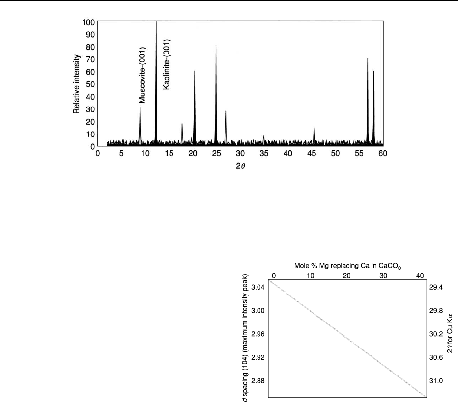
imum-intensity trace. The result for each pure min-
eral is a fingerprint of XRD peaks on a chart
of intensity on the y-axis and 2y on the x-axis
(Figure 11). This can be compared with collections
of standards to identify the mineral.
Figure 9 Basic elements of an X-ray diffraction device. An
X-ray source is directed at a sample at a controlled and variable
angle (y). The X-ray detector is at the equivalent angle on the
opposite side of the pivot point. The source and detector are at an
angle of 180
2y to one another. The source and detector are
thus simultaneously rotated about the pivot point. When diffrac-
tion occurs the X-ray detector records a signal above the back-
ground. The sample is usually a powder and preferably randomly
orientated.
Figure 10 Diffraction from a crystal. The incident X-rays are in
phase as they hit the mineral surface. The grey line shows the
path-length difference between the two X-rays. Constructive
interference occurs when the extra path length (2
d siny)isan
integral multiple (typically one) of the wavelength of the X-rays.
Constructive interference leads to an X-ray diffraction peak set
against a low level of background noise.
62 ANALYTICAL METHODS/Geochemical Analysis (Including X-ray)
The intensity of the collection of diffraction peaks
from a given mineral in a mixture (e.g. rock, soil, or
sediment) is broadly a function of the proportion of the
mineral in the mixture. This is a subject of ongoing
research since the issue is complicated by different
minerals having different efficiencies at diffracting
X-rays. In simple terms, the intensities of the max-
imum-intensity peaks from a mixture of minerals
give a guide to the relative proportions of the different
minerals (Figure 11).
There is a range of problems for XRD when dealing
with natural Earth materials. The most obvious is the
simple identification of minerals when each one has its
own collection of diffraction peaks. These must be
carefully deconvoluted by a process of elimination.
Computer-based programs can be of great help in
this task. Many rock-forming minerals do not conform
to a perfectly defined composition. The set of interlat-
tice d-spacings in minerals is a function of the way
atoms are packed together in the lattice, so that com-
positional variation affects the precise 2y position of a
given crystal orientation. In some cases, this variability
can be put to good use since it can be used to identify
the composition of a given mineral (Figure 12).
Another issue with XRD analysis, especially of
sediments, soils, and sedimentary rocks is that many
minerals have poorly defined crystal structure. This
problem is typical of clay minerals, pedogenic oxides,
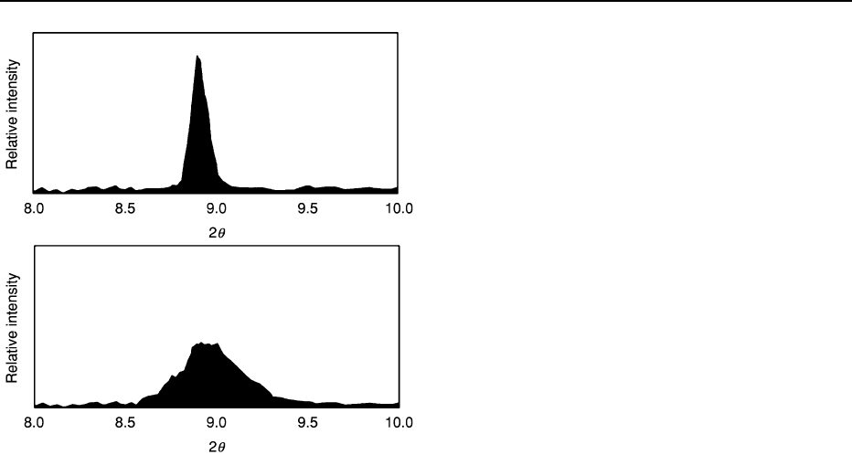
etc. Poorly defined crystal structure translates into
wider XRD peaks. This presents problems for quan-
tification and results in a need to measure the area
under an entire peak rather than the height of the
maximum-intensity peak. However, this too can be
put to good use by enabling the quantification of
the transformation of a poorly defined crystal struc-
ture into a well-defined structure (typically as a funct-
ion of time and temperature) during diagenesis or
metamorphism (Figure 13).
Optical Techniques
Spectroscopy is the use of the absorption, emission, or
scattering of electromagnetic radiation by atoms or
molecules (or atomic or molecular ions) to study the
atoms or molecules qualitatively or quantitatively.
Isolated atoms can absorb and emit packets of
electromagnetic radiation with discrete energies that
Figure 11 The X-ray diffraction output from a rock composed of the clay minerals muscovite (pale grey peaks) and kaolinite (black
peaks). The two minerals have discrete d spacings, which are reflected in their discrete peaks on the chart. The peaks at the low 2y end
have high
d-spacings, and vice versa. The dominant basal spacings of these two sheet silicates (the (001) planes) are labelled. The
maximum-intensity diffraction peaks from these minerals are produced by the (001) planes. From this diagram it would appear that
there is much more kaolinite than muscovite in the mixture (approximately three times as much) since its maximum-intensity peak is
much more intense. The mixture therefore has about 25% muscovite and 75% kaolinite.
Figure 12 X-ray diffraction for the same peak from the same
mineral may occur at a different 2y value if there is solid solution
and the substituted element has a different ionic radius. For
example, in calcite the dominant (104) diffraction peak moves
systematically as magnesium replaces calcium. Magnesium
ions are smaller than calcium ions so the structure collapses
slightly. In favourable circumstances, this approach can be
used to help determine mineral composition.
ANALYTICAL METHODS/Geochemical Analysis (Including X-ray) 63
are dictated by the detailed electronic structure of
the atoms. When the resulting altered light is passed
through a prism or spectrograph it is separated
according to wavelength. Emission spectra are pro-
duced by high-temperature gases. The emission lines
correspond to photons of discrete energies, which are
emitted when excited atomic states in the gas make
transitions back to lower-lying levels. Generally,
solids, liquids, and dense gases emit light at all wave-
lengths when heated. An absorption spectrum occurs
when light passes through a cold dilute gas and atoms
in the gas absorb light at characteristic frequencies.
Since the re-emitted light is unlikely to be emitted
in the same direction as the absorbed photon was
travelling, this gives rise to dark lines (absence of
light) in the spectrum.
Emission and absorption spectroscopy have been
used widely in geochemical analysis for many years
(Table 3). They are commonly used to analyse waters
but can also be employed to analyse rock and other
solid samples that have been quantitatively dissolved
(e.g. in acid solutions).
Atomic Absorption and Atomic Emission
Spectroscopy
Atomic-absorption (AA) spectroscopy uses the absorp-
tion of light to measure the concentration of gas-phase
atoms. Since samples are usually liquids or solids, the
sample atoms or ions must be vaporized in a flame
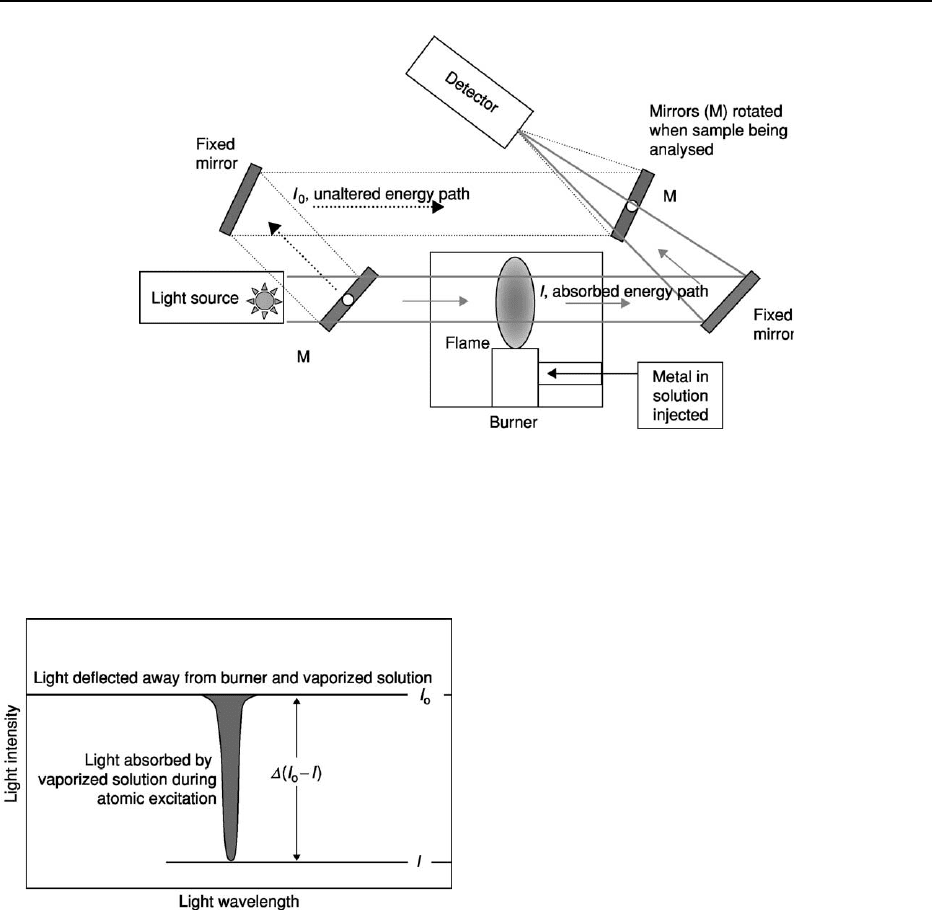
or graphite furnace (Figure 14). The atoms absorb
ultraviolet or visible light. Concentrations are usually
determined from a working curve after calibrating the
instrument with standards of known concentration
(Figure 15). The Lambert–Beer Law is the relationship
between the change in light intensity for a given
wavelength and the relative incident light energy:
log I
o
/I ¼ aLc,whereI is the light intensity after the
metal is added, I
o
is the initial light intensity, a is a
machine-dependent constant, L is the path length of
light through the torch, and c is the concentration.
The light source is usually a hollow-cathode lamp
of the element that is being measured. A major disad-
vantage of these narrow-band light sources is that
only one element can be measured at a time. AA
spectroscopy requires that the target atoms be in the
gas phase. Ions or atoms in a sample must be desol-
vated and vaporized in a high-temperature source
such as a flame or graphite furnace. Flame AA can
analyse only solutions. Sample solutions are usually
aspirated with the gas flow into a nebulizing and
mixing chamber to form small droplets before
entering the flame. AA spectrometers use monochro-
mators and detectors for UV and visible light. The
main purpose of the monochromator is to isolate the
absorption line from background light due to inter-
ferences. Photomultiplier tubes are the most common
detectors for AA spectroscopy.
This technique can be used to analyse aqueous
samples with negligible sample preparation. It can
also be used for rock and mineral samples if they are
quantitatively dissolved (typically using acids of vari-
ous strengths). Flame AA spectroscopy can typically
detect concentrations as low as mg l
1
(ppm), al-
though graphite-furnace AA spectroscopy has been
shown to be able to detect concentrations that are
orders of magnitude below this.
Inductively Coupled Plasma Optical Emission
Spectroscopy
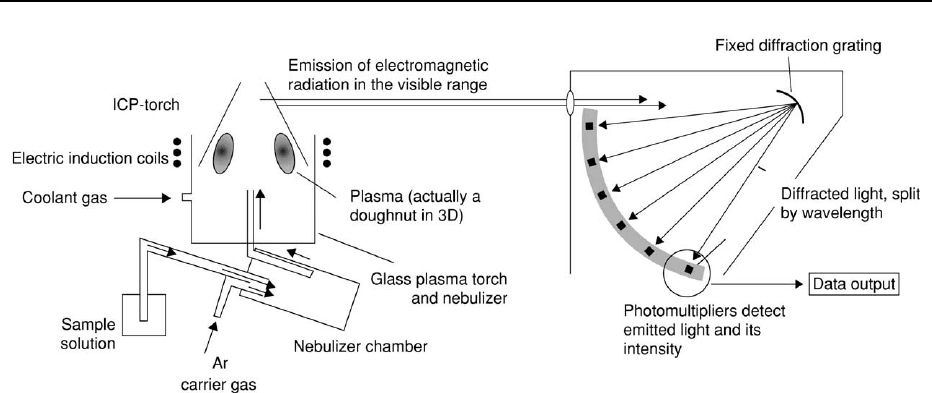
ICP-OES is short for optical (or atomic) emission
spectrometry with inductively coupled plasma.
Plasma is a luminous volume of atoms and gas at
extremely high temperature in an ionized state. The
plasma is formed by argon flowing through a radio
frequency field, where it is kept in a state of partial
ionization, i.e. the gas consists partly of electrically
charged particles. This allows it to reach very high
temperatures of up to approximately 10 000
C. At
these high temperatures, most elements emit light of
characteristic wavelengths, which can be measured and
used to determine the concentration of the elements
in the solution.
Figure 13 X-ray diffraction outputs from a mineral (illite) with
variable degrees of crystallinity. The top image represents a
well-crystalline sample, e.g. a slate. The lower image represents
a poorly crystalline material, e.g. from a juvenile sedimentary
rock, soil, or weathering horizon. The degree of crystallinity can
be quantified by measuring the peak width at half the peak height.
64 ANALYTICAL METHODS/Geochemical Analysis (Including X-ray)

Table 3 Optical spectroscopic techniques commonly used in geochemical analysis
Technique Output-1 Output-2 Output-3 Sample type Advantages Disadvantages
Atomic absorption
spectroscopy (AAS)
Element concentrations
in water
Element
concentration in
quantitatively
dissolved rock
samples
Water sample from the
natural or altered
environment (or
solid rocks and
minerals dissolved
in acid)
Well-established
technique; relatively low
costs
Relatively high detection
limit; relatively slow –
one element at a time;
limited range of
elements
Inductively coupled plasma
optical emission
spectroscopy (ICP-OES)
Element concentrations
in water
Element
concentration in
quantitatively
dissolved rock
samples
Water sample from the
natural or altered
environment (or
solid rocks and
minerals dissolved
in acid)
Many elements analysed
simultaneously;
relatively fast; linear
calibration over wide
concentration range;
wide concentration
range; one calibration
suitable for most
material types; good for
water samples; excellent
(ppb) resolution for
some trace elements
Expensive equipment;
technique not for the
novice analyst; carrier
gases cause
interference with some
elements; minerals and
rocks must be dissolved
prior to analysis;
relatively new technique
still undergoing
development
Infrared spectroscopy Mineral proportions Water content in
quartz
Clay and other
minerals
Ultra-small samples;
quantitative; relatively
low cost; simple sample
preparation
Complex to interpret
absorption spectra;
adsorbed water can
interfere with diagnostic
peaks
Ultraviolet spectroscopy Presence of organic
inclusions in
minerals and rocks
Presence of liquid
organics in
porous
materials (e.g.
pollution or oil)
Approximate
indication of
the density
and
maturity of
petroleum
Organic-bearing rock,
sediment, or soil
Simple to make qualitative
observations; easily
repeated; can be used
on bulk and microscopic
samples
Some minerals have
masking fluorescence;
difficult to quantity
ANALYTICAL METHODS/Geochemical Analysis (Including X-ray) 65
The sample being analysed is introduced into the
plasma as an aerosol of fine droplets (Figure 16). Light
from the different elements is separated into different
wavelengths by means of a grating and is captured
simultaneously by light-sensitive detectors. This per-
mits the simultaneous analysis of up to 40 elem-
ents, and ICP-OES is consequently a multi-element
technique. In terms of sensitivity, ICP-OES is gener-
ally comparable with graphite furnace AA, i.e. detec-
tion limits are typically at the mgl
1
level in aqueous
solutions.
Ultraviolet Spectroscopy
Many organic-derived materials fluoresce under
ultraviolet (UV) light – that is, they absorb light from
the incident ultraviolet beam and release light, often
in the visible range, that has a different wavelength
from the primary UV light. UV spectroscopy can be
used qualitatively to determine whether complex or-
ganic molecules are present in sediment or a rock.
UV spectroscopy is commonly used to determine the
presence of diffuse oil-shows in petroleum-reservoir
cores.
There is a loose correspondence between the pre-
cise wavelength of the fluorescent light and the nature
of the organic material. This relationship has been
developed into an analytical technique to determine
the thermal maturity of petroleum in rock samples.
This technique has reached its apotheosis in its appli-
cation to petroleum trapped in inclusions in mineral
cements and healed fractures. These fluid inclusions
have been used to track the petroleum generation of
source rocks and the petroleum migration history into
reservoirs (see Fluid Inclusions).
Infrared Spectroscopy
The electrical bonds between molecules continually
vibrate as a function of interaction between neigh-
bouring molecules. These bonds can be excited by
infrared radiation, resulting in higher amplitudes of
vibration. Only discrete (quantile) increases in vibra-
tion energy are possible, and this results in an
Figure 14 Atomic absorption spectroscopy equipment. The aqueous sample for analysis is injected into the burner and light
is shone through the flame. The mirrors (labelled M) are rotated to measure original and the absorbed light characteristics. The
path that is deflected away from the flame (labelled as unaltered energy path) measures the unaffected light intensity. The path that
goes through the flame (labelled as absorbed energy path) measures the intensity of the light after it has passed through a burner
containing the co-injected dissolved sample.
Figure 15 Light absorption due to atomic excitation with base-
line and post-excitation light intensities indicated. The frequency
of the absorbed light depends on the element. Concentration is
proportional to
I
o
/I, all other things being equal.
66 ANALYTICAL METHODS/Geochemical Analysis (Including X-ray)
absorption spectrum. The molecular environment is
different in all minerals, even in minerals of similar
structure (e.g. the clay family), so that infrared spec-
troscopy can be used to identify minerals in rock and
sediment samples. The strength of the absorption
spectrum depends on concentration, so the technique
can be used for quantitative analysis of minerals
under favourable circumstances. Infrared spectros-
copy has been used to identify clay minerals in sand-
stones but is most useful for identifying organic
inclusions and non-hydrocarbon gases trapped in
fluid inclusions.
Chromatography
Chromatography has proved invaluable in geochem-
ical analysis (Table 4). It is an analytical technique
used to quantify and separate mixtures of fluid chem-
ical compounds. There are many different kinds of
chromatography, among them gas chromatography,
organic liquid chromatography, and ion-exchange
chromatography. All chromatographic methods share
the same basic principles and mode of operation. In
every case, a sample of the mixture to be analysed is
applied to some stationary fixed material (the adsorb-
ent or stationary phase) and then a second material
(the eluent or mobile phase) is passed through or over
the stationary phase. The compounds contained in the
sample are then partitioned between the stationary
phase and the mobile phase. The success of the ap-
proach depends on the fact that different materials
adhere to the adsorbent with different forces. Those
that adhere more strongly are moved through the
adsorbent more slowly as the mobile phase flows
over them. Other components of the sample that are
less strongly adsorbed on the stationary phase and
moved along more quickly by the moving phase.
Thus as the mobile phase flows through the column,
components in the sample move down the column at
different rates and therefore separate from one an-
other. At the end of the column, molecules or ions of
the fastest-moving substance (least tightly bound to
the stationary phase) emerge first, usually with each
compound emerging over a well-defined time inter-
val. A suitable detector analyses the output at the end
of the column. Each time molecules or ions of the
sample emerge from the chromatography column
the detector generates a measurable signal, which is
recorded as a peak on the chromatogram. The chro-
matogram is thus a record of detector output as a
function of time and consists of a series of peaks
corresponding to the different times at which com-
ponents of the sample mixture emerge from the
column.
By running standards and mixtures of known con-
centration, it is possible to relate the arrival time to
species type and the size of the peak to concentration,
making chromatography a valuable quantitative
technique. There are three main types of chromatog-
raphy employed in geochemical laboratories: liquid
chromatography, gas chromatography, and ion chro-
matography. Gas chromatography is used to separate
mixtures of gases or vaporized liquids. Ion chroma-
tography is used to separate and analyse ions, typic-
ally but not exclusively anions, in aqueous solutions.
Liquid chromatography is included in Table 4 but is
mainly used as a sample-preparation procedure prior
to gas chromatography to split petroleum, e.g. into
Figure 16 Essential components of an inductively coupled plasma optical emission spectrometer. The induction coils produce a ring
of plasma at temperatures of ca. 10 000
C. The water sample, drawn in by the flowing carrier gas, is dispersed and drawn into the
plasma. Elements emit light due to thermal excitation. A wide range of light frequencies can be analysed simultaneously using a
diffraction grating to disperse the light.
ANALYTICAL METHODS/Geochemical Analysis (Including X-ray) 67

Table 4 Chromatographic techniques commonly used in geochemical analysis
Technique Output-1 Output-2 Output-3 Sample type Advantages Disadvantages
Liquid chromatography Physical separation of
different components of
complex liquid mixture
(petroleum)
Whole petroleum
or extracted
bitumen samples
Excellent pre-separation
technique for GC and
GC-MS analyses; Gives
quantities of groups of
petroleum compounds
Limited separation
capability
Gas chromatography Physical separation of
different components of
complex gas-phase
mixture (petroleum gas
or heated volatilized
liquid)
Quantitative
measure of
proportions of
different
compounds in
gas and liquid
petroleum
Either whole
petroleum or
separate parts
(achieved
using liquid
chromatography)
Splits gas and liquid range
compounds; easily
quantified; well-
established technology;
good for samples with
dominant alkanes
Co-elution of different
compounds; requires
sample preparation;
unknown GC-peaks can
give ambiguous
interpretation
Ion chromatography Physical separation of
different charged
(aqueous) components
in complex natural
solutions
Quantitative
measure of
proportions of
different anions
in water
(common
application)
Quantitative
measure of
proportions of
different cations
in water (less
common
application)
Water sample or
solid sample
quantitatively
dissolved in
water
Splits a range of anions in
water; high resolution;
relatively fast and
simultaneously analyses
all anions; no real
alternative
Unsuitable for bicarbonate
analysis
68 ANALYTICAL METHODS/Geochemical Analysis (Including X-ray)
saturated, aromatic, and resin groups, and will not be
covered further here.
Gas Chromatography
Gas chromatography (GC) – specifically gas–liquid
chromatography – involves a sample being vaporized
and injected onto the head of the chromatographic
column (Table 4). The sample is transported through
the column by the flow of an inert gaseous mobile
phase. The column itself contains a liquid stationary
phase that is adsorbed onto the surface of an inert
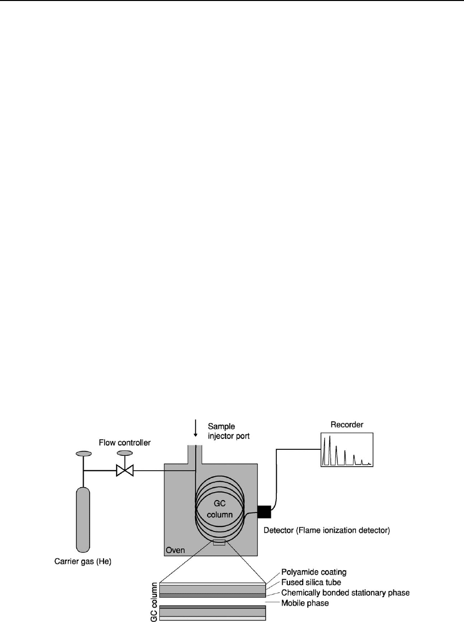
solid (Figure 17).
The carrier gas is chemically inert (e.g. helium).
A sample of gas or petroleum is injected into the
column quickly as a slug to prevent peak broadening
and loss of resolution. The temperature of the sample
port is somewhat higher than the boiling point of the
least-volatile component of the sample. Sample sizes
typically range from tenths of a microlitre to 20 ml.
The carrier gas enters a mixing chamber, where the
sample vaporizes to form a mixture of carrier gas,
vaporized solvent, and vaporized solutes. A propor-
tion of this mixture passes onto the chromatography
column. Chromatography columns have an internal
diameter of a few tenths of a millimetre and walls
coated with a liquid but stationary phase. The opti-
mum column temperature depends on the boiling
point of the sample; typically a temperature slightly
above the average boiling point of the sample results
in an elution time of 2–30 min. As the carrier gas
containing the chromatographically separated sample
passes out of the end of the column, it is passed
into one of a number of detectors such as a flame
ionization detector, which has high sensitivity, a large
linear response range, and low noise. An example
of a flame-ionization-detector signal from a whole-
petroleum sample injected onto a GC column is given
in Figure 18.
One of the problems of GC analysis of geochemical
samples is that different compounds can have similar
elution times, rendering identification and quantifica-
tion difficult. However, the output stream from a gas
chromatograph can be passed into other types of
analytical instrument (e.g. a mass spectrometer) for
further analysis of the separated compounds over and
above simple quantification of compounds with a
common elution time. GC is useful for analysing
organic compounds and can be used to quantify
mixtures if suitable standards have been employed.
Ion Chromatography
Ion chromatography can be used for both cations and
anions. However, it is in the analysis of non-metal
ions that the technique has proved most useful mainly
because there are no real alternatives for the simul-
taneous quantitative analysis of these important
species in waters or synthetic solutions.
Ion chromatography is used to analyse aqueous
samples containing ppm quantities of common anions
(such as fluoride, chloride, nitrite, nitrate, and sul-
phate). Ion chromatography is a form of liquid chro-
matography that uses ion-exchange resins to separate
atomic or molecular ions based on their interaction
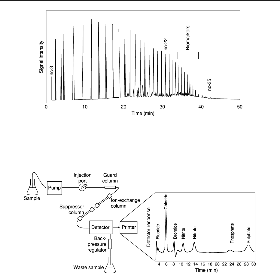
with the resin (Figure 19). Its greatest utility is for the
Figure 17 Essential components of a gas chromatograph. The sample injection port is heated to volatilize liquid-phase organics.
The GC column is held in an oven at a temperature above the boiling point of the compounds of interest. The inert carrier gas drives the
sample through the capillary with its stationary phase. The stationary phase retards larger molecules more efficiently than small
molecules. Smaller molecules thus pass out of the column more rapidly than large molecules.
ANALYTICAL METHODS/Geochemical Analysis (Including X-ray) 69
analysis of anions for which there are no other rapid
analytical methods. For anion chromatography the
mobile phase is a dilute aqueous solution of sodium
bicarbonate and sodium carbonate prepared with
pure water.
The ion-exchange column is tightly packed with
the stationary adsorbent. This adsorbent is usually
composed of tiny polymer beads that have positively
charged centres. These become coated with bicarbon-
ate and carbonate anions if no sample is passing
through the column. As anions in the sample enter
the column, they are attracted to the positive centres
on the polymer surface and may replace (exchange
with) the bicarbonate and carbonate ions stuck to the
surface. Usually, the greater the charge on the anion
the more strongly it is attracted to the surface of the
polymer bead. Also, larger anions generally move
more slowly through the column than smaller anions.
The result is that the sample separates into bands of
different kinds of ion as it travels through the column.
The detector, usually a conductivity cell, measures
the conductance of the solution passing through it.
The conductance is proportional to the concentration
of ions dissolved in the solution. It is essential to
Figure 18 Typical GC output from a black oil sample. Most of the peaks correspond to normal alkanes. The longest-chain alkane
detected is C
35
H
72
. The biomarkers, molecules of clear biological origin, form an area of low-level noise that is difficult to discern with
this technique. On the figure nc-3, nc-22, nc-35 refer to normal alkanes with 3, 22 and 35 carbon atoms. The three labelled peaks are
thus from C
3
H
8
,C
22
H
46
and C
35
H
72
.
Figure 19 Essential components of an ion-exchange chromatograph, and a typical output trace for a low-concentration standard. The
suppressor column removes the bicarbonate and carbonate that are released from the ion-exchange chromatography column to avoid
the real sample signal being swamped. The trace has 0.02 ppm fluoride and 0.1 ppm of all the other anions. Note that the transit time
through the column increases with atomic number for the halogens and is greatest for the multivalent anions phosphate and sulphate.
70 ANALYTICAL METHODS/Geochemical Analysis (Including X-ray)
