Egerton R.F. Physical Principles of Electron Microscopy. An Introduction to TEM, SEM, and AEM
Подождите немного. Документ загружается.

122 Chapter 4
All of the above methods involve the application of a mechanical force to
achieve thinning. Unfortunately, for all but the most well-behaved materials
(e.g., biological tissue, layer materials, silicon), the specimen becomes
extremely fragile and cannot be mechanically thinned below about 1 Pm. In
addition, the mechanical forces involved may leave a damaged surface layer,
containing a high density of defects such as dislocations. This damage is
undesirable if the TEM is being used to study the original defect structure of
the specimen. Therefore, some non-mechanical method is commonly used
for the final thinning.
One such method is chemical thinning, in which a chemical solution
dissolves the original surface and reduces the specimen thickness to a value
suitable for TEM imaging. In the simplest case, a thin piece of material is
floated onto the surface of a chemical solution that attacks its lower surface;
the sample is retrieved (e.g, by picking up by a TEM grid held in tweezers)
before it dissolves completely. More commonly, a jet of chemical solution is
directed at one or both surfaces of a thin disk. As soon as a small hole forms
in the center (detected by the transmission of a light beam), the polishing
solution is replaced by rinse water. If the procedure is successful, regions of
the specimen surrounding the hole are thin enough for TEM examination.
Alternatively, electrochemical thinning is carried out with a direct
current flowing between the specimen (at a negative potential) and a positive
electrode, immersed in a chemical solution. In the original window-frame
method (Fig. 4-20f), the specimen is in the form of a thin sheet (1 cm or
more in height and width) whose four edges are previously painted with
protective lacquer to prevent erosion at the edge. When partially immersed
in the electrolytic solution, thinning is most rapid at the liquid/air interface,
which perforates first. Small pieces of foil are cut adjacent to the perforated
edge and mounted on a TEM grid. Nowadays, electrochemical thinning is
usually done using the jet-thinning geometry (Fig. 4-20e), by applying a dc
voltage between the specimen and jet electrodes. Recipes for the solutions
used in chemical and electrochemical thinning are given in Goodhew (1985).
When thinning metals, glycerin is sometimes added to the solution to make
the liquid more viscous, helping to give the thinned specimen a polished
(microscopically smooth) surface.
Increasingly, ion-beam thinning is used for the final thinning stage,
particularly of materials that are chemically inert. Following mechanical
thinning (if necessary), a 3mm-diameter thin disk of the material is placed in
a vacuum system, where it is bombarded by argon ions produced by a gas
discharge within an ion gun. These ions transfer energy to surface atoms and
remove the material by the process of sputtering; see Fig. 4-20g. A focused
ion beam (FIB) machine uses a beam of gallium ions to cut thin slices, often

TEM Specimens and Images 123
of a semiconductor material. This instrument also gives an electron or ion-
beam image of the surface of the specimen, allowing the ion beam to be
positioned and scanned along a line in order to cut a slice of material at a
precise location. In this way, it is possible to prepare a TEM specimen that
represents a cross section through a particular component; see Fig. 4-21.
Instead of thinning a bulk material, TEM specimens are sometimes built
up in thickness by thin-film deposition. Typically, the material is placed in
a tungsten “boat” that is electrically heated in vacuum, causing the material
to evaporate and then condense onto a substrate; see Fig. 4.20h. If the
substrate is soluble (for example, alkali halides dissolve in water), the film is
floated onto the liquid surface and captured on a TEM grid, similar to thin
sections of biological tissue. Vacuum-deposited thin films are used in the
electronics and optical industries but are usually on insoluble substrates such
as silicon or glass. For TEM examination, the substrate must be thinned
down, for example by dimple grinding or jet thinning from its back surface.
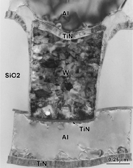
Figure 4-21. Cross-sectional TEM image showing a polycrystalline tungsten “via” that makes
electrical connection between adjacent layers of aluminum in a multilayer integrated circuit.
From such images, the thickness and grain structure of each layer can be determined. From
Zhang (2000), by courtesy of Elsevier Ltd.
124 Chapter 4
The procedures outlined in Fig. 4-20 yield plan-view images, in which
the sample is viewed perpendicular to its original surface. For some
purposes, a cross-sectional image is needed, for example to show the growth
mechanism of a film. The required specimen is made by using a diamond
wheel to cut the film (on its substrate) into several thin strips, which are then
turned through 90 degrees about their long axis and glued together by epoxy
cement. Dimple grinding followed by ion milling then produces a specimen
that is thin at the center, resulting in a TEM image of the substrate and film
seen in cross section.
Cross-sectional specimens are invaluable for analyzing problems that
arise in the manufacture of integrated circuits on a silicon chip. As device
sizes shrink, it becomes necessary to make use of the high spatial resolution
of the TEM for this kind of failure analysis. The problem of producing a
cross section containing a specific component is solved by using a FIB
machine, in which Ga+ ions are focused (by electrostatic lenses) into a beam
as small as 10 nm in diameter. The machine produces a scanned image of the
surface of the chip, allowing the ion beam to be precisely positioned and
then scanned in a line in order to cut into the silicon on either side of the
component. This leaves a slice only 100 nm in thickness, which is then lifted
out and viewed in cross section in the TEM, as in Fig. 4-21.
Chapter 5
THE SCANNING ELECTRON MICROSCOPE
As we discussed in Chapter 1, the scanning electron microscope (SEM) was
invented soon after the TEM but took longer to be developed into a practical
tool for scientific research. As happened with the TEM, the spatial resolution
of the instrument improved after magnetic lenses were substituted for
electrostatic ones and after a stigmator was added to the lens column. Today,
scanning electron microscopes outnumber transmission electron microscopes
and are used in many fields, including medical and materials research, the
emiconductor industry, and forensic-science laboratories.s
Figure 1-15 (page 19) shows one example of a commercial high-
resolution SEM. Although smaller (and generally less expensive) than a
TEM, the SEM incorporates an electron-optical column that operates
according to the principles already discussed in Chapter 2 and Chapter 3.
Accordingly, our description will be shorter than for the TEM, as we can
make use of many of the concepts introduced in these earlier chapters.
5.1 Operating Principle of the SEM
The electron source used in the SEM can be a tungsten filament, or else a
LaB
6
or Schottky emitter, or a tungsten field-emission tip. Because the
maximum accelerating voltage (typically 30 kV) is lower than for a TEM,
the electron gun is smaller, requiring less insulation. Axially-symmetric
magnetic lenses are used but they are also smaller than those employed in
the TEM; for electrons of lower kinetic energy, the polepieces need not
generate such a strong magnetic field. There are also fewer lenses; image
formation uses the scanning principle that was outlined in Chapter 1, and as
a result imaging lenses are not required.

126 Chapter 5
electron gun
condenser
lenses
specimen
scan
coils
specimen
detector
signal
amplifier
display
device
magnification
control
image
scan
scan
generators
objective
lens
x
y
x
z
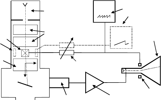
Figure 5-1. Schematic diagram of a scanning electron microscope with a CRT display.
Above the specimen, there are typically two or three lenses, which act
somewhat like the condenser lenses of a TEM. But whereas the TEM, if
operating in imaging mode, has a beam of diameter | 1 Pm or more at the
specimen, the incident beam in the SEM (also known as the electron probe)
needs to be as small as possible: a diameter of 10 nm is typical and 1 nm is
possible with a field-emission source. The final lens that forms this very
small probe is named the objective; its performance (including aberrations)
largely determines the spatial resolution of the instrument, as does the
objective of a TEM or a light-optical microscope. In fact, the resolution of an
SEM can never be better than its incident-probe diameter, as a consequence
of the method used to obtain the image.
Whereas the conventional TEM uses a stationary incident beam, the
electron probe of an SEM is scanned horizontally across the specimen in two
perpendicular (x and y) directions. The x-scan is relatively fast and is
generated by a sawtooth-wave generator operating at a line frequency f
x
; see
Fig. 5-2a. This generator supplies scanning current to two coils, connected in
series and located on either side of the optic axis, just above the objective
lens. The coils generate a magnetic field in the y-direction, creating a force
on an electron (traveling in the z-direction) that deflects it in the x-direction;
see Fig. 5-1.
The y-scan is much slower (Fig. 5-2b) and is generated by a second
sawtooth-wave generator running at a frame frequency f
y
= f
x
/n where n is
an integer. The entire procedure is known as raster scanning and causes the
beam to sequentially cover a rectangular area on the specimen (Fig. 5-2d).

The Scanning Electron Microscope 127
m pixels
(a)
(b)
(c)
(d)
n lines
E
C
A
D
B
Y'
Y
Z
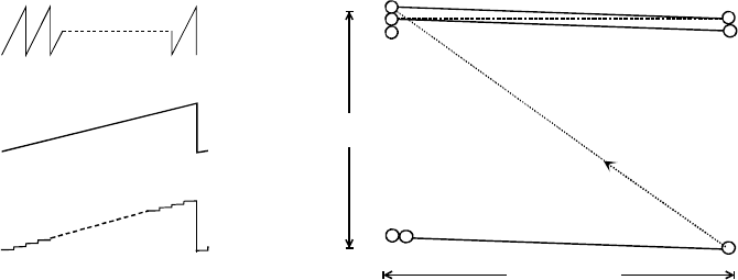
Figure 5-2. (a) Line-scan waveform (scan current versus time),. (b) frame-scan waveform,
and (c) its digital equivalent. (d) Elements of a single-frame raster scan: AB and YZ are the
first and last line scans in the frame, Y and Y’ represent adjacent pixels.
During its x-deflection signal, the electron probe moves in a straight line,
from A to B in Fig. 5-2d, forming a single line scan. After reaching B, the
beam is deflected back along the x-axis as quickly as possible (the flyback
portion of the x-waveform). But because the y-scan generator has increased
its output during the line-scan period, it returns not to A but to point C ,
displaced in the y-direction. A second line scan takes the probe to point D, at
which point it flies back to E and the process is repeated until n lines have
been scanned and the beam arrives at point Z. This entire sequence
constitutes a single frame of the raster scan. From point Z, the probe quickly
returns to A, as a result of the rapid flyback of both the line and frame
generators, and the next frame is executed. This process may run
continuously for many frames, as happens in TV or video technology.
The outputs of the two scan generators are also applied to a display
device, such as a TV-type cathode-ray tube (CRT), on which the SEM image
will appear. The electron beam in the CRT scans exactly in synchronism
with the beam in the SEM, so for every point on the specimen (within the
raster-scanned area) there is a equivalent point on the display screen,
displayed at the same instant of time. Maxwell's first rule of imaging is
therefore obeyed (although approximately because the electron beams are
not points but circles of small diameter). In order to introduce contrast into
the image, a voltage signal must be applied to the electron gun of the CRT,
to vary the brightness of the scanning spot. This voltage is derived from a
detector that responds to some change in the specimen induced by the SEM
incident probe.
In a modern SEM, the scan signals are generated digitally, by computer-
controlled circuitry, and the x- and y-scan waveforms are actually staircase
128 Chapter 5
functions with m and n levels respectively; see Fig. 5-2c. This procedure
divides the image into a total of mn picture elements (pixels) and the SEM
probe remains stationary for a certain dwell time before jumping to the next
pixel. One advantage of digital scanning is that the SEM computer “knows”
the (x, y) address of each pixel and can record the appropriate image-
intensity value (also as a digitized number) in the corresponding computer-
memory location. A digital image, in the form of position and intensity
information, can therefore be stored in computer memory, on a magnetic or
optical disk, or transmitted over data lines such as the Internet.
Also, the modern SEM uses a flat-panel display screen in which there is
no internal electron beam. Instead, computer-generated voltages are used to
sequentially define the x- and y-coordinates of a screen pixel and the SEM
detector signal is applied electronically to that pixel, to change its brightness.
In other respects, the raster-scanning principle is the same as for a CRT
display.
Image magnification in the SEM is achieved by making the x- and y-scan
distances on the specimen a small fraction of the size of the displayed image,
as by definition the magnification factor M is given by:
M = (scan distance in the image) /(scan distance on the specimen) (5.1)
It is convenient to keep the image a fixed size, just filling the display screen,
so increasing the magnification involves reducing the x- and y-scan currents,
each in the same proportion (to avoid image distortion). Consequently, the
SEM is actually working hardest (in terms of current drawn from the scan
generator) when operated at low magnification.
The scanning is sometimes done at video rate (about 60 frames/second)
to generate a rapidly-refreshed image that is useful for focusing the specimen
or for viewing it at low magnification. At higher magnification, or when
making a permanent record of an image, slow scanning (several seconds per
frame) is preferred; the additional recording time results in a higher-quality
image containing less electronic noise.
The signal that modulates (alters) the image brightness can be derived
from any property of the specimen that changes in response to electron
bombardment. Most commonly, the emission of secondary electrons
(atomic electrons ejected from the specimen as a result of inelastic
scattering) is used. However, a signal derived from backscattered electrons
(incident electrons elastically scattered through more than 90 degrees) is also
useful. In order to understand these (and other) possibilities, we need to
The Scanning Electron Microscope 129
consider what happens when an electron beam enters a thick (often called
bulk) specimen.
5.2 Penetration of Electrons into a Solid
When accelerated electrons enter a solid, they are scattered both elastically
(by electrostatic interaction with atomic nuclei) and inelastically (by
interaction with atomic electrons), as already discussed in Chapter 4. Most of
this interaction is “forward” scattering, which implies deflection angles of
less than 90q. But a small fraction of the primaries are elastically
backscattered (T > 90q) with only small fractional loss of energy. Due to
their high kinetic energy, these backscattered electrons have a reasonable
probability of leaving the specimen and re-entering the surrounding vacuum,
n which case they can be collected as a backscattered-electron (BSE) signal. i
Inelastic scattering involves relatively small scattering angles and so
contributes little to the backscattered signal. However, it reduces the kinetic
energy of the primary electrons until they are eventually brought to rest and
absorbed into the solid; in a metal specimen they could become conduction
electrons. The depth (below the surface) at which this occurs is called the
penetration depth or the electron range. The volume of sample containing
(most of) the scattered electrons is called the interaction volume and is
often represented as pear-shaped in cross section (Fig. 5-3), because
scattering causes the beam to spread laterally as the electrons penetrate the
solid and gradually lose energy.
The electron range R for electrons of incident energy E
0
is given by the
following approximate formula (Reimer 1998):
UR | aE
0
r
(5.2)
where b | 1.35 and U is the density of the specimen. If E
0
is given in keV in
Eq. (5.2), a | 10 Pg/cm
2
. Expressing the range as a mass-thickness UR
makes the coefficient a roughly independent of atomic number Z. However,
this implies that the distance R itself decreases with Z, as the densities of
solids tend to increase with atomic number. For carbon (Z = 6), U| 2 g/cm
3
and R | 1 Pm for a 10-keV electron. But for gold (Z = 79), U| 20 g/cm
3
and
R | 0.2 Pm at E
0
= 10 keV. This strong Z-dependence arises mainly because
backscattering depletes the number of electrons moving forward into the
solid, the probability of such high-angle elastic scattering being proportional
to Z
2
, as seen from Eq. (4.15). The interaction volume is therefore smaller
for materials of higher atomic number (Fig. 5-3).
According to Eq. (5.2), the range decreases substantially with decreasing
incident energy, not surprising because lower-energy electrons require fewer

130 Chapter 5
inelastic collisions to bring them to rest and also the probability of inelastic
scattering is inversely proportional to E
0
. A 1-keV electron penetrates only
about 50 nm into carbon and less than 10 nm into gold. The interaction
volume therefore becomes very small at low incident energy; see Fig. 5-3.
increasing E
0
increasing Z
penetration
depth
interaction
volume
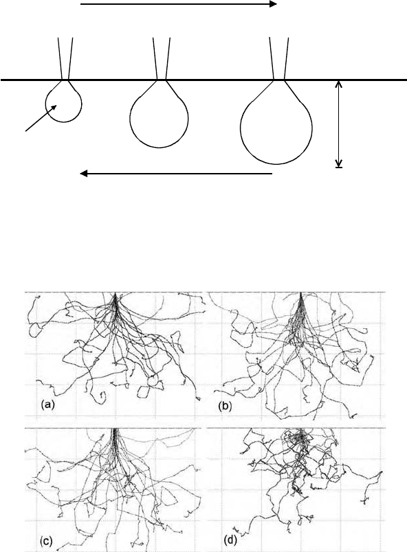
Figure 5-3. Schematic dependence of the interaction volume and penetration depth as a
function of incident energy E
0
and atomic number Z of the incident (primary) electrons.
Figure 5-4. Penetration of (a) 30-keV, (b) 10-keV and (c) 3-keV electrons into aluminum (Z =
13) and (d) 30-keV electrons into gold (Z = 79). Note that the dimensional scales are
different: the maximum penetration is about 6.4 Pm, 0.8 Pm, and 0.12 Pm in (a), (b), and (c),
and 1.2 Pm in (d). These Monte Carlo simulations were carried out using the CASINO
program (Gauvin et al., 2001) with 25 primary electrons and an incident-beam diameter equal
to 10 nm in each case.
The Scanning Electron Microscope 131
Penetration depth and interaction volume are macroscopic quantities,
averaged over a large number of electrons, but the behavior of an individual
electron is highly variable. In other words, scattering is a statistical process.
It can be simulated in a computer by running a Monte Carlo program that
contains a random-number generator and information about the angular
distributions of elastic and inelastic scattering. Figure 5-4 shows the
trajectories of 25 primary electrons entering both aluminum and gold.
Sudden changes in direction represent the elastic or inelastic scattering.
Although the behavior of each electron is different, the very dissimilar
length scales needed to represent the different values of E
0
and Z in Fig. 5-4
are a further illustration of the overall trends represented by Eq. (5.2).
5.3 Secondary-Electron Images
From the principle of conservation of energy, we know that any energy lost
by a primary electron will appear as a gain in energy of the atomic electrons
that are responsible for the inelastic scattering. If these are the outer-shell
(valence or conduction) electrons, weakly bound (electrostatically) to an
atomic nucleus, only a small part of this acquired energy will be used up as
potential energy, to release them from the confines of a particular atom. The
remainder will be retained as kinetic energy, allowing the escaping electrons
to travel through the solid as secondary electrons (abbreviated to SE or
secondaries). As moving charged particles, the secondaries themselves will
interact with other atomic electrons and be scattered inelastically, gradually
losing their kinetic energy. In fact, most SEs start with a kinetic energy of
less than 100 eV and, because the probability of inelastic scattering depends
inversely on kinetic energy as in Eq. (4.16), the average distance that a
econdary travels in the solid is very small, typically one or two nm.s
As a result, most secondaries are brought to rest within the interaction
volume. But those created close to the surface may escape into the vacuum,
especially if they are initially traveling toward the surface. On average, the
escaping secondaries are generated only within very small depth (< 2 nm)
below the surface, called the escape depth. Because the SE signal used in
the SEM is derived from secondaries that escape into the vacuum, the SE
image is a property of the surface structure (topography) of the specimen
rather than any underlying structure; the image is said to display
opographical contrast.t
The average number of escaping secondaries per primary electron is
called the secondary-electron yield, G, and is typically in the range from 0.1
to 10; the exact value depends on the chemical composition of the specimen
