Egerton R.F. Physical Principles of Electron Microscopy. An Introduction to TEM, SEM, and AEM
Подождите немного. Документ загружается.

92 Chapter 3
Frequently, liquid nitrogen is used to help in achieving adequate vacuum
inside the TEM, through a process known as cryopumping. With a boiling
point of 77 K, liquid nitrogen can cool internal surfaces sufficiently to cause
water vapor and organic vapors (hydrocarbons) to condense onto the surface.
These vapors are therefore removed from the vacuum, so long as the surface
remains cold. In a diffusion-pumped vacuum system, liquid nitrogen is often
poured into a container (known as a trap) above the DP to minimize
“backstreaming” of diffusion-pump vapor into the TEM column.
Organic vapors can also enter the vacuum from components such as the
rubber o-ring seals. When a surface is irradiated with electrons, hydrocarbon
molecules that arrive at the surface can become chemically bonded together
to form a solid polymer. This electron-beam contamination may occur on
both the top and bottom surfaces of a TEM specimen; the resulting
contamination layers cause additional scattering of the electrons, reducing
the contrast of the TEM image. To combat this process, liquid nitrogen is
added to a decontaminator trap attached to the side of the TEM column. By
conduction through metal rods or braiding, it cools metal plates located just
above or below the specimen, thereby condensing water and organic vapors
and giving a low partial pressure of these components in the immediate
vicinity of the specimen.
Chapter 4
TEM SPECIMENS AND IMAGES
In the transmitted-light microscope, variation of intensity within an image is
caused by differences in the absorption of photons within different regions
of the specimen. In the case of a TEM, however, essentially all of the
incoming electrons are transmitted through the specimen, provided it is
suitably thin. Although not absorbed, these electrons are scattered (deflected
in their path) by the atoms of the specimen. To understand the formation and
interpretation of TEM images, we need to examine the nature of this
cattering process.s
Many of the concepts involved can be understood by considering just a
single “target” atom, which we represent in the simplest way as a central
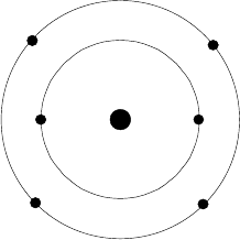
nucleus surrounded by orbiting particles, the atomic electrons; see Fig. 4-1.
This is the classical planetary model, first established by Ernest Rutherford
from analysis of the scattering of alpha particles. In fact, our analysis will
largely follow that of Rutherford. Instead of positive alpha particles, we have
negative incident electrons, but the cause of the scattering remains the same,
namely the electrostatic (Coulomb) interaction between charged particles.
Interaction between the incoming fast electron and an atomic nucleus
gives rise to elastic scattering where (as we will see) almost no energy is
transferred. Interaction between the fast electron and atomic electrons results
in inelastic scattering, in which process the transmitted electron can lose an
appreciable amount of energy. We will consider these two types of event in
turn, dealing first with the kinematics of scattering, where we consider the
momentum and energy of the fast electron, without needing to know the
nature of the forces involved. As the nature of the force does not matter, the
kinematics of electron scattering employs the same principles of classical
mechanics as are used to treat the collision of larger (macroscopic) objects.
94 Chapter 4
+Ze
-e
-e
-e
-e
-e
-e
Figure 4-1. Rutherford model of an atom (in this case, carbon with an atomic number Z = 6).
The electrostatic charge of the central nucleus (whose mass is M ) is +Ze ; the balancing
negative charge is provided by Z atomic electrons, each with charge e .
4.1 Kinematics of Scattering by an Atomic Nucleus
The nucleus of an atom is truly miniscule: about 3 u 10
-15
m in diameter,
whereas the whole atom has a diameter of around 3 u 10
-10
m. The fraction
of space occupied by the nucleus is therefore of the order (10
-5
)
3
= 10
-15
, and
the probability of an electron actually “hitting” the nucleus is almost zero.
However, the electrostatic field of the nucleus extends throughout the atom,
and incoming electrons are deflected (scattered) by this field.
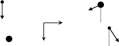
Applying the principle of conservation of energy to the electron-nucleus
system (Fig. 4-2) gives:
mv
0
2
/ 2 = mv
1
2
/ 2 + M V
2
/2 (4.1)
where v
0
and v
1
represent the speed of the electron before and after the
“collision,” and V is the speed of the nucleus (assumed initially at rest)
immediately after the interaction. Because v
0
and v
1
apply to an electron that
is distant from the nucleus, there is no need to include potential energy in
Eq. (4.1). For simplicity, we are using the classical (rather than relativistic)
formula for kinetic energy, taking m and M as the rest mass of the electron
and nucleus. As a result, our analysis will be not be quantitatively accurate
for incident energies E
0
above 50 keV. However, this inaccuracy will not
ffect our general conclusions.a
Applying conservation of momentum to velocity components in the z- and
x-directions (parallel and perpendicular to the original direction of travel of
he fast electron) gives: t
mv
0
= mv
1
cosT + MV cosI (4.2)
0 = mv
1
sinTMV sinI (4.3)
TEM Specimens and Images 95
(a) before
(b) after
T
I
M
m
m
M
z
x
V
v
1
v
0
Figure 4-2. Kinematics of electron-nucleus scattering, showing the speed and direction of the
motion of the electron (mass m) and nucleus (mass M ) before and after the collision.
In the TEM we detect scattered electrons, not recoiling atomic nuclei, so
we need to eliminate the nuclear parameters V and I from Eqs. (4.1) – (4.3).
The angle I can removed by introducing the formula (equivalent to the
Pythagoras rule): sin
2
I + cos
2
I = 1. Taking sin I from Eq. (4.3) and cos I
rom Eq. (4.2) gives: f
M
2
V
2
= M
2
V
2
cos
2
I+ M
2
V
2
sin
2
I
= m
2
v
0
2
+ m
2
v
1
2
2m
2
v
0
v
1
cosT (4.4)
where we have used sin
2
T + cos
2
T = 1 to further simplify the right-hand side
of Eq. (4.4). By using Eq. (4.4) to substitute for MV
2
/2 in Eq. (4.1), we can
liminate V and obtain: e
mv
0
2
/2 mv
1
2
/2 = MV
2
/2 = (1/M)[ m
2
v
0
2
+ m
2
v
1
2
- 2m
2
v
0
v
1
cosT ] (4.5)
The left-hand side of Eq. (4.5) represents kinetic energy lost by the electron
(and gained by the nucleus) as a result of the Coulomb interaction. Denoting
this energy loss as E, we can evaluate the fractional loss of kinetic energy
E/E
0
) as: (
E/E
0
= 2E/(mv
0
2
) = (m/M) [ 1 + v
1
2
/v
0
2
2 (v
1
/v
0
) cosT ] (4.6)
The largest value of E/E
0
occurs when cosT|1, giving:
E/E
0
| (m/M) [ 1 + v
1
2
/v
0
2
+ 2 (v
1
/v
0
) ] = (m/M) [1+v
1
/v
0
]
2
(4.7)
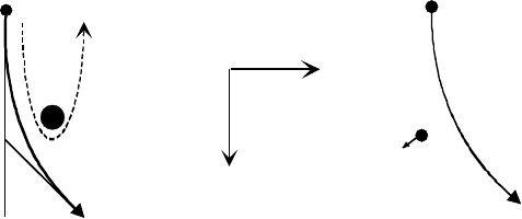
This situation corresponds to an almost head-on collision, where the electron
makes a 180-degree turn behind the nucleus, as illustrated by the dashed
trajectory in Fig. 4-3a. (Remember that a direct collision, in which the
electron actually collides with the nucleus, is very rare).
Kinematics cannot tell us the value of v
1
but we know from Eq. (4.1) that
v
1
< v
0
, so from Eq. (4.7) we can write:
E/E
0
< (m/M) [1+1]
2
= 4m/M = 4m /(Au) (4.8)
where u is the atomic mass unit and A is the atomic mass number (number of
96 Chapter 4
(b) inelastic
m
T
(a) elastic
m
z
x
Figure 4-3. Hyperbolic trajectory of an incident electron during (a) elastic scattering (the case
of a large-angle collision is indicated by the dashed curve) and (b) inelastic scattering.
protons + neutrons in the nucleus), essentially the atomic weight of the atom.
For a hydrogen atom (A = 1), m/M | 1/1836 and Eq. (4.8) gives E/E
0
<
0.002, so for 180-degree scattering only 0.2% of the kinetic energy of the
electron is transmitted to the nucleus. For other elements, and especially for
smaller scattering angles, this percentage is even lower. Scattering in the
electrostatic field of a nucleus is therefore termed elastic, implying that the
scattered electron retains almost all of its kinetic energy.
4.2 Electron-Electron Scattering
If the incident electron passes very close to one of the atomic electrons that
surround the nucleus, both particles experience a repulsive force. If we
ignore the presence of the nucleus and the other atomic electrons, we have a
two-body encounter (Fig. 4-3b) similar to the nuclear interaction depicted in
Figs. 4-2 and 4-3a. Equations (4.1) to (4.6) should still apply, provided we
replace the nuclear mass M by the mass m of an atomic electron (identical to
the mass of the incident electron, as we are neglecting relativistic effects). In
his case, Eq. (4.6) becomes:t
E/E
0
= 1 + v
1
2
/v
0
2
2 (v
1
/v
0
) cosT (4.9)
Because Eq. (4.9) no longer contains the factor (m/M), the energy loss E can
be much larger than for an elastic collision, meaning that scattering from the
electrons that surround an atomic nucleus is inelastic. In the extreme case of
a head-on collision, v
1
| 0 and E | E
0
; in other words, the incident electron
loses all of its original kinetic energy. In practice, E is in the range 10 eV to
50 eV for the majority of inelastic collisions. A more correct treatment of
inelastic scattering is based on wave-mechanical theory and includes the
ollective response of all of the electrons contained within nearby atoms.c
TEM Specimens and Images 97
In practice, an electron traveling through a solid must experience both
repulsive forces (from other electrons) and attractive forces (from atomic
nuclei). But in scattering theory, it turns out to be justified to imagine that
either one or the other effect predominates, and to divide the scattering that
occurs into elastic and inelastic components.
4.3 The Dynamics of Scattering
Because the force acting on a moving electron is electrostatic, its magnitude
F is described by Coulomb’s law. So for elastic scattering of the electron by
he electrostatic field of a single nucleus: t
F = K (e)(Ze)/r
2
(4.10)
where K = 1/(4SH
0
) = 9.0 u 10
9
Nm
2
C
-2
is the Coulomb constant. Applying
Newtonian mechanics (F = ma) to this problem, the electron trajectory can
be shown to be a hyperbola, as depicted in Fig. 4-3a.
The angular distribution of elastic scattering can be expressed in terms
of a probability P(T) for an electron to be scattered through a given angle T.
But to understand TEM contrast, we are more interested in the probability
P(>D) that an electron is scattered through an angle that exceeds the semi-
angle D of the objective aperture. Such an electron will be absorbed within
the diaphragm material that surrounds the aperture, resulting in a reduction
in intensity within the TEM image.
We can think of the nucleus as presenting (to each incident electron) a
target of area V, known as a scattering cross section. Atoms being round,
this target takes the form of a disk of radius a and of area V = S a
2
. An
electron is scattered through an angle T that exceeds the aperture semi-angle
D only if it arrives at a distance less than a from the nucleus, a being a
function of D . But relative to any given nucleus, electrons arrive randomly
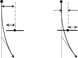
with various values of the impact parameter b, defined as the distance of
closest approach if we neglect the hyperbolic curvature of the trajectory, as
shown in Fig. 4-4. So if b < a, the electron is scattered through an angle
greater than D and is subsequently absorbed by the objective diaphragm.
By how much the scattering angle T exceeds D depends on just how close
the electron passes to the center of the atom: small b leads to large T and vice
versa. Algebraic analysis of the force and velocity components, making use
of Eq. (4.7) and Newton's second law of motion (see Appendix), gives:
T|K Z e
2
/(E
0
b) (4.11)
Equation (4.11) specifies an inverse relation between b and T, as expected
because electrons with a smaller impact parameter pass closer to the nucleus,
98 Chapter 4
T
a
b
T
a
b
(a)
(b)
Figure 4-4. Electron trajectory for elastic scattering through an angle T which is (a) less than
and (b) greater than the objective-aperture semi-angle D. The impact parameter b is defined as
the distance of closest approach to the nucleus if the particle were to continue in a straight-
line trajectory. It is approximately equal to the distance of closest approach when the
scattering angle (and the curvature of the trajectory) is small.
so they experience a stronger attractive force. Because Eq. (4.11) represents
a general relationship, it must hold for T = D, which corresponds to b = a.
Therefore, we can rewrite Eq. (4.11) for this specific case, to give:
D = KZ e
2
/(E
0
a) (4.12)
As a result, the cross section for elastic scattering of an electron through any
ngle greater than D can be written as: a
V
e
= S a
2
= S [KZe
2
/(DE
0
)]
2
= Z
2
e
4
/(16SH
0
2
E
0
2
D
2
) (4.13)
Because V
e
has units of m
2
, it cannot directly represent scattering probability;
we need an additional factor with units of m
2
to provide the dimensionless
number P
e
(>D). In addition, our TEM specimen contains many atoms, each
capable of scattering an incoming electron, whereas V
e
is the elastic cross
section for a single atom. Consequently, the total probability of elastic
cattering in the specimen is: s
P
e
(>D) = N V
e
(4.14)
where N is the number of atoms per unit area of the specimen (viewed in the
direction of an approaching electron), sometimes called an areal density of
toms.a
For a specimen with n atoms per unit volume, N = nt where t is the
specimen thickness. If the specimen contains only a single element of atomic
number A, the atomic density n can be written in terms of a physical density:
U = (mass/volume) = (atoms per unit volume) (mass per atom) = n (Au),
where u is the atomic mass unit (1.66 u 10
-27
kg). Therefore:
P
e
(>D) = [U/(Au)] t V = (U t) (Z
2
/A) e
4
/(16SH
0
2
uE
0
2
D
2
) (4.15)
TEM Specimens and Images 99
P
e
(
>D
)
D
with screening
no screening
0
Figure 4-5. Elastic-scattering probability P
e
(>D), as predicted from Eq. (4.15) (solid curve)
and after including screening of the nuclear field (dashed curve). P
e
(>0) represents the total
probability of elastic scattering, through any angle.
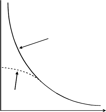
Equation (4.15) indicates that the number of electrons absorbed at the angle-
limiting (objective) diaphragm is proportional to the mass-thickness (Ut) of
he specimen and inversely proportional to the square of the aperture size. t
Unfortunately, Eq. (4.15) fails for the important case of small D; our
formula predicts that P
e
(>D) increases toward infinity as the aperture angle is
reduced to zero. Because any probability must be less than 1, this prediction
shows that at least one of the assumptions used in our analysis is incorrect.
By using Eq. (4.10) to describe the electrostatic force, we have assumed that
the electrostatic field of the nucleus extends to infinity (although diminishing
with distance). In practice, this field is terminated within a neutral atom, due
to the presence of the atomic electrons that surround the nucleus. Stated
another way, the electrostatic field of a given nucleus is screened by the
atomic electrons, for locations outside that atom. Including such screening
complicates the analysis but results in a scattering probability that, as D falls
to zero, rises asymptotically to a finite value P
e
(>0), the probability of elastic
cattering through any angle; see Fig. 4-5. s
Even after correction for screening, Eq. (4.15) can still give rise to an
unphysical result, for if we increase the specimen thickness t sufficiently,
P
e
(>D) becomes greater than 1. This situation arises because our formula
represents a single-scattering approximation: we have tried to calculate the
100 Chapter 4
fraction of electrons that are scattered only once in a specimen. For very thin
specimens, this fraction increases in proportion to the specimen thickness, as
implied by Eq. (4.15). But in thicker specimens, the probability of single
scattering must decrease with increasing thickness because most electrons
become scattered several times. If we use Poisson statistics to calculate the
probability P
n
of n-fold scattering, each probability comes out less than 1, as
expected. In fact, the sum 6P
n
over all n (including n = 0) is equal to 1, as
would be expected because all electrons are transmitted (except for
pecimens that are far too thick for transmission microscopy).s
The above arguments can be repeated for the case of inelastic scattering,
taking the electrostatic force as F = K(e)(e)/r
2
because the incident electron
is now scattered by an atomic electron rather than by the nucleus. The result
is an equation similar to Eq. (4.15) but with the factor Z
2
missing. However,
this result would apply to inelastic scattering by only a single atomic
electron. Considering all Z electrons within the atom, the inelastic-scattering
probability must be multiplied by Z, giving:
P
i
(>D) = (Ut) (Z/A) e
4
/(16SH
0
2
uE
0
2
D
2
) (4.16)
Comparison of Eq. (4.16) with Eq. (4.15) shows that the amount of inelastic
cattering, relative to elastic scattering, is s
P
i
(>D) /P
e
(>D) = 1/Z (4.17)
Equation (4.17) predicts that inelastic scattering makes a relatively small
contribution for most elements. However, it is based on Eq. (4.15), which is
accurate only for larger aperture angles. For small D, where screening of the
nuclear field reduces the amount of elastic scattering, the inelastic/elastic
ratio is much higher. More accurate theory (treating the incoming electrons
as waves) and experimental measurements of the scattering show that P
e
(>0)
is typically proportional to Z
1.5
(rather than Z
2
), that P
i
(>0) is proportional
o Z
0.5
(rather than Z ) and that, considering scattering through all angles,t
P
i
(>0)/P
e
(>0) | 20/Z (4.18)
Consequently, inelastic scattering makes a significant contribution to the
otal scattering (through any angle) in the case of light (low-Z) elements.t
Although our classical-physics analysis turns out to be rather inaccurate
in predicting absolute scattering probabilities, the thickness and material
parameters involved in Eq. (4.15) and Eq. (4.16) do provide a reasonable
explanation for the contrast (variation in intensity level) in TEM images
obtained from non-crystalline (amorphous) specimens such as glasses,
certain kinds of polymers, amorphous thin films, and most types of
biological material.
TEM Specimens and Images 101
4.4 Scattering Contrast from Amorphous Specimens
Most TEM images are viewed and recorded with an objective aperture
(diameter D) inserted and centered about the optic axis of the TEM objective
lens (focal length f ). As represented by Eq. (3.9), this aperture absorbs
electrons that are scattered through an angle greater than D| 0.5D/f.
However, any part of the field of view that contains no specimen (such as a
hole or a region beyond the specimen edge) is formed from electrons that
remain unscattered, so that part appears bright relative to the specimen. As a
esult, this central-aperture image is referred to as a bright-field image. r
Biological tissue, at least in its dry state, is mainly carbon and so, for this
common type of specimen, we can take Z = 6, A = 12, and U| 2 g/cm
2
=
2000 kg/m
3
. For a biological TEM, typical parameters are E
0
= 100 keV =
1.6 u 10
-14
J and D| 0.5D/f = 10 mrad = 0.01 rad, taking an objective-lens
focal length f = 2 mm and objective-aperture diameter D = 40 Pm. With
these values, Eq. (4.15) gives P
e
(D) | 0.47 for a specimen thickness of t = 20
nm. The same parameters inserted into Eq.(4.16) give P
i
(>D) | 0.08, and the
total fraction of electrons that are absorbed by the objective diaphragm is
(>10 mrad) | 0.47 + 0.08 = 0.54 . P
We therefore predict that more than half of the transmitted electrons are
intercepted by a typical-size objective aperture, even for a very thin (20 nm)
specimen. This fraction might be even larger for a thicker specimen, but our
single-scattering approximation would not be valid. In practice, the specimen
thickness must be less than about 200 nm, assuming an accelerating potential
of 100 kV and a specimen consisting mainly of low-Z elements. If the
specimen is appreciably thicker, only a small fraction of the transmitted
electrons pass through the objective aperture, and the bright-field image is
ver dim on the TEM screen.y
Because (A/Z) is approximately the same (|2) for all elements, Eq.(4.16)
indicates that P
i
(>D) increases only slowly with increasing atomic number,
due to the density term U, which tends to increase with increasing Z. But
using the same argument, Eq. (4.15) implies that P
e
(>D) is approximately
proportional to UZ. Therefore specimens that contain mainly heavy elements
scatter electrons more strongly and would have to be even thinner, placing
unrealistic demands on the specimen preparation. Such specimens are
usually examined in a “materials science” TEM that employs an accelerating
voltage of 200 kV or higher, taking advantage of the reduction in V
e
and P
e
with increasing E
0
; see Eqs. (4.13) and (4.15).
For imaging non-crystalline specimens, the main purpose of the objective
aperture is to provide scattering contrast in the TEM image of a specimen
