Egerton R.F. Physical Principles of Electron Microscopy. An Introduction to TEM, SEM, and AEM
Подождите немного. Документ загружается.

82 Chapter 3
A more correct procedure for finding the minimum is to differentiate 'r
with respect to D and set the total derivative to zero, corresponding to zero
slope at the minimum of the curve, which gives: D* = 0.67 (O/C
s
)
1/4
.
An even better procedure is to treat the blurring terms r
s
and 'x like
statistical errors and combine their effect in quadrature:
('r)
2
| (r
s
)
2
+ ('x)
2
| (C
s
D
3
)
2
+ (0.6 O/D)
2
(3.12)
Taking the derivative and setting it to zero then results in:
D* = 0.63 (O/C
s
)
1/4
(3.13)
As an example, let us take E
0
= 200 keV, so that O = 2.5 pm, and C
s
| f /4 =
0.5 mm, as in Fig. 2-13. Then Eq. (3.13) gives D* = 5.3 mrad, corresponding
to an objective aperture of diameter D | 2D*f | 20 Pm. Using Eq.(3.12), the
optimum resolution is 'r* | 0.29 nm.
Inclusion of chromatic aberration in Eq. (3.12) would decrease D* a little
and increase 'r*. However, our entire procedure is somewhat pessimistic: it
assumes that electrons are present in equal numbers at all scattering angles
up to the aperture semi-angle D. Also, a more exact treatment would be
based on wave optics rather than geometrical optics. In practice, a modern
200 kV TEM can achieve a point resolution below 0.2 nm, allowing atomic
resolution under suitable conditions, which include a low vibration level and
low ambient ac magnetic field (Muller and Grazul, 2001). Nevertheless, our
calculation has illustrated the importance of lens aberrations. Without these
aberrations, large values of D would be possible and the TEM resolution,
limited only by Eq. (3.10), would (for sin D| 0.6) be 'x |O| 0.025 nm,
well below atomic dimensions.
In visible-light microscopy, lens aberrations can be made negligible.
Glass lenses of large aperture can be used, such that sin D approaches 1 and
Eq. (3.10) gives the resolution as 'x | 300 nm as discussed in Chapter 1. But
as a result of the much smaller electron wavelength, the TEM resolution is
better by more than a factor of 1000, despite the electron-lens aberrations.
Objective stigmator
The above estimates of resolution assume that the imaging system does not
suffer from astigmatism. In practice, electrostatic charging of contamination
layers (on the specimen or on an objective diaphragm) or hysteresis effects
in the lens polepieces give rise to axial astigmatism that may be different for
each specimen. The TEM operator can correct for axial astigmatism in the
objective (and other imaging lenses) using an objective stigmator located just
below the objective lens. Obtaining the correct setting requires an adjustment
The Transmission Electron Microscope 83
of amplitude and orientation, as discussed in Section 2.6. One way of setting
these controls is to insert an amorphous specimen, whose image contains
small point-like features. With astigmatism present, there is a “streaking
effect” (preferred direction) visible in the image, which changes in direction
through 90 degrees when the objective is adjusted from underfocus to
overfocus of the specimen image (similar to Fig. 5.17). The stigmator
controls are adjusted to minimize this streaking effect.
Selected-area aperture
As indicated in Fig. 3-12c and Fig. 3-14, a diaphragm can be inserted in the
plane that contains the first magnified (real) image of the specimen, the
image plane of the objective lens. This selected-area diffraction (SAD)
diaphragm is used to limit the region of specimen from which an electron
diffraction pattern is recorded. Electrons are transmitted through the aperture
only if they fall within its diameter D, which corresponds to a diameter of
D/M at the specimen plane. In this way, diffraction information can be
obtained from specimen regions whose diameter can be as small as 0.2 Pm
(taking D | 20 Pm and an objective-lens magnification M | 100).
As seen from Fig. 3-12c, sharp diffraction spots are formed at the
objective back-focal plane (for electrons scattered at a particular angle) only
if the electron beam incident on the specimen is almost parallel. This
condition is usually achieved by defocusing the second condenser lens,
giving a low convergence angle but a large irradiation area at the specimen
(Fig. 3-9). The purpose of the SAD aperture is therefore to provide
diffraction information with good angular resolution, combined with good
spatial resolution.
Intermediate lens
A modern TEM contains several lenses between the objective and the final
(projector) lens. At least one of these lenses is referred to as the intermediate,
and the combined function of all of them can be described in terms of the
action of a single intermediate lens, as shown in Fig. 3-14. The intermediate
serves two purposes. First of all, by changing its focal length in small steps,
its image magnification can be changed, allowing the overall magnification
of the TEM to be varied over a large range, typically 10
3
to 10
6
.
Second, by making a larger change to the intermediate lens excitation, an
electron diffraction pattern can be produced on the TEM viewing screen. As
depicted in Fig. 3-14, this is achieved by reducing the current in the
intermediate so that this lens produces, at the projector object plane, a
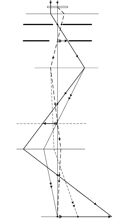
84 Chapter 3
diffraction pattern (solid ray crossing the optic axis) rather than an image of
the specimen (dashed ray crossing the axis).
Because the objective lens reduces the angles of electrons relative to the
optic axis, intermediate-lens aberrations are not of great concern. Therefore,
the intermediate can be operated as a weak lens (f | several centimeters)
without degrading the image or diffraction pattern.
specimen
objective
intermediate
lens(es)
projector
lens
viewing screen
objective
aperture
SAD
aperture
projector
object plane
Figure 3-14. Thin-lens ray diagram of the imaging system of a TEM. As usual, the image
rotation has been suppressed so that the electron optics can be represented on a flat plane.
Image planes are represented by horizontal arrows and diffraction planes by horizontal dots.
Rays that lead to a TEM-screen diffraction pattern are identified by the double arrowheads.
(Note that the diagram is not to scale: the final image magnification is only 8 in this example).
The Transmission Electron Microscope 85
Projector lens
The purpose of the projector lens is to produce an image or a diffraction
pattern across the entire TEM screen, with an overall diameter of several
centimeters. Because of this requirement, some electrons (such as the solid
single-arrow ray in Fig. 3-14) arrive at the screen at a large distance from the
optic axis, introducing the possibility of image distortion (Chapter 2). To
minimize this effect, the projector is designed to be a strong lens, with a
focal length of a few millimeters. Ideally, the final-image diameter is fixed
(the image should fill the TEM screen) and the projector operates at a fixed
excitation, with a single object distance and magnification v/u | 100.
However, in many TEMs the projector-lens strength can be reduced in order
to give images of relatively low magnification (< 1000) on the viewing
screen. As in the case of light optics, the final-image magnification is the
algebraic product of the magnification factors of each of the imaging lenses.
TEM screen and camera
A phosphor screen is used to convert the electron image to a visible form. It
consists of a metal plate coated with a thin layer of powder that fluoresces
(emits visible light) under electron bombardment. The traditional phosphor
material is zinc sulfide (ZnS) with small amounts of metallic impurity added,
although alternative phosphors are available with improved sensitivity
(electron/photon conversion efficiency). The phosphor is chosen so that light
is emitted in the middle of the spectrum (yellow-green region), to which the
human eye is most sensitive. The TEM screen is used mainly for focusing a
TEM image or diffraction pattern, and for this purpose, light-optical
binoculars are often mounted just outside the viewing window, to provide
some additional magnification. The viewing window is made of special glass
(high lead content) and is of sufficient thickness to absorb the x-rays that are
produced when the electrons deposit their energy at the screen.
To permanently record a TEM image or diffraction pattern, photographic
film can be used. This film has a layer of a silver halide (AgI and/or AgBr)
emulsion, similar to that employed in black-and-white photography; both
electrons and photons produce a subtle chemical change that can be
amplified by immersion in a developer solution. In regions of high image
intensity, the developer reduces the silver halide to metallic silver, which
appears dark. The recorded image is a therefore a photographic negative,
whose contrast is reversed relative to the image seen on the TEM screen. An
optical enlarger can be used to make a positive print on photographic paper,
which also has a silver-halide coating.
86 Chapter 3
Nowadays, photographic film has been largely replaced by electronic
image-recording devices, based on charge-coupled diode (CCD) sensors.
They contain an array of a million or more silicon photodiodes, each of
which provides an electrical signal proportional to the local intensity level.
Because such devices are easily damaged by high-energy electrons, they are
preceded by a phosphor screen that converts the electron image to variations
in visible-light intensity. Electronic recording has numerous advantages. The
recorded image can be inspected immediately on a monitor screen, avoiding
the delay (and cost) associated with photographic processing. Because the
CCD sensitivity is high, even high-magnification (with low-intensity)
images can be viewed without eyestrain, making focusing and astigmatism
correction a lot easier. The image information is stored digitally in computer
memory and subsequently on magnetic or optical disks, from which previous
images can be rapidly retrieved for comparison purposes. The digital nature
of the image also allows various forms of image processing, as well as rapid
transfer of images between computers by means of the Internet.
Depth of focus and depth of field
As a matter of convenience, the film or CCD camera is located several
centimeters below (or sometimes above) the TEM viewing screen. If a
specimen image (or a diffraction pattern) has been brought to exact focus on
the TEM screen, it is strictly speaking out of focus at any other plane. At a
plane that is a height h below (or above) the true image plane, each point in
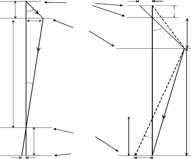
the image becomes a circle of confusion whose radius is s = h tan E , as
illustrated in Fig. 3-15a. This radius is equivalent to a blurring:
's = s/M = (h/M) tan E (3.14)
in the specimen plane, where M is the combined magnification of the entire
imaging system and E is the convergence angle at the screen, corresponding
to scattering through an angle D in the specimen, as shown in Fig. 3-15a.
However, this blurring will be significant only if 's is comparable to or
greater that the resolution 'r of the in-focus TEM image. In other words, the
additional image blurring will not be noticeable if 's << 'r.
To compute 's we first note, from trigonometry of the two large triangles
in Fig. 3-15a, that x = utanD = v tanE, giving tanE = (u/v)tanD = (1/M)tanD.
If an objective diaphragm is in place to remove electrons above D| 5 mrad,
then E| tan E|D/ M = 5 u 10
-6
rad for M = 1000, decreasing to 5 u 10
-9
rad
for M =10
6
. These are extremely small angles, giving rise to correspondingly
small values of 's: for h = 10 cm, Eq. (3.14) gives 's = 0.5 nm for M = 1000
and 's = 5 u 10
-16
m for M = 10
6
. Therefore, for typical TEM magnifications,
the final image recorded at a different plane is equally sharp.
The Transmission Electron Microscope 87
Another way of expressing this result is as follows. Suppose we take 's =
'r, so that the image resolution is only slightly degraded (by a factor |2)
by being out-of-focus. The value of h that would give rise to this 's, for a
given M and D, is known as the depth of focus of the image (sometimes
called depth of image). Taking D = 5 mrad and 'r = 0.2 nm, our depth of
focus is h = M 'r/E| M
2
'r/D = 4 cm for M = 1000, increasing to h = 40 km
for M = 10
6
. In practice, a magnification as low as 1000 would be unsuitable
for recording a high-resolution image, because image detail on a scale M'r |
0.2 Pm would be below the resolution limit of a phosphor, film, or CCD
sensor. Therefore, practical values of the depth of focus are so large that a
magnified image of the specimen is equally sharp at all recording planes.
v
u
h
s
D
E
x
'
u
specimen
viewing
screen
recording
plane
equivalent
lens
D
u
(a) (b)
v
'
v
E
'
r
R
Figure 3-15. (a) Ray diagram illustrating depth of focus; the TEM imaging lenses have been
replaced by a single lens of large magnification M. (b) Ray diagram illustrating depth of field.
The radius of blurring (caused by raising the specimen through a distance 'u) is R in the
image-recording plane, equivalent to 'r = R / M in the specimen plane.
88 Chapter 3
A related concept is depth of field: the distance 'u (along the optic axis)
that the specimen can be moved without its image (focused at a given plane)
becoming noticeably blurred. Replacing the TEM imaging system with a
single lens of fixed focal length f and using 1/u + 1/v = 1/f , the change in
image distance is 'v = (v/u)
2
'u = M
2
'u (the minus sign denotes a
decrease in v, as in Fig. 3-15b). The radius of the disk of confusion in the
image plane (due to defocus) is R = E_'v_ = (D/M) ('v) = D M 'u,
equivalent to a radius 'r = R/M = D'u at the specimen plane. This same
result can be obtained more directly by extrapolating backwards the solid ray
in Fig. 3-15b to show that the electrons that arrive at a single on-axis point in
the image could have come from anywhere within a circle whose radius
(from geometry of the topmost right-angle triangle) is 'r = D'u.
If we take D = 5 mrad and set the loss or resolution 'r equal to the TEM
resolution (| 0.2 nm), we get 'u = (0.2 nm)/(0.005) = 40 nm as the depth of
field. Most TEM specimens are of the order 100 nm or less in thickness. If
the objective lens is focused on features at the mid-plane of the specimen,
features close to the top and bottom surfaces will still be acceptably in focus.
In other words, the small value of D ensures that a TEM image is normally a
projection of all structure present in the specimen. It is therefore difficult or
impossible to obtain depth information from a single TEM image. On the
other hand, this substantial depth of field avoids the need to focus on several
different planes within the specimen, as is sometimes necessary in a light-
optical microscope where D may be large and the depth of field can be
considerably less than the specimen thickness.
3.6 Vacuum System
It is essential to remove most of the air from the inside of a TEM column, so
that the accelerated electrons can follow the principles of electron optics,
rather than being scattered by gas molecules. In addition, the electron gun
requires a sufficiently good vacuum in order to prevent a high-voltage
discharge and to avoid oxidation of the electron-emitting surface.
A mechanical rotary pump (RP) is used in many vacuum systems. This
pump contains a rotating assembly, driven by an electric motor and equipped
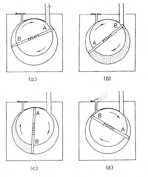
with internal vanes (A and B in Fig. 3-16) separated by a coil spring so that
they press against the inside cylindrical wall of the pump, forming an airtight
seal. The interior of the pump is lubricated with a special oil (of low vapor
pressure) to reduce friction and wear of the sliding surfaces. The rotation
axis is offset from the axis of the cylinder so that, as gas is drawn from the
inlet tube (at A in Fig. 3-16a), it expands considerably in volume before
being sealed off by the opposite vane (B in Fig. 3-16b).
The Transmission Electron Microscope 89
Figure 3-16. Schematic diagram of a rotary vacuum pump, illustrating one complete cycle of
the pumping sequence. Spring-loaded vanes A and B rotate clockwise, each drawing in gas
molecules and then compressing them toward the outlet.
During the remainder of the rotation cycle, the air is compressed (Fig. 3-16c)
and driven out of the outlet tube (Fig. 3-16d). Meanwhile, air in the opposite
half of the cylinder has already expanded and is about to be compressed to
the outlet, giving a continuous pumping action. The rotary pump generates a
“rough” vacuum with a pressure down to 1 Pa, which is a factor | 10
5
less
than atmospheric pressure but still too high for a tungsten-filament source
and totally inadequate for other types of electron source.
To produce a “high” vacuum (| 10
-3
Pa), a diffusion pump (DP) can be
added. It consists of a vertical metal cylinder containing a small amount of a
special fluid, a carbon- or silicon-based oil having very low vapor pressure
and a boiling point of several hundred degrees Celsius. At the base of the
pump (Fig. 3-17), an electrical heater causes the fluid to boil and turn into a
vapor that rises and is then deflected downwards through jets by an internal
baffle assembly. During their descent, the oil molecules collide with air
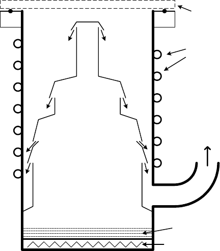
90 Chapter 3
molecules, imparting a downward momentum and resulting in a downward
flow of gas from the inlet of the pump. When the oil molecules arrive at the
cooled inside wall of the DP, they condense back into a liquid that falls
under gravity to the base of the pump, allowing the oil to be continuously
recycled. To prevent oxidation of the oil and multiple collisions between oil
and gas molecules, the pressure at the base of the diffusion pump must be
below about 10 Pa before the heater is turned on. A rotary pump is therefore
connected to the bottom end of the diffusion pump, to act as a “backing
pump” (Fig. 3-17). This rotary pump (or a second one) is also used to
remove most of the air from the TEM column before the high-vacuum valve
(connecting the DP to the column) is opened.
Sometimes a tubomolecular pump (TMP), essentially a high-speed
turbine fan, is used in place of (or to supplement) a diffusion pump. Usually
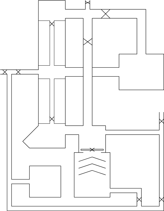
an ion pump (IP in Fig. 3-18) is used to achieve pressures below 10
-4
Pa, as
required to operate a LaB
6
, Schottky, or field-emission electron source. By
applying a potential difference of several kilovolts between large electrodes,
a low-pressure discharge is set up (aided by the presence of a magnetic field)
which removes gas molecules by burying them in one of the electrodes.
fluid
heater
water
cooling
to rotary
backing
pump
high-vacuum
valve
Figure 3-17. Cross section through a diffusion pump. The arrows show oil vapor leaving jets
within the baffle assembly. Water flowing within a coiled metal tube keeps the walls cool.
The Transmission Electron Microscope 91
These different pumps must work together in the correct sequence. If the
TEM column has been at atmospheric pressure (perhaps for maintenance),
most of the air is removed by a rotary pump, which can then function as a
backing pump for the DP, TMP, or IP. The pumping sequence is controlled
by opening and closing vacuum valves in response to the local pressure,
monitored by vacuum gauges located in different parts of the system. In a
modern TEM, the vacuum system is automated and under electronic control;
in fact, its circuit board can be the most complicated in the entire electronics
of the microscope.
DP
RP
viewing
chamber
gun
IP
G
G
G
G
Figure 3-18. Simplified diagram of the vacuum system of a typical TEM. Vacuum valves are
indicated by
u and vacuum gauges by G. Sometimes a turbomolecular pump is used in place
of the diffusion pump, resulting in a cleaner “oil-free” vacuum system.
