Egerton R.F. Physical Principles of Electron Microscopy. An Introduction to TEM, SEM, and AEM
Подождите немного. Документ загружается.

72 Chapter 3
The second condenser (C2) lens is a weak magnetic lens (f | several
centimeters) that provides little or no magnification ( M | 1) but allows the
diameter of illumination (d) at the specimen to be varied continuously over a
wide range. The C2 lens also contains the condenser aperture (the hole in
the condenser diaphragm) whose diameter D can be changed in order to
control the convergence semi-angle D of the illumination, the maximum
angle by which the incident electrons deviate from the optic axis.
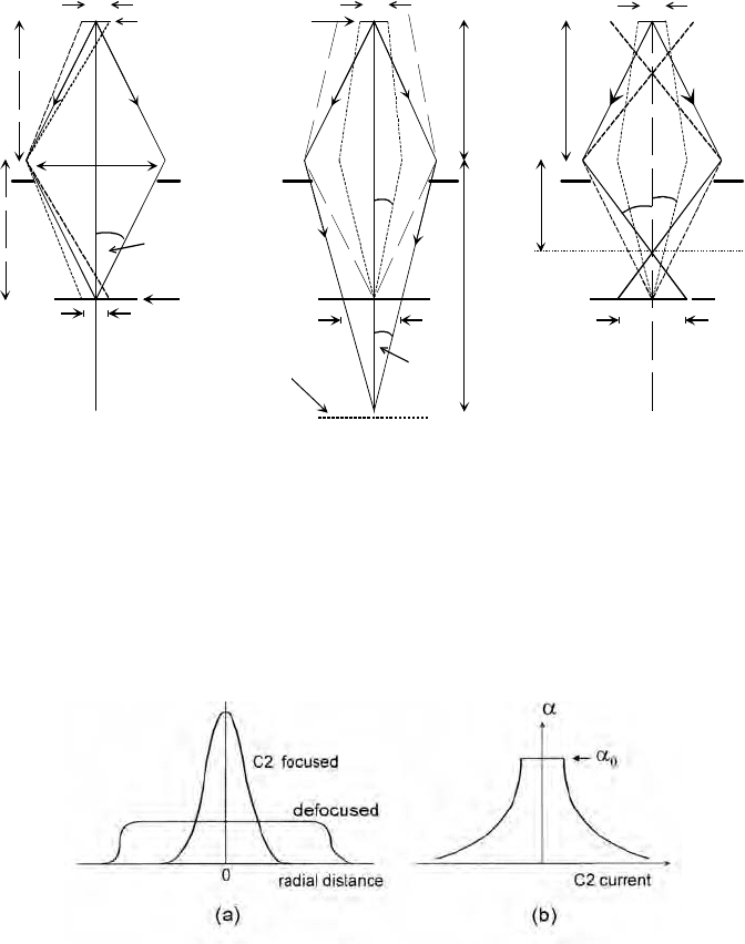
The case of fully-focused illumination is shown in Fig. 3-8a. An image
of the electron source is formed at the specimen plane (image distance v
0
),
and the illumination diameter at that plane is therefore d
0
= M d
1
(| d
1
if
object distance u | v
0
). This condition provides the smallest illumination
diameter (below 1 Pm), as required for high-magnification imaging. Because
the condenser aperture is located close to the principal plane of the lens, the
illumination convergence angle is given by 2D
0
| D/v
0
| 10
-3
rad = 1 mrad
for = 100 Pm and v
0
= 10 cm.D
Figure 3-8b shows the case of underfocused illumination, in which the
C2 lens current has been decreased so that an image of the electron source is
formed below the specimen, at a larger distance v from the lens. Because the
specimen plane no longer contains an image of the electron source, the
diameter of illumination at that plane is no longer determined by the source
diameter but by the value of v. Taking v = 2v
0
, for example, simple geometry
gives the convergence semi-angle at the image as T|D/v |D
0
/2 and the
illumination diameter as d | (2T)(v v
0
) |D
0
v
0
= 50 Pm. As shown by the
dashed lines in Fig. 3-8b, electrons arriving at the center of the specimen at
the previous angle D
0
relative to the optic axis (as in Fig. 3-8a) would have
to originate from a region outside the demagnified source, and because there
are no such electrons, the new convergence angle D of the illumination must
be smaller than D
0
. Using the brightness-conservation theorem, Eq. (3.4), the
product (Dd) must be the same at the new image plane and at the specimen,
giving D = D
0
(d
0
/d) | (0.5mrad)(1Pm/50Pm) | 0.010 mrad. Defocusing the
illumination therefore ensures that the incident electrons form an almost
parallel beam. This condition is useful for recording electron-diffraction
patterns in the TEM or for maximizing the contrast in images of crystalline
specimens and is obtained by defocusing the C2 lens or using a small C2
perture, or both.a
The situation for overfocused illumination, where the C2 current has
been increased so that the image occurs above the specimen plane, is shown
in Fig. 3-8c. In comparison with the fully-focused condition, the illumination
diameter d is again increased and the convergence semi-angle D at the
specimen plane is reduced in the same proportion, in accordance with the
brightness theorem. Note that this low convergence angle occurs despite an

The Transmission Electron Microscope 73
increase in the beam angle T at the electron-source image plane. In this
context, it should be noted that the convergence angle of the illumination is
always defined in terms of the variation in angle of the electrons that arrive
at a single point in the specimen.
(a) (b) (c)
demagnified source
C2
aperture
D
D
specimen
D
v
0
u
d
1
dd
d
0
v'
T
T
v'
u
image
plane
image plane
u
D
d
1
d
1
Figure 3-8. Operation of the second condenser (C2) lens; solid rays represent electrons
emitted from the center of the C1-demagnified source. (a) Fully-focused illumination whose
diameter d
0
is comparable to the diameter d
1
of the demagnified source (see dashed rays) and
whose convergence angle (2D
0
) depends on the diameter of the C2 aperture. (b) Underfocused
illumination whose diameter d depends on the image distance v and whose convergence angle
D depends on v and on d
1
. (c) Overfocused illumination, also providing large d and small D .
Figure 3-9. (a) Current density at the specimen as a function of distance from the optic axis,
for illumination fully focused and for defocused (underfocused or overfocused) illumination.
(b) Convergence semi-angle of the specimen illumination, as a function of C2-lens excitation.
74 Chapter 3
Figure 3-9 summarizes these conclusions in terms of the current-density
profile at the specimen plane, which is directly observable (with the radial
distance magnified) as a variation in image intensity on the TEM screen.
Condenser aperture
The condenser aperture is the small hole in a metal diaphragm located just
below the polepieces of the C2 lens. In order to center the aperture on the
optic axis, the diaphragm is mounted at the end of a support rod that can be
moved precisely in the horizontal plane (x and y directions) by turning knobs
located outside the microscope column, or by an electric-motor drive. The
aperture is correctly aligned (on the optic axis) when variation of the C2
current changes the illumination diameter but does not cause the magnified
disk of illumination to move across the viewing screen. In practice, there are
three or four apertures of different diameter (D | 20 200 Pm), arranged
along the length of the support rod, so that moving the rod in or out by
several mm places a different aperture on the optic axis. Choosing a larger
size increases the convergence angle D of the illumination but allows more
electrons to reach the specimen, giving higher intensity in the TEM image.
Condenser stigmator
The condenser-lens system also contains a stigmator to correct for residual
astigmatism of the C1 and C2 lenses. When such astigmatism is present and
the amplitude control of the stigmator is set to zero, the illumination (viewed
on the TEM screen, with or without a TEM specimen) expands into an
ellipse (rather than a circle) when the C2 lens excitation is increased or
decreased from the focused-illumination setting; see Fig. 3-10. To correctly
adjust the stigmator, its amplitude control is first set to maximum and the
orientation control adjusted so that the major axis of the ellipse lies
perpendicular to the zero-amplitude direction. The orientation is then correct
but the lens astigmatism has been overcompensated. To complete the
process, the amplitude setting is reduced until the illumination ellipse
becomes a circle, now of smaller diameter than would be possible without
astigmatism correction (Fig. 3-10e). In other words, the illumination can be
focused more tightly after adjusting the stigmator. To check that the setting
is optimum, the C2 current can be varied around the fully-focused condition;
the illumination should contract or expand but always remain circular.
Condenser-lens astigmatism does not directly affect the resolution of a
TEM-specimen image. However, it does reduce the maximum intensity
(assuming focused illumination) of such an image on the TEM screen and

The Transmission Electron Microscope 75
therefore the ability of the operator to focus on fine details. Therefore, the
condenser stigmator is routinely adjusted as part of TEM-column alignment.
(a) (b) (c) (d) (e)
Figure 3-10. TEM-screen illumination when axial astigmatism is present and the C2 lens is
(a) underfocused, (b) fully focused, and (c) overfocused. Also shown: effect of the condenser
stigmator with (d) its correct orientation (for overfocus condition) but maximum amplitude,
and (e) correct orientation and amplitude; note that the focused illumination now has smaller
diameter than with no astigmatism correction.
Illumination shift and tilt controls
The illumination system contains two pairs of coils that apply uniform
magnetic fields in the horizontal (x and y) directions, in order to shift the
electron beam (incident on the specimen) in the y and x directions,
respectively. The current in these coils is varied by two illumination-shift
controls, which are normally used to center the illumination on the TEM
screen, correcting for any horizontal drift of the electron gun or slight
misalignment of the condenser lenses.
A second pair of coils is used to adjust the angle of the incident beam
relative to the optic axis. They are located at the plane of the C1 image so
that their effect on the electron rays does not shift the illumination. The
currents in these coils are adjusted using (x and y) illumination-tilt controls,
which are often adjusted to align the illumination parallel to the optic axis
(to minimize aberrations of the TEM imaging lenses) but can also be used to
set up the illumination to give dark-field images, as discussed in Chapter 4.
3.4 The Specimen Stage
To allow observation in different brands or models of microscope, TEM
specimens are always made circular with a diameter of 3 mm. Perpendicular
to this disk, the specimen must be thin enough (at least in some regions) to
allow electrons to be transmitted to form the magnified image. The specimen
stage is designed to hold the specimen as stationary as possible, as any drift
or vibration would be magnified in the final image, impairing its spatial
76 Chapter 3
resolution (especially if the image is recorded by a camera over a period of
several seconds). But in order to view all possible regions of the specimen, it
is also necessary to move the specimen horizontally over a distance of up to
3 mm if necessary.
The design of the stage must also allow the specimen to be inserted into
the vacuum of the TEM column without introducing air. This is achieved by
inserting the specimen through an airlock, a small chamber into which the
specimen is placed initially and which can be evacuated before the specimen
enters the TEM column. Not surprisingly, the specimen stage and airlock are
the most mechanically complex and precision-machined parts of the TEM.
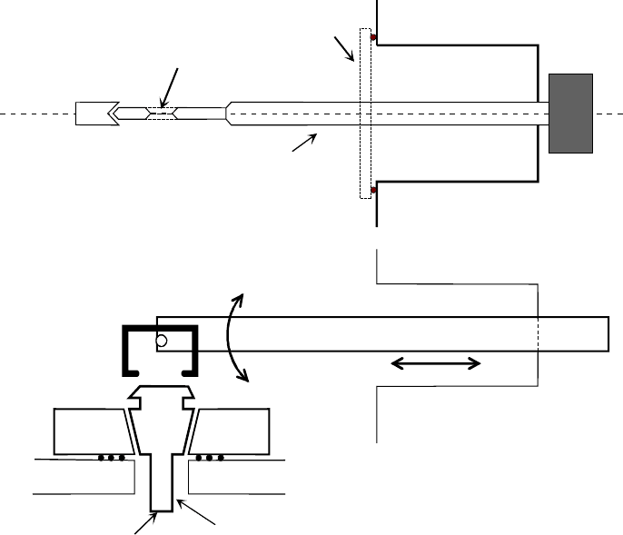
There are two basic designs of the specimen stage: side-entry and top-entry.
In a side-entry stage, the specimen is clamped (for example, by a
threaded ring) close to the end of a rod-shaped specimen holder and is
inserted horizontally through the airlock. The airlock-evacuation valve and a
high-vacuum valve (at the entrance to the TEM column) are activated by
rotation of the specimen holder about its long axis; see Fig. 3-11a.
One advantage of this side-entry design is that it is easy to arrange for
precision motion of the specimen. Translation in the horizontal plane (x and
y directions) and in the vertical (z) direction is often achieved by applying
the appropriate movement to an end-stop that makes contact with the pointed
end of the specimen holder. Specimen tilt (rotation to a desired orientation)
about the long axis of the rod is easily achieved by turning the outside end of
the specimen holder. Rotation about a perpendicular (horizontal or vertical)
axis can be arranged by mounting the specimen on a pivoted ring whose
orientation is changed by horizontal movement of a rod that runs along the
inside of the specimen holder. Precise tilting of the specimen is sometimes
required in order to examine the shape of certain features or to characterize
the nature of microscopic defects in a crystalline material.
A further advantage of the side-entry stage is that heating of a specimen
is easy to arrange, by installing a small heater at the end of the specimen
holder, with electrical leads running along the inside of the holder to a power
supply located outside the TEM. The ability to change the temperature of a
specimen allows structural changes in a material (such as phase transitions)
to be studied at the microscopic level.
Specimen cooling can also be achieved, by incorporating (inside the side-
entry holder) a heat-conducting metal rod whose outer end is immersed in
liquid nitrogen (at 77 K). If the temperature of a biological-tissue specimen
is lowered sufficiently below room temperature, the vapor pressure of ice
becomes low enough that the specimen can be maintained in a hydrated state
during its examination in the TEM.

The Transmission Electron Microscope 77
One disadvantage of the side-entry design is that mechanical vibration,
picked up from the TEM column or from acoustical vibrations in the
external air, is transmitted directly to the specimen. In addition, any thermal
expansion of the specimen holder can cause drift of the specimen and of the
TEM image. These problems have been largely overcome by careful design,
including choice of materials used to construct the specimen holder. As a
result, side-entry holders are widely used, even for high-resolution imaging.
In a top-entry stage, the specimen is clamped to the bottom end of a
cylindrical holder that is equipped with a conical collar; see Fig. 3-11b. The
holder is loaded into position through an airlock by means of a sliding and
tilting arm, which is then detached and retracted. Inside the TEM, the cone
of the specimen holder fits snugly into a conical well of the specimen stage,
which can be translated in the (x and y) horizontal directions by a precision
gear mechanism.
(a) Side-entry stage
end-stop
specimen
specimen holder
high-
vacuum
valve
airlock
specimen
specimen holder
movable
stage
top of objective lens
specimen loading rod
(b) Top-entry stage
airlock
Figure 3-11. Schematic diagrams of (a) a side-entry and (b) a top-entry specimen holder.
78 Chapter 3
The major advantage of a top-entry design is that the loading arm is
disengaged after the specimen is loaded, so the specimen holder is less liable
to pick up vibrations from the TEM environment. In addition, its axially
symmetric design tends to ensure that any thermal expansion occurs radially
about the optic axis and therefore becomes small close to the axis. However,
it is more difficult to provide tilting, heating, or cooling of the specimen.
Although such facilities have all been implemented in top-entry stages, they
require elaborate precision engineering, making the holder fragile and
expensive. Because the specimen is held at the bottom of its holder, it is
difficult to collect more than a small fraction of the x-rays that are generated
by the transmitted beam and emitted in the upward direction, making this
design less attractive for high-sensitivity elemental analysis (see Chapter 6).
3.5 TEM Imaging System
The imaging lenses of a TEM produce a magnified image or an electron-
diffraction pattern of the specimen on a viewing screen or camera system.
The spatial resolution of the image is largely dependent on the quality and
design of these lenses, especially on the first imaging lens: the objective.
Objective lens
As in the case of a light-optical microscope, the lens closest to the specimen
is called the objective. It is a strong lens, with a small focal length; because
of its high excitation current, the objective must be cooled with temperature-
controlled water, thereby minimizing image drift that could result from
thermal expansion of the specimen stage. Because focusing power depends
on lens excitation, the current for the objective lens must be highly
stabilized, using negative feedback within its dc power supply. The power
supply must be able to deliver substantially different lens currents, in order
to retain the same focal length for different electron-accelerating voltages.
The TEM also has fine controls that enable the operator to make small
fractional adjustments to the objective current, to allow the specimen image
to be accurately focused on the viewing screen.
The objective produces a magnified real image of the specimen (M | 50
to 100) at a distance v | 10 cm below the center of the lens. Because of the
small value of f , Eq. (2.2) indicates that the object distance u is only slightly
greater than the focal length, so the specimen is usually located within the
pre-field of the lens (that part of the focusing field that acts on the electron
before it reaches the center of the lens). By analogy with a light microscope,
the objective is therefore referred to as an immersion lens.

The Transmission Electron Microscope 79
(b)
S
BFP
PP
(c)
u ~ f
f
D
objective
aperture
specimen
image
R
D
v
(a)
S
BFP
PP
selected-area
aperture
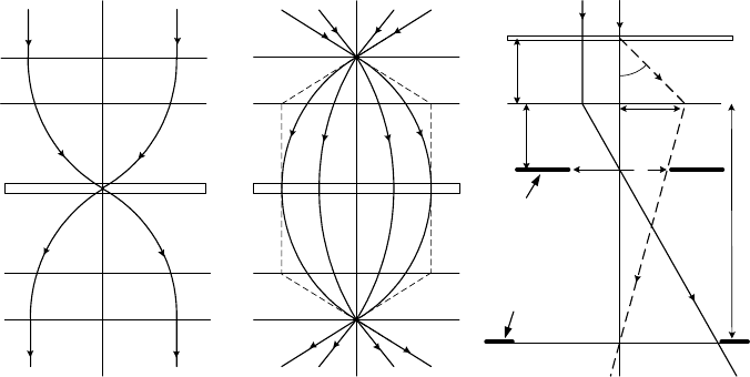
Figure 3-12. Formation of (a) a small-diameter nanoprobe and (b) parallel illumination at the
specimen, by means of the pre-field of the objective lens. (c) Thin-lens ray diagram for the
objective post-field, showing the specimen (S), principal plane (PP) of the objective post-field
and back-focal plane (BFP).
In fact, in a modern materials-science TEM (optimized for high-
resolution imaging, analytical microscopy, and diffraction analysis of non-
biological samples), the specimen is located close to the center of the
objective lens, where the magnetic field is strong. The objective pre-field
then exerts a strong focusing effect on the incident illumination, and the lens
is often called a condenser-objective. When the final (C2) condenser lens
produces a near-parallel beam, the pre-field focuses the electrons into a
nanoprobe of typical diameter 1 – 10 nm; see Fig. 3-12a. Such miniscule
electron probes are used in analytical electron microscopy to obtain chemical
information from very small regions of the specimen. Alternatively, if the
condenser system focuses electrons to a crossover at the front-focal plane of
the pre-field, the illumination at the specimen is approximately parallel, as
required for most TEM imaging (Fig. 3-12b). The post-field of the objective
then acts as the first imaging lens with a focal length f of around 2 mm. This
small focal length provides small coefficients of spherical and chromatic
aberration and optimizes the image resolution, as discussed in Chapter 2.
In a biological TEM, atomic-scale resolution is not required and the
objective focal length can be somewhat larger. Larger f gives higher image
80 Chapter 3
contrast (for a given objective-aperture diameter), which is usually of prime
concern because the contrast in tissue-sample images can be very low.
Objective aperture
An objective diaphragm can be inserted located at the back-focal plane
(BFP) of the post-field of the objective lens, the plane at which a diffraction
pattern of the specimen is first produced. In this plane, distance from the
optic axis represents the direction of travel (angle relative to the optic axis)
of an electron that has just left the specimen.
Although in practice the objective behaves as a thick lens, we will discuss
its properties using a thin-lens ray diagram in which the focusing deflection
is considered to occur at a single plane: the principal plane of the lens.
Accordingly, an electron that leaves the specimen parallel to the optic axis is
deflected at the principal plane and crosses the axis at the BFP, a distance f
below the principal plane, as illustrated by the solid ray in Fig. 3-12c.
Assuming parallel illumination and correctly-adjusted tilt controls, such an
electron will have arrived at the specimen parallel to the axis and must have
rem ined undeflected (unscattered) during its passage through the specimen.a
The dashed ray in Fig. 3.12c represents an electron that arrives along the
optic axis and is scattered (diffracted) through an angle D by interaction with
one or more atoms in the specimen. It therefore leaves the specimen on the
optic axis but at angle D relative to it, arriving at the principal plane at a
radial distance from the axis equal to R = u tan D|f tan D. After deflection
by the objective, this electron crosses the optic axis at the first image plane, a
relatively large distance (v | 10 cm) from the lens. Below the principal
plane, the dashed ray is therefore almost parallel to the optic axis, and its
displacement from the axis at the back-focal plane is approximately R . By
inserting an aperture of diameter D (centered around the optic axis) at the
BFP, we can therefore ensure that the electrons that pass through the rest of
the imaging system are those with scattering angles between zero and D,
where
D| tanD| R/f = D/(2f ) (3.9)
Electrons scattered through larger angles are absorbed by the diaphragm
surrounding the aperture and do not contribute to the final image. By making
D small, we can ensure that almost all scattered electrons are absorbed by the
diaphragm. As a result, regions of specimen that scatter electrons strongly
will appear as dark areas (due to fewer electrons) in the corresponding final
image, which is said to display scattering contrast or diffraction contrast.

The Transmission Electron Microscope 81
Besides producing contrast in the TEM image, the objective aperture
limits the amount of image blurring that arises from spherical and chromatic
aberration. By restricting the range of scattering angles to values less than D,
given by Eq. (3.9), the loss of spatial resolution is limited to an amount r
s
|
C
s
D
3
due to spherical aberration and r
c
| C
c
D ('E/E
0
) due to chromatic
aberration. Here, C
s
and C
c
are aberration coefficients of the objective lens
(Chapter 2); 'E represents the spread in kinetic energy of the electrons
emerging from the specimen, and E
0
is their kinetic energy before entering
the specimen. Because both r
s
and r
c
decrease with aperture size, it might be
thought that the best resolution corresponds to the smallest possible aperture
dia eter.m
But in practice, objective diaphragm gives rise to a diffraction effect that
becomes more severe as its diameter decreases, as discussed in Section 1.1,
leading to a further loss of resolution 'x given by the Rayleigh criterion:
'x | 0.6 O/sinD| 0.6 O/D (3.10)
where we have assumed that D is small and is measured in radians.
Ignoring chromatic aberration for the moment, we can combine the effect
of spherical aberration and electron diffraction at the objective diaphragm by
adding the two blurring effects together, so that the image resolution 'r
(measured in the object plane) is
'r | r
s
+ 'x | C
s
D
3
+ 0.6 O/D (3.11)
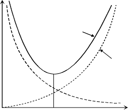
Because the two terms in Eq. (3.11) have opposite D-dependence, their sum
is represented by a curve that displays a minimum value; see Fig. 3-13. To a
first approximation, we can find the optimum aperture semi-angle D*
(corresponding to smallest 'r) by supposing that both terms make equal
contributions at D = D*. Equating both terms gives (D*)
4
= 0.6 O/C
s
and
results in D* = 0.88 (O/C
s
)
1/4
.
D
'r
'x
C
s
D
D
0
blurring
measured
in the
object
plane
Figure 3-13. Loss or resolution due to spherical aberration (in the objective lens) and
diffraction (at the objective aperture). The solid curve shows the combined effect.
