Cohen I.M., Kundu P.K. Fluid Mechanics
Подождите немного. Документ загружается.

812 Introduction to Biofluid Mechanics
as p
2
/Z from equation (17.151). Now we could calculate the incident, reflected and
transmitted quantities at the junction. Thus,
Rate of energy transmission by incident wave =
p
2
I
Z
1
, (17.161)
Rate of energy transmission by reflected wave =
(
Rp
I
)
2
Z
1
= R
2
p
2
I
Z
1
(17.162)
The quantity R
2
is called the energy reflection coefficient. Similarly, the energy
transmission coefficient which is the rate of energy transfer in the two transmitted
waves compared with that in the incident wave may be defined by,
p
2
T 2
Z
2
+
p
2
T 3
Z
3
p
2
I
Z
1
=
Z
−1
2
+ Z
−1
3
Z
−1
1
p
T 2
p
I
2
=
Z
−1
2
+ Z
−1
3
Z
−1
1
T
2
, (17.163)
where we have noted that in our case, p
T 2
= p
T 3
.
A comparison of equations (17.159) and (17.160) shows that, if we include
reflection at bifurcations, the pressure and flow wave forms are no longer of the
same shape. Pedley (1980) has offered interesting discussions about the behavior
of the waves at the junction. From equation (17.153), for real values of c
j
and Y
j
,
if
Y
j
<Y
1
, then the reflected wave has the same sign as the incident wave, and
the pressures in the two waves are in phase at x = 0. They combine additively to
form a large-amplitude fluctuation at the junction, and the effect of the junction is
similar to that of a closed end (P
R
= P
I
). If
Y
j
>Y
1
, there is a phase change
at x = 0, the smallest-amplitude pressure fluctuation occurs there, and the junction
resembles an open end (P
R
=−P
I
). If
Y
j
= Y
1
, there is no reflected wave,
and the junction is said to be perfectly matched. Pedley (2000) has noted that the
increase in the pressure wave amplitude in the aorta with distance down the vessel
may indicate that there is a closed end type of reflection at (or beyond) the iliac
bifurcation. Peaking of the pressure pulse is a consequence of closed end type of
reflection in a blood vessel.
Waves in more complex systems consisting of many branches may be analyzed
by repeated application of the results presented above.
Next, we will study blood flow in curved tubes. Almost all blood vessels have
curvature and the curvature affects both the nature (stability) and volume rate of flow.
Flow in a Rigid Walled Curved Tube
Blood vessels are typically curved and the curvature effects have to be accounted
for in modelling in order to get a realistic understanding. The aortic arch is a 3D
bend twisting through more than 180
◦
, Ku (1997). In a curved tube, fluid motion is
not everywhere parallel to the curved axis of the tube (see Fig. 17.17), secondary
motions are generated, the velocity profile is distorted, and there is increased energy
3. Modelling of Flow in Blood Vessels 813
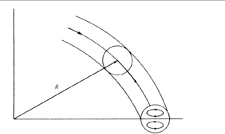
Figure 17.17 Schematic of flow in a curved tube.
dissipation. However, curving of a tube increases the stability of flow, and the critical
Reynolds number increases significantly, and a critical Reynolds number of 5,000 is
easily obtained (see, McDonald (1974)). Flows in curved tubes are discussed in detail
by McConalogue and Srivastava (1968), Singh (1974), Pedley (1980) and by Berger
et al. (1983). In this Section, we will concentrate on some of the most important
aspects and will focus on the flow in a uniformly curved vessel of small curvature.
The wall is considered to be rigid. Pulsatile flow through a curved tube can induce
complicated secondary flows with flow reversals and is very difficult to analyze. It
may be noted that steady viscous flow in a symmetrical bifurcation resembles that in
two curved tubes stuck together. Thus, an understanding gained in studying curved
flows will be beneficial in that regard as well.
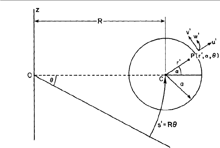
Consider fully developed, steady, laminar, viscous flow in a curved tube of radius
a and a uniform radius of curvature R. Let us employ the toroidal coordinate system
(r
,α,θ), where r
denotes the distance from the center of the circular cross section
of the pipe, α is the angle between the radius vector and the plane of symmetry, and θ
is the angular distance of the cross section from the entry of the pipe (see Fig. 17.18).
Let the corresponding dimensional velocity components be (u
,v
,w
). As a fluid
particle traverses a curved path of radius R (radius of curvature) with a (longitudinal)
speed w
along the θ direction, it will experience a lateral (centrifugal) acceleration
of w
2
/R, and a lateral force equal to m
p
w
2
/R, where m
p
is the mass of the particle.
The radii of curvature of the particle paths near the inner bend, the central axis, and
the outer bend will be of increasing magnitude as we move away from the inner bend.
Also, due to the no-slip condition, the velocities, w
, of particles near the inner and
outer bends will be lower, while that of the particle at the central axis will be the
highest. The particle at the central axis will experience the highest centrifugal force
814 Introduction to Biofluid Mechanics
Figure 17.18 Toroidal coordinate system.
while that near the outer bend will experience the least. A lateral pressure gradient
will cause the faster flowing fluid near the center to be swept towards the outside of
the bend and be replaced at the inside by the slower moving fluid near the wall. In
effect, a secondary circulation will be set up resulting in two vortices, called Dean
vortices because Dean (1928) was the first to systematically study these secondary
motions in curved tubes (see Fig. 17.17). Dean vortices significantly influence the
axial flow. The wall shear near the outside of the bend is relatively higher than the
(much reduced) wall shear on the inside of the bend. Fully developed flow upstream
of or through curved tubes exhibits velocity that skews toward the outer wall of the
bend. For most arterial flows, skewing will be toward the outer wall. If the flow into
the entrance region of a curved tube is not developed, then the inviscid core of the
fluid in the curve can act like a potential vortex with velocity skewing toward the
inner wall.
Secondary flow in curved tubes is utilized in heart-lung machines to promote
oxygenation of blood ( Fung (1997)). In the machine, blood flows inside the curved
tube and oxygen flows on the outside. The tube is permeable to oxygen. The secondary
flow in the tube stirs up the blood and results in faster oxygenation.
Let us now analyze the flow in a curved tube so as to understand the salient
features. Introduce non-dimensional variables, r = r
/a, s = Rθ/a, u = u
/
¯
W
0
,
and p = p
/ρ
¯
W
2
0
, where u = (u, v, w) is the velocity vector, p is the pressure, ρ is
the density, and
¯
W
0
is the mean axial velocity in the pipe. Restrict consideration to

3. Modelling of Flow in Blood Vessels 815
the case where the flow is fully developed (∂u/∂s = 0). Introduce the dimensionless
ratio,
δ =
radius of tube cross section
radius of curvature of the centerline
=
a
R
, (17.164)
We restrict consideration to a uniformly curved tube, δ = constant, and with a
slight curvature (weakly curved), δ 1. Since δ is a constant, the velocity field is
independent of s, the components are functions only of r and θ , and the pressure
gradient ∂p/∂s is independent of s. With δ constant, the only way that the transverse
velocities are affected by the axial velocity is through the centrifugal force, and
it is the centrifugal force that drives the secondary motion. This means that the
centrifugal force terms must be of the same order of magnitude as the viscous and
inertial terms in the momentum equation, and this requires rescaling the velocities.
The transformation that accomplishes this is (u, v, w) → (
√
δ ˆu,
√
δ ˆv, ˆw ). We will
also let s = Rθ/a =
√
1/δ ˜s for convenience.
In the following, we shall omit writing the “ ˆ”onu, v, w, and the “˜”ons for
convenience. When δ 1, the major contribution to the axial pressure gradient may
be separated from the transverse component, and we may write,
p = p
0
(s) + δp
1
(r,α,s)+ ..., (17.165)
Under all these restrictions, the governing equations become,
∂u
∂r
+
u
r
+
1
r
∂v
∂α
= 0, (17.166)
u
∂u
∂r
+
v
r
∂u
∂α
−
v
2
r
− w
2
cos α =−
∂p
1
∂r
−
2
κ
1
r
∂
∂α
∂v
∂r
+
v
r
−
1
r
∂u
∂α
, (17.167)
u
∂v
∂r
+
v
r
∂v
∂α
+
uv
r
+ w
2
sin α =−
1
r
∂p
1
∂α
+
2
κ
∂
∂r
∂v
∂r
+
v
r
−
1
r
∂u
∂α
, (17.168)
u
∂w
∂r
+
v
r
∂w
∂α
=−
∂p
0
∂s
+
2
κ
∂
2
w
∂r
2
+
1
r
∂w
∂r
+
1
r
2
∂
2
w
∂α
2
. (17.169)
The boundary conditions are:
u = v = w = 0atr = 1, no singularity at r = 0 . (17.170 )
The flow is governed by just one parameter κ in the equations, and it is called
the Dean number. It is given by,
κ =
√
δ
2 a
¯
W
0
ν
=
√
δ 2 Re, (17.171)
where
¯
W
0
is the mean axial velocity in the pipe. The Dean number is the Reynolds
number modified by the pipe curvature. The appearance of the numerical constant 2
816 Introduction to Biofluid Mechanics
in the definition of the Dean number is by convention. At higher Dean numbers, the
flow can separate along the inner boundary curve.
There are many different definitions of Dean number in the literature and the
reader must be careful to see which particular form is being used in any given
discussion.
From equation (17.169), ∂p
0
/∂s is independent of s, and p
0
can be written as
p
0
(s) =−Gs, where G is a constant. The equation (17.166) admits the existence of
a stream function for the secondary flow, ψ, defined by
u =
1
r
∂ψ
∂α
,v=−
∂ψ
∂r
. (17.172)
Substitution of equation (17.172) into equation (17.169) yields,
∇
2
1
w −
κ
2
∂p
0
∂s
=
κ
2r
∂ψ
∂α
∂w
∂r
−
∂ψ
∂r
∂w
∂α
, (17.173)
while elimination of pressure from equations (17.167) and (17.168) yields,
2
κ
∇
4
1
ψ −
1
r
∂ψ
∂r
∂
∂α
−
∂ψ
∂α
∂
∂r
∇
2
1
ψ =−2w
sin α
∂w
∂r
+
cos α
r
∂w
∂α
, (17.174)
where,
∇
2
1
ψ =
∂
2
∂r
2
+
1
r
∂
∂r
+
1
r
2
∂
2
∂α
2
. (17.175)
The boundary conditions are:
ψ =
∂ψ
∂r
= w = 0,atr= 1. (17.176)
The equations (17.173) and (17.174) subject to conditions (17.176) have to be solved.
For small values of Dean number, following Dean (1928), we expand w and ψ
in terms of a series in powers of the Dean number as follows:
w =
∞
n=0
κ
2n
w
n
(r, α), and,ψ= κ
∞
n=0
κ
2n
ψ
n
(r, α). (17.177)
The w
0
term corresponds to Poiseuille flow in a straight tube with rigid walls. The ψ
0
term is O(κ). The series expansion in κ is equivalent to the successive approximation
of inertia terms in lubrication theory. The leading term in the secondary flow takes the
form of a pair of counter rotating helical vortices, placed symmetrically with respect
to the plane of symmetry. This flow pattern arises because of a centrifugally induced
pressure gradient, approximately uniform over the cross section. The dimensionless
volume flux is,
Q
πa
2
¯
W
= 1 − 0.0306
K
576
2
+ 0.0120
K
576
4
+ O
K
6
, (17.178)

3. Modelling of Flow in Blood Vessels 817
where K = (2a/R)(W
max
a/ν)
2
= 2(κ)
2
, is another frequently used definition of
Dean’s number. Here, W
max
= 2
¯
W ; W
max
and
¯
W are the maximum and mean
velocities, respectively, in a straight pipe of the same radius under the same axial
pressure gradient and under fully developed flow conditions. The first term corre-
sponds to the Poiseuille straight pipe solution. The effect of curvature is seen to
reduce the flux.
Many other authors define Dean’s number by,
D =
√
2δ
ˆ
Ga
2
µ
a
ν
, (17.179)
where, −
ˆ
G is the dimensional pressure gradient,
ˆ
G =−
8µ
¯
W
a
2
. (17.180)
In terms of D, equation (17.178) becomes,
Q
πa
2
¯
W
= 1 − 0.0306
D
96
4
+ 0.0120
D
96
8
+ O
D
12
, (17.181)
Next, consider the friction factor for flow in a curved tube. Let λ
c
and λ
s
denote
the flow resistance in a curved and a straight pipe, respectively, while the flows in
them are subject to pressure gradients equal in magnitude. The ratio λ is,
λ =
λ
c
λ
s
=
Q
c
Q
s
−1
= 1 + 0.0306
K
576
2
− 0.0110
K
576
4
+ ..., (17.182)
where, Q
c
and Q
s
are the fluxes in straight and curved pipes, respectively. The flow
resistance in a curved tube is not affected by the first order terms and is increased
only by higher order terms. With regard to shear stress, the curvature increases axial
wall shear on the outside wall and decreases it on the inside, and it also generates a
positive secondary shear in the α direction.
The size of the coefficients suggests that the small D expansion is valid for values
of D up to about 100 or K ≈ 600, and the results here are useful only for smaller
blood vessels. Pedley points out that in the canine aorta, where δ ≈ 0.2, the mean D
is greater than 2000. As mentioned earlier, flow in a curved tube is much more stable
than that in a straight tube and the critical Reynolds number could be as high as 5000
which corresponds to K ≈ 1.6 × 10
6
.
For intermediate values of D, only numerical solutions are possible due to the
importance of non-linear terms. Numerical results of Collins and Dennis (1975) for
developed flow up to a D of 5000 are stated to compare very well with experimental
results. At intermediate values of D, a boundary layer develops on the outside wall of
the bend where the axial shear is high. The secondary flow in the core is approximately
uniform and continues to manifest a two vortex structure. At higher values of D, there
is greater distortion of the secondary streamlines. The wall shear at r = 1, α = 0, is
proportional to D (≈ 0.85D); see Pedley (2000).

818 Introduction to Biofluid Mechanics
At large Dean numbers, the centers of the two vortices move toward the outer
bend, α = 0, and the flow is very much reduced compared with a straight pipe
for equal magnitude pressure gradients. Detailed studies using advanced computa-
tional methods are required to resolve the flow structure at large D. They are as yet
unavailable in the published literature.
Pedley (2000) discusses nonuniqueness of curved tube flow results. When D
is sufficiently small, the steady flow equations have just one solution and there is a
single secondary flow vortex in each half of the tube. However, there is a critical value
of D, above which more than one steady solution exists and these may correspond to
four vortices, two in each half. Again, detailed computational studies are necessary
to resolve these features.
We will next study the flow of blood in collapsible tubes. The role of pressure
difference,
(
p
e
− p(x)
)
, on the vessel wall will be significant in such flows.
Flow in Collapsible Tubes
At large negative values of the transmural pressure difference (the difference between
the pressure inside and the pressure outside), the cross sectional area of a blood vessel
is either very small, the lumen being reduced to two narrow channels separated by a
flat region of contact between the opposite walls or it may even fall to zero. There is an
intermediate range of values of transmural pressure difference in which the cross sec-
tion is very compliant and even the small viscous or inertial pressure drop of the flow
may be enough to cause a large reduction in area, that is, collapse. Collapse occurs in
a number of situations and a listing is given by Kamm and Pedley (1989). Collapse
occurs, for example, in systemic veins above the heart (and outside the skull), as a
result of the gravitational decrease in internal pressure with height; intramyocardial
coronary blood vessels during systole; systemic arteries compressed by a sphygmo-
manometer cuff, or within the chest during cardiopulmonary resuscitation; pulmonary
blood vessels in the upper levels of the lung; large intrathoracic airways during forced
expiration or coughing; the urethra during micturition and in the ureter during peri-
staltic pumping. Collapse, therefore occurs both in small and large blood vessels, and
as a result both at low and high Reynolds numbers. In certain cases, at high Reynolds
number, collapse is accompanied by self-excited, flow-induced oscillations. There is
audible sound. For example, Korotkoff sounds heard during sphygmomanometry are
associated with this.
A Note on Korotkoff Sounds
Korotkoff sounds, named after Dr. Nikolai Korotkoff, a physician who described them
in 1905, are sounds that physicians listen for when they are taking blood pressure.
When the cuff of a sphygmomanometer is placed around the upper arm and inflated
to a pressure above the systolic pressure, there will be no sound audible because the
pressure in the cuff would be high enough to completely occlude the blood flow. If
the pressure is now dropped, the first Korotkoff sound will be heard. As the pressure
in the cuff is the same as the pressure produced by the heart, some blood will be able
to pass through the upper arm when the pressure in the artery rises during systole.

3. Modelling of Flow in Blood Vessels 819
This blood flows in spurts as the pressure in the artery rises above the pressure in the
cuff and then drops back down, resulting in turbulence that results in audible sound.
As the pressure in the cuff is allowed to fall further, thumping sounds continue to be
heard as long as the pressure in the cuff is between the systolic and diastolic pressures,
as the arterial pressure keeps on rising above and dropping back below the pressure
in the cuff. Eventually, as the pressure in the cuff drops further, the sounds change in
quality, then become muted, then disappear altogether when the pressure in the cuff
drops below the diastolic pressure. Korotkoff described 5 types of Korotkoff sounds.
The first Korotkoff sound is the snapping sound first heard at the systolic pressure.
The second sounds are the murmurs heard for most of the area between the systolic
and diastolic pressures. The third and the fourth sounds appear at pressures within
10 mmHg above the diastolic blood pressure, and are described as “thumping” and
“muting”. The fifth Korotkoff sound is silence as the cuff pressure drops below the
diastolic pressure. Traditionally, the systolic blood pressure is taken to be the pressure
at which the first Korotkoff sound is first heard and the diastolic blood pressure is the
pressure at which the fourth Korotkoff sound is just barely audible. There has recently
been a move towards the use of the 5th Korotkoff sound (i.e. silence) as the diastolic
pressure, as this has been felt to be more reproducible.
Starling Resistor: A Motivating Experiment for Flow in Collapsible Tubes
The study of flows in collapsible tubes is facilitated by a well known experiment
carried out under varying conditions by different researchers. In the experiment, a
length of uniform collapsible tube is mounted at each end to a shorter length of rigid
tube and is enclosed in a chamber whose pressure p
e
can be adjusted. Fluid, say water,
flows through the tube. The inlet and outlet pressures at the ends of the collapsible
tube are p
1
and p
2
. The volume rate of flow is Q. The pressures and the flow rate are
next varied in a systematic way and the results are noted. The set up described is called
a Starling resistor after physiologist Starling (see Fung (1997)). This experiment will
enable us understand some aspects of actual flows in physiological systems. There are
many different versions of the description of the Starling resistor experiment in the
literature. The experiments have been carried out under both steady flow and unsteady
flow conditions. We will describe the experiments as reported by Kamm and Pedley
(1989).
Case (1): (p
1
− p
2
) is increased while (p
1
− p
e
) is held constant.
This is accomplished either by reducing p
2
with p
1
and p
e
fixed, or by simulta-
neously increasing p
1
and p
e
while p
2
is held constant. With either procedure, Q at
first increases, but above a critical value it levels off and the condition of flow limi-
tation is reached. In this condition, however much the driving pressure is increased
the flow rate remains constant, or may even fall as a result of increasingly severe tube
collapse. This experiment is relevant to forced expiration from the lung, to venous
return, and to micturition.
Case (2): (p
1
− p
2
) or Q is increased while (p
2
− p
e
) is held constant at some
negative value.

820 Introduction to Biofluid Mechanics
In this case, the tube is collapsed at low flow rates, but starts to open up from the
upstream end as Q increases above a critical value, so that the resistance falls and
(p
1
− p
2
) ceases to rise. This is termed pressure-drop limitation. This experiment
does not seem to apply to any particular physiological condition.
Case (3): (p
1
− p
2
) is held constant while (p
2
− p
e
) is decreased from a large
positive value.
In this case, the tube first behaves as though it were rigid and the flow rate is
nearly constant. Then as (p
2
− p
e
) becomes sufficiently negative to produce partial
collapse, the resistance rises and Q begins to fall. This experiment is relevant to
pulmonary capillary flows.
Case (4): p
e
fixed. The outlet end is connected to a flow resistor. The pressure
downstream of the flow resistor is fixed (flow is exposed to atmosphere). Thus p
2
is
equal to atmospheric pressure plus Q times the fixed resistance. p
1
is varied.
In this case, p
2
varies with Q due to the presence of a fixed downstream resistance.
The degree of tube collapse (progressive collapse) also varies with Q for the same
reason. At high flow rates, the tube is distended and its resistance is low. As the flow
rate is reduced below a critical value the tube starts to collapse. Its resistance and
(p
1
−p
2
) both increase as Q is decreased. Only when the tube is severely collapsed
along most of its length does (p
1
−p
2
) start to decrease again as Q approaches zero.
When p
1
is approximately equal to p
e
, virtually the entire tube is collapsed (Fung
(1997)). The tube often flutters in Case 4 (see discussions in Fung).
Case (5): Unsteady flow experiments
Excepting at small Reynolds numbers, there is always some parameter range
where flow oscillations occur. The oscillations have a wide variety of modes.
The experiments reveal the importance of a tube law relating transmural pressure
difference with the area of cross section of the collapsible tube and the flow and
pressure drop limitations when analyzing collapsible tubes. Shapiro (1977a, 1977b)
has developed a comprehensive one dimensional theory for steady flow based on a
suitable tube law. Kamm and Shapiro (1979) have extended it to unsteady flow in a
collapsible tube. In the following, we shall discuss the steady flow theory.
One-Dimensional Flow Treatment
The equations describing one-dimensional flow in a collapsible tube are similar to
those in gas dynamics or channel flow of a liquid with a free surface (see,Shapiro
(1977a)). Here, we will study the one dimensional steady flow formulation for the
collapsible tube. However, before that let us recapitulate the traditional basic equations
for one dimensional flow in a smoothly varying elastic tube. (see Section 3.3.2 on
Pulse wave propagation in an elastic tube: Inviscid theory).
We had studied flow in an elastic tube with cross section A(x, t) and longitudinal
velocity u(x, t). The constant external pressure on the tube was set at p
e
. The primary
mechanism of unsteady flow in the tube was wave propagation. The transmural pres-
sure difference, (p −p
e
) was related to the local cross sectional area by a “tube law”
which involved hoop tension, and it may be expressed as,
(p − p
e
) =
ˆ
P (A) (17.183)
3. Modelling of Flow in Blood Vessels 821
where the functional form
ˆ
P depends on data. For disturbances of small amplitude
and long wave length compared to the tube diameter,
A = A
0
+ A
,p− p
e
=
ˆ
P(A
0
) + p
, |A
|A
0
, |p
|
ˆ
P(A
0
) (17.184)
and the wave speed is given by,
c
2
=
A
ρ
d
ˆ
P
dA
=
A
ρ
d(p − p
e
)
dA
. (17.185)
Tube collapse is associated with negative transmural pressure difference and the
pressure difference is supported by bending stiffness of the tube wall (see Fig. 17.19).
Contrast this with positive transmural pressure difference discussed earlier which was
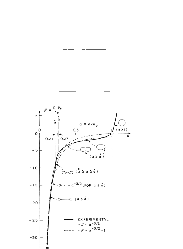
supported by hoop tension. Following Shapiro (1977a), introduce,
P =
(p − p
e
)
K
p
, and,α=
A
A
0
, (17.186)
Figure 17.19 Behavior of a collapsible tube. (Reproduced with permission from the American Society
of Mechanical Engineers, NY.).