Buschow K.H.J. (Ed.) Concise Encyclopedia of Magnetic and Superconducting Materials
Подождите немного. Документ загружается.


a practical possibility, however. Of course, head
gaps of these dimensions make it necessary to aban-
don some of the major advantages of optical disk
technology. The large separations between head
and media, and between the point of focus on ac-
tual recording media and the disk surface adjacent
to the environment, can no longer be supported.
The SIL must be flown like the head in a conven-
tional magnetic hard drive, but considerably closer
to the recording surface. In these circumstances, in-
tegration of the write coil on the same slider car-
rying the SIL is a natural step. Optical access to the
actual recording medium can no longer be protected
through the substrate and front-surface media a thin
dielectric overcoat must be used. Fortunately, the
refractive indices of the common protective over-
coats such as silicon nitride match reasonably well
to those of SILs. In general, it is also necessary,
because of the extreme intensity in the very small
spot, to desensitize the medium. A fuller treat-
ment of near-field optics is given elsewhere in this
encyclopedia.
See also: Magneto-optical Effects: Enhancement of;
Magneto-optic Recording: Overwrite and Associated
Problems; Magneto-optic Recording: Total Film
Stack, Layer Configuration; Optical and Magneto-
optic Data Storage: Channels and Coding
Bibliography
Awano H, Ohnuki S, Shirai H, Ohta N, Yamaguchi A, Sumi S,
Torazawa K 1996 Appl. Phys. Lett. 69 (27), 4257–9
Betzig E, Isaacson M, Lewis A 1987 Appl. Phys. Lett. 51, 2088
Callan H, Josephs R M 1971 J. Appl. Phys. 42, 1977
Honda S, Ueda K, Kusuda T 1981 J. Appl. Phys. 52, 2295
Hosaka S, Shintani T, Kikukawa A, Imura R 1996 J. Magn.
Soc. Jpn. 20 (S1), 79–82
Kaneko M, Nakaoki A 1996 J. Magn. Soc. Jpn. 20 (S1), 7–12
Murakami Y, Maeda S, Takhashi A, Tanaka Y, Watanabe T
1996 J. Magn. Soc. Jpn. 23 (S1), 181–4
News Letter no. 4 (Jan 1998) www.oitda.or.jp/Optoelectronic
Industry and Technology Development Association (OIT-
DA), Japan
Nichia Corporation, Japan, Blue Laser model NLHV500A
October 1999
Nussbaum A, Phillips R A 1976 Contemporary Optics for Sci-
entists and Engineers. Prentice-Hall, Englewood Cliffs, NJ,
pp. 88–90
Ohta M, Fukumoto A, Aratani K, Kaneko M, Watanabe K
1991 J. Magn. Soc. Jpn. 15 (S1), 319–22
Saito J, Sato M, Matsumoto H, Akasaka H 1987 Jpn. J. Appl.
Phys. 26 (suppl. 4), 155
Shono K 1999 J. Magn. Soc. Jpn. 23 (S1), 177–80
Takagi N, Mitani K, Awano H, Shimazaki K, Ohta N 1999 J.
Magn. Soc. Jpn. 23 (S1), 161–4
Tokunaga T, Fujii Y, Yamada K 1999 J. Magn. Soc. Jpn. 23
(S1), 169–72
D. M. Newman
Centre for Data Storage Materials
Coventry University, UK
1220
Super-resolution: Optical and Magnetic Techniques
Textured Magnets: Deformation-induced
There are two different processes for producing
anisotropic rare earth–transition metal–boron
(RE–TM–B) magnets with high energy density. One
is the conventional technique of powder metallurgy
(see Magnets: Sintered). The crystal alignment is
achieved by applying an external magnetic field dur-
ing the cold-pressing stage. The alternative processing
route is hot deformation of cast alloys or of fine-
grained powder, the latter consolidated previously by
hot pressing. The hot working process produces grain
alignment along the c -axis perpendicular to the plas-
tic flow direction. The key for understanding the
formability of the inherently brittle alloys is the pres-
ence of the RE-rich grain boundary phase, which is
liquid at the deformation temperature (4700 1C). It
is conceivable that the liquid grain boundary phase
can annihilate microcracks created during the defor-
mation. However, a liquid cannot resist high tensile
stresses. Therefore, the deformation must be per-
formed under compressive stress.
1. Deformation of Cast Alloys
Permanent magnet production by hot deformation of
cast alloys promises to be a potentially simpler and
more economical option in comparison with the sin-
tering route because of the troublesome handling of
magnetic powder that is extremely sensitive to oxygen
contamination.
The simplest deformation method is upsetting of
cylindrical specimens between two punches. Extensive
hot-pressing experiments performed by Shimoda et al.
(1989) revealed that Pr–Fe–B alloys with the compo-
sition Pr
17
Fe
79
B
4
are particularly suitable for hot de-
formation. Hot pressing at 1000 1C with strain rates of
10
4
–10
3
s
1
introduces a preferred magnetization
direction parallel to the press direction. Additionally,
the magnetic alignment is closely related to the struc-
ture of the cast ingots and the direction of principal
stress. The appropriate structure is a columnar struc-
ture in which the c-axis of the hard magnetic Pr
2
Fe
14
B
phase (2:14:1 or F phase) is in the plane perpendicular
to the crystal growth direction. Therefore, the prin-
cipal stress during deformation should also be per-
pendicular to the growth direction. Inserting the
cylindrical specimens into an iron ring prevents crack-
ing up to strain rates of 10
1
s
1
as well as squeezing
out the liquid intergranular phase. Additionally, in-
creasing the strain rate results in a grain refinement
and with a consequent enhancement of the coercivity.
Studies concerning additive elements show that cop-
per addition is effective in increasing coercivity and
in improving the magnetic alignment. Microstructural
studies reveal that copper (i) refines the as-cast grain
size and (ii) reduces the melting point of the praseo-
dymium-rich phase. Copper probably leads to a better
wetting behavior and separation of the hard magnetic
grains. In the studies of Shimoda et al.(1989)the
magnetic properties of hot-pressed and annealed
Pr
17
Fe
76.5
B
5
Cu
1.5
magnets were as follows: remanence,
B
r
¼1.25 T; intrinsic coercivity,
i
H
c
¼796 kA m
1
;and
maximum energy product, (BH)
max
¼288 kJ m
3
.
Hot rolling of RE–TM–B alloys has been studied
by Ohki et al. (1989). The essential feature of the hot
rolling is a high strain rate (1–10 s
1
), which makes this
process very suitable for mass production. The opti-
mum rolling temperature is about 950 1C. Regarding
the optimum structure, it is found that the primary
crystallization of iron is necessary for the formation
of fine-grained F crystallites during the following pe-
ritectic reaction. Optimum magnetic properties de-
mand a postdeformation heat treatment that is
beneficial to both the coercivity and the remanence.
Regarding Pr–Fe–B alloys, the following maximum
values have been achieved: (BH)
max
¼340 kJ m
3
and
i
H
c
¼1194 kA m
1
. Concerning neodymium-based al-
loys, the maximum published values are: (BH)
max
¼
269 kJm
3
and
i
H
c
¼525 kA m
1
. Hot rolling of ingots
is also used for the preparation of anisotropic powder
suited for bonded magnets (Hinz et al. 1994).
Extrusion of RE–TM–B alloys results in magnets
with radial c-axis orientation. Nozieres et al. (1988)
carried out extrusion experiments with large ingots,
cast in an iron sheath. Energy products in the range
120–160 kJ m
3
have been obtained.
Regarding the mechanism of deformation and tex-
turing, the microstructural feature of the hot defor-
mation of cast alloys is the marked grain refinement.
It is assumed that the crystal alignment is related to
rotation of the grain debris in the liquid intergranular
phase. The start of the alignment is supported by the
plate-like shape anisotropy with its c-axis perpendic-
ular to the larger and flat surface of the dendritic
grains (Yuri and Ohki 1994). During the deformation
process the cracking occurs preferentially along the
c-plane of the main phase, resulting in an enhance-
ment of the plate-like shape anisotropy and the ro-
tation of the grains. In a later stage the rotation of the
grains is accompanied by an irregular cracking at the
surface of the grains. This generates significant
amounts of small and unaligned grain debris. Post-
deformation heat treatment results in grain growth
with dissolution of the small and unaligned grains
and in blunting of the sharp edges of the large grains.
2. Deformation of Powder Precursors
The possibility of an alignment of compacted
melt-spun RE–TM–B powder precursors by hot
T
1221
deformation was discovered by Lee et al. (1985). In
the first step a fully dense isotropic precursor must be
produced by hot pressing. The high densification is
possible because the RE-rich grain boundary phase is
liquid in the applied temperature range from 700 1C
to 750 1C. In the second step the deformation of the
precursor takes place at temperatures between 750 1C
and 800 1C. In the case of the die-upset method, this
step can be accomplished by transferring the sample
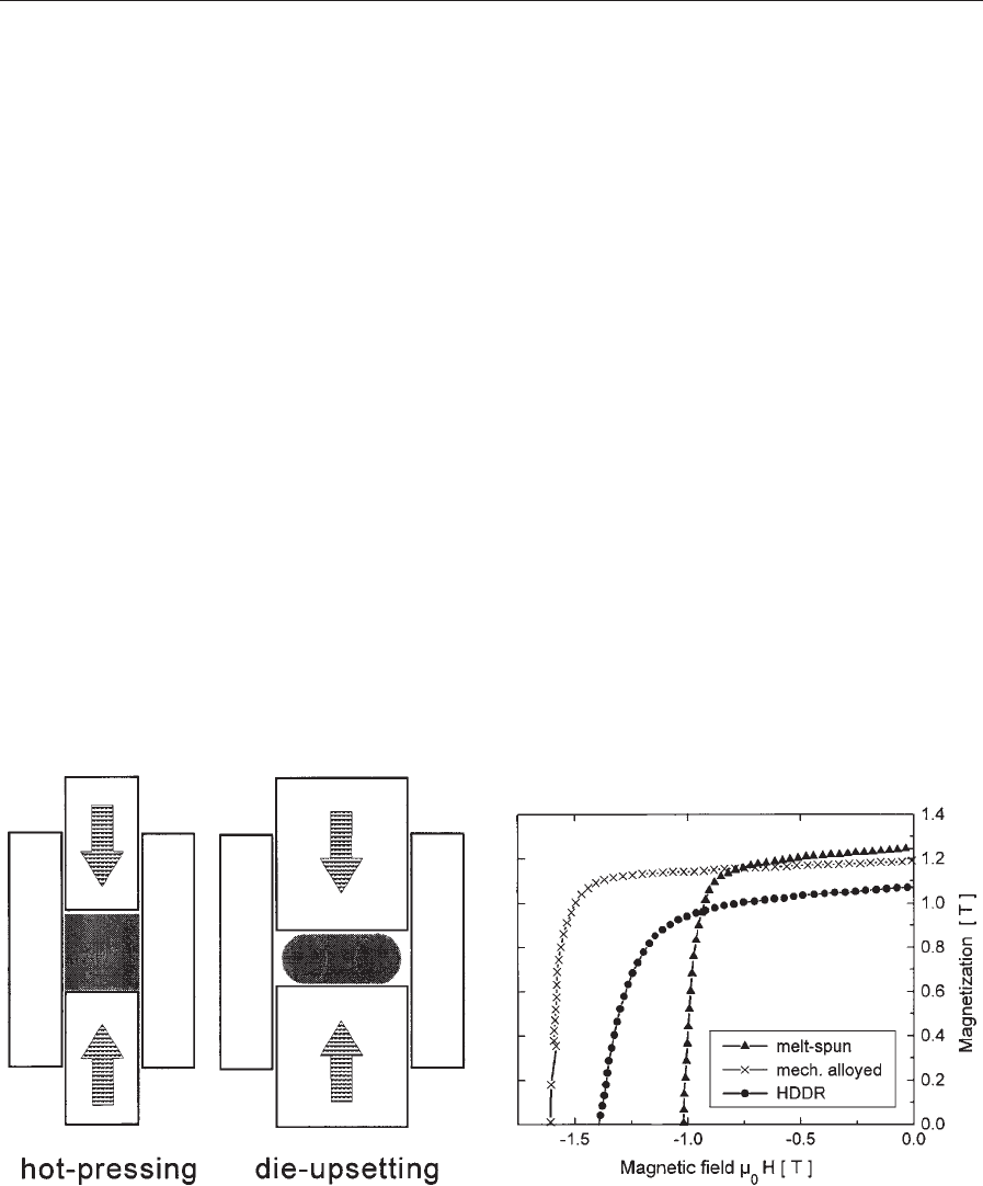
to an oversized die cavity (Fig. 1). The deformation
leads to an anisotropic magnet with the alignment
parallel to the pressing direction.
Optimizing the composition in the Nd–Fe–B ter-
nary system involves a compromise between maxi-
mizing the volume fraction of the Nd
2
Fe
14
B phase
and providing excess neodymium for the grain
boundary phase. According to Nozawa et al. (1988)
the optimum composition of die-upset magnets is in
the range Nd
13.5–14
Fe
80.5–80
B
6
. Because the coercivity
is reduced after hot deformation of ternary alloys,
the effect of additional elements has been investi-
gated. A 0.5 at.% gallium addition proves to be fa-
vorable leading to a coercivity enhancement of up to
1600 kA m
1
and also a relatively small reduction in
remanence (Nozawa et al. 1988). The partial substi-
tution of cobalt for iron leads to an enhancement of
the Curie temperature of the magnetic main phase
and, therefore, to better thermomagnetic stability of
the magnets. According to Yoshida et al. (1991) the
addition of cerium or silicon has a positive effect on
the workability.
For melt spinning, a high quench rate should pre-
vent the formation of coarse grains on the wheel-free
surface of the ribbons. Surface velocities of
30–34 m s
1
lead to an optimum alignment after up-
setting (Fuerst and Brewer 1993). Concerning the
deformation conditions, increasing the die-upset level
results in an enhancement of the alignment up to a
height reduction of about 75%. The optimum defor-
mation temperature is in the range 750–800 1C and
the optimum strain rate is 10
2
–10
1
s
1
. Die-upset
magnets are commercially available. The energy
product of the different grades ranges from about
255 kJ m
3
to 340 kJ m
3
and the intrinsic coercivity
ranges from 955 kA m
1
to 1670 kA m
1
(Panchana-
than 1995). The published maximum energy product
is about 400 kJ m
3
. Saito et al. (1998) investigated
the magnetic properties of amorphous powder con-
solidated by shock compression. Die-upset magnets
with the composition Nd
13.5
(Fe
0.975
Co
0.025
)
80
Ga
0.5
B
6
show a maximum energy product of 433 kJ m
3
and
an intrinsic coercivity of 995 kA m
1
offer optimal
deformation.
Mechanically alloyed powder (see Magnets: Me-
chanically Alloyed) and HDDR powder (see Magnets:
HDDR Processed) compacts can also be used as pre-
cursors for the successful production of die-upset
magnets (e.g., Schultz et al. 1991). A comparison of
deformation and magnetic properties has been given
by Kirchner et al. (1998) (Fig. 2).
The hot deformation of fine-grained powder pre-
cursors is also suited to produce radially oriented ring
magnets by backward extrusion (Croat 1989). Mag-
nets with different dimensions and energy products of
more than 280 kJ m
3
are commercially available.
The process seems to be especially attractive because
the magnetic properties of rings surpass those of ring
Figure 1
Schematic representation of the preparation of
RE–TM–B magnets from fine-grained powder by hot
pressing (isotropic magnet) and subsequent hot die-
upsetting (anisotropic magnet).
Figure 2
Demagnetization curves measured along the texture axis
of die-upset Nd–Fe–B magnets produced from melt-
spun (m), mechanically alloyed ( ), and HDDR
powders () (after Kirchner et al. 1998).
1222
Textured Magnets: Deformation-induced

magnets made from sintered material, especially at
small dimensions. Backward extruded ring magnets
do not quite reach the maximum properties of die-
upset magnets. The reason is the inherent inhomoge-
neity of the deformation process, which results in
inhomogeneous properties in the cross-section of the
rings (Gru
¨
nberger et al. 1996).
Contrary to cast ingots, microstructural investiga-
tions in fine-grained alloys show the formation of flat
platelet-shaped grains which are larger than those of
the starting material (e.g., Lee et al. 1985). Therefore,
it is consequent to discuss diffusion-supported mech-
anisms for deformation and texturing. It is believed
that deformation and alignment are produced by a
combination of grain boundary sliding and aniso-
tropic grain growth caused by the inherent aniso-
tropy of the crystal growth of the tetragonal F phase
or by the anisotropy of the elastic properties. Lewis
et al. (1997) published a survey of the existing liter-
ature in this field. Gru
¨
nberger (1998) investigated the
applicability of the model of solution precipitation
creep. This is based on the transport of matter in the
liquid intergranular phase caused by the local de-
pendence of the solution equilibrium between solid
and liquid on the local stress state. Because the local
stress state depends on the elastic properties, the an-
isotropy of the latter can contribute to the develop-
ment of texture. The kinetic analysis of the strain rate
stress relationships of die-upset experiments indicates
an interface-controlled process.
See also: Magnetic Materials: Hard; Magnets:
Bonded Permanent Magnets; Magnets: Rema-
nence-enhanced; Magnets: Sintered
Bibliography
Croat J J 1989 Current status of rapidly solidified Nd–Fe–B
permanent magnets. IEEE Trans. Magn. 25, 3550–4
Fuerst C D, Brewer E G 1993 High-remanence rapidly solid-
ified Nd–Fe–B: die-upset magnets. J. Appl. Phys. 73, 5751–6
Gru
¨
nberger W 1998 The solution-precipitation creep—a model
for deformation and texturing mechanisms of nanocrystalline
NdFeB alloys. In: Proc. 15th Int. Workshop on Rare-Earth
Magnets and Their Applications. Dresden, Germany, pp.
333–48
Gru
¨
nberger W, Hinz D, Schla
¨
fer D, Schultz L 1996 Microstruc-
ture, texture, and magnetic properties of backward extruded
NdFeB ring magnets. J. Magn. Magn. Mater. 157/158,41–2
Hinz D, Handstein A, Harris I R 1994 Microstructure and
magnetic properties of anisotropic NdFeB powders from hot
rolled ingots by HD process. IEEE Trans. Magn. 30, 601–3
Kirchner A, Gru
¨
nberger W, Gutfleisch O, Neu V, Mu
¨
ller K-H,
Schultz L 1998 A comparison of the magnetic properties and
deformation behaviour of Nd–Fe–B magnets made from
melt-spun, mechanically alloyed and HDDR powders. J.
Phys. D: Appl. Phys. 31, 1660–6
Lee R W, Brewer E G, Schaffel N A 1985 Processing of neo-
dymium–iron–boron melt-spun ribbons to fully dense mag-
nets. IEEE Trans. Magn. 21, 1958–63
Lewis L H, Thurston T R, Panchanathan V, Wildgruber U,
Welch D O 1997 Spatial texture distribution in thermome-
chanically deformed 2-14-1-based magnets. J. Appl. Phys. 82,
3430–41
Nozawa Y, Iwasaki K, Tanigawa S, Tokunaga M, Harada H
1988 Nd–Fe–B die-upset and anisotropic bonded magnets.
J. Appl. Phys. 64, 5285–9
Nozieres J P, Perrier de la Bathie R, Gavinet J 1988 A novel
process for rare earth–iron–boron permanent magnets prep-
aration. J. Physique 49 (C8), 667–8
Ohki T, Yuri T, Miyagawa M, Takahashi Y, Yoshida C,
Kambe S, Higashi M, Itayama K 1989 A new method of
producing Pr–Fe–B base hard magnetic materials. In: Proc.
10th Int. Workshop on Rare-Earth Magnets and Their Appli-
cations. pp. 399–408 Kyoto, Japan
Panchanathan V 1995 Magnequench magnets status overview.
J. Mater. Eng. Perform. 4, 423–9
Saito T, Fujita, M, Kuji T, Fukuoka K, Syono Y 1998 The
development of high performance Nd–Fe–Co–Ga–B die up-
set magnets. J. Appl. Phys. 83, 6390–2
Schultz L, Schnitzke K, Wecker J, Katter M, Kuhrt C 1991
Permanent magnets by mechanical alloying. J. Appl. Phys.
70, 6339–44
Shimoda T, Akioka K, Kobayashi O, Yamagami T, Ohki T,
Miyagawa M, Yuri T 1989 Hot-working behavior of cast
Pr–Fe–B magnets. IEEE Trans. Magn. 25, 4099–104
Yoshida Y, Kasai Y, Watanabe T, Shibata S, Panchanathan V,
Croat J J 1991 Hot workability of melt-spun Nd–Fe–B mag-
nets. J. Appl. Phys. 69, 5841–3
Yuri T, Ohki T 1994 In: Crystal alignment in Pr–Fe–B hot-rolled
magnet. Proc. 14th Int. Workshop on Rare-Earth Magnets and
Their Applications, pp. 645–54 Birmingham, UK
W. Gru
¨
nberger
Institut fu
¨
r Festko
¨
rper- und Werkstofforschung
Dresden, Germany
Thin Film Magnetism: Band Calculations
The understanding and design of complex magnetic
nanostructures is one of the current frontiers in mag-
netism. Research in this field is stimulated by its in-
teresting fundamental physics as well as applications
today and in the future in the field of data storage
and magnetoelectronics. New experimental tech-
niques, like molecular beam epitaxy and spin-polar-
ized electron spectroscopy techniques, for growth and
characterization of the nanostructures go hand-in-
hand with powerful theoretical methods and provide
a great deal of insight into these phenomena. Ab in-
itio calculations based on density functional theory
play a crucial role for the microscopic understanding
of fundamental magnetic properties, such as magnet-
ization and magnetic order.
1. Spin-density Functional Theory
Ab initio calculation means that the system under
consideration is described without any free parameter.
1223
Thin Film Magnetism: Band Calculations

The problem that needs to be solved is the many-body
problem of a system formed by interacting electrons
and nuclei. With the Born–Oppenheimer approxima-
tion (Born and Oppenheimer 1927) the problem can
be reduced to a system of N interacting electrons
moving in the electrostatic potential V
ext
caused by
the nuclei
H ¼
X
N
i¼1
_
2
2m
@
2
@r
2
i
þ
X
N
i¼1
V
ext
ðr
i
Þþ
1
2
X
N
iaj
e
2
7r
i
r
j
7
ð1Þ
where e
2
¼e
2
/4pe
o
. e
o
is the dielectric constant. r
i
is
the position of electron i while all other constants
have their traditional meaning. Depending on the
boundary conditions used for the external potential,
systems with arbitrary dimensions like atoms or clus-
ters, wires, slabs, or bulk materials can be described
(Fig. 1) Atoms and clusters are considered to be quasi
one-dimensional and are finite in all spatial direc-
tions. Wires are confined in two directions and are
translational invariant in the third, slabs are finite in
one direction and periodically repeated in the other
two directions, and bulk systems are translationally
invariant in all spatial directions.
Density functional theory (Hohenberg and Kohn
1964, Kohn and Sham 1965, Sham and Kohn 1966)
constitutes the main underlying basis for the solution
of this problem. In the generalization of density
functional theory to magnetic systems (Rajagopal
1980, Hedin and Lundqvist 1971, Barth and Hedin
1972, Moruzzi et al. 1978, Gunnarsson and Lundq-
vist 1976, Gunnarsson et al. 1972), the so-called spin
density functional theory, one distinguishes between
two types of electrons, namely the majority electrons
(s ¼þ) and the minority electrons (s ¼) whose
spin is in and opposite to the direction of magneti-
zation, respectively. In addition to the Coulomb po-
tential of the nuclei the electrons experience an extra
contribution 7m
B
H due to an external field H. the
spin degeneracy is lifted. As a consequence the ma-
jority electrons are more attracted than the minority
electrons. Contributions from orbital momenta and
from spin–orbit coupling are neglected, giving an
adequate approximation for itinerant magnetism.
The two types of particles are represented by spin-
dependent densities n
þ
(r) and n
(r), leading accord-
ingly to the following functional for the ground state
energy
E½n
þ
; n
¼T½nþ
Z
d
3
rV
ext
ðrÞnðrÞ
þ U½nþE
xc
½n
þ
; n
ð2Þ
where T[n] is the functional of the kinetic energy for a
set of N noninteracting electrons of density n(r). U[n]
is the classical Hartree energy
U½n¼
1
2
Z
d
3
rnðrÞ
Z
d
3
r
0
nðr
0
Þ
e
2
7r r
0
7
ð3Þ
and E
xc
[n
þ
, n
] the exchange-correlation energy
which includes all many-body interactions, exchange
and correlation, in an effective way. The total density
is given by
nðrÞ¼n
þ
ðrÞþn
ðrÞð4Þ
and the magnetization density is given by
mðrÞ¼n
þ
ðrÞn
ðrÞð5Þ
Clearly, the ground state energy is a functional of
both spin-dependent densities, E[n
þ
, n
]. Using spin-
dependent densities in terms of single-particle wave
functions j
s
k
(r)
n
s
ðrÞ¼
X
k
7j
s
k
ðrÞ7
2
ð6Þ
the variation of the ground state energy yields the
spin-dependent Kohn–Sham equations
_
2
2m
@
2
@r
2
þ V
s
ðrÞ
j
s
k
ðrÞ¼E
s
k
j
s
k
ðrÞð7Þ
and, by analogy with the spin-independent case an
effective though spin-dependent potential follows as
V
s
¼ V
ext
ðrÞþ
Z
d
3
r
0
nðr
0
Þ
e
2
7r r
0
7
þ V
s
xc
ðrÞð8Þ
with
V
s
xc
¼
dE
xc
dn7ðrÞ
Under the local spin density approximation (LSDA),
the exchange-correlation energy (Barth and Hedin
1972, Moruzzi et al. 1978) is written as Eqn. (9)
E
xc
½n
þ
; n
E
Z
d
3
rðn
þ
ðrÞþn
ðrÞÞ
e
hom
xc
ðn
þ
ðrÞ; n
ðrÞÞ ð9Þ
and
V
s
xc
E
@
@n
7
ðn
þ
þ n
Þ e
hom
xc
ðn
þ
; n
Þ
n
s
¼n
s
ðrÞ
ð10Þ
Figure 1
Dimension of the systems under consideration. Zero-
dimensional clusters, one-dimensional wires, two-
dimensional slabs, and three-dimensional bulk systems.
1224
Thin Film Magnetism: Band Calculations
Different approximations of the local density and the
local spin density functional (Hedin and Lundqvist
1971, Barth and Hedin 1972, Moruzzi et al. 1978,
Vosko et al. 1980) have been used. They lead to
variations within the error of the local density or spin
density approximation itself.
2. Stoner Model of Ferromagnetism
The one-particle nature of the Kohn–Sham equation
(Eqn.(7)) allows a Stoner-like model to be derived for
ferromagnetism (Vosko and Perdew 1975, Gunnars-
son 1976, Janak 1977, Stoner 1938, Blu
¨
gel 1995).
Starting from the assumption that the magnetization
density m(r) is a small parameter compared to the
density n(r), the exchange and correlation potential
V
s
xc
can be expanded in powers of m(r). The linear
approximation yields
V
7
xc
¼ V
0
xc
8mðrÞ
*
VðnðrÞÞ;
*
V40 ð11Þ
where V
s
xc
is the exchange-correlation potential for
the nonmagnetic case. As a consequence, the majority
of electrons experience a more attractive potential as
minority electrons. In the Stoner model this potential
shift is expressed in terms of a constant
V
7
xc
¼ V
0
xc
8
1
2
IM; M ¼
Z
V
atom
drmðrÞð12Þ
with the local atomic moment M and the Stoner pa-
rameter I. In the case of ferromagnetism M is the
same for all atoms. As a consequence of the constant
potential shift in Eqn. (12), the single particle wave
functions j
s
k
(r) (Eqn. (7)) do not change with respect
to the nonmagnetic wavefunctions, while the eigen-
values E
k
are shifted rigidly downwards or upwards
j
7
k
ðrÞ¼j
0
k
ðrÞ and E
7
k
¼ E
k
8
1
2
IM ð13Þ
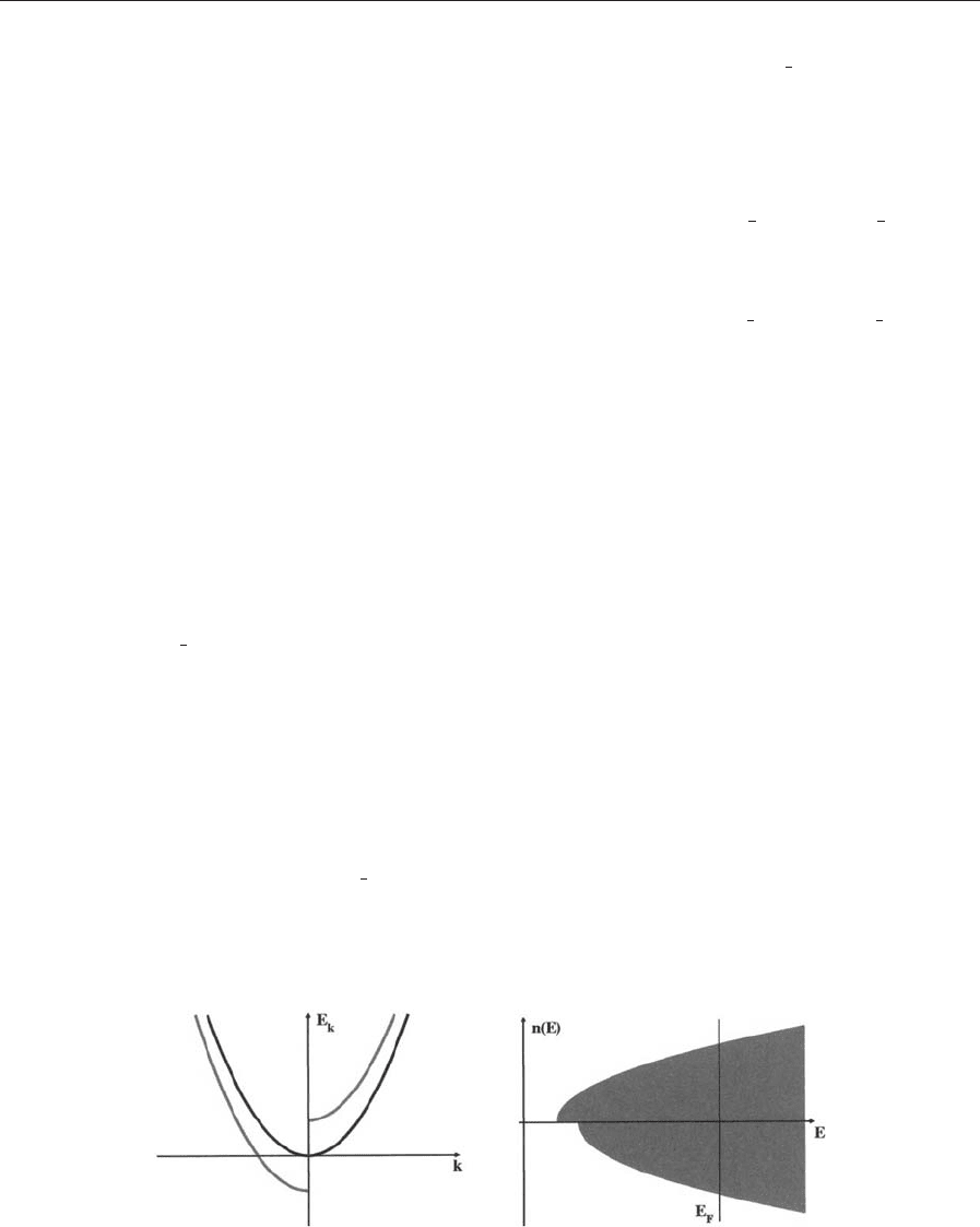
This is the so-called spin–split bandstructure (Fig. 2).
The symmetric energy shift is transferred to the
spin-dependent density of states
n
s
ðEÞ¼nðE7
1
2
IMÞð14Þ
with respect to the density of states of the nonmagnetic
system n(E) (Fig. 2). Integration over all occupied
states up to the Fermi energy E
F
gives the number of
electrons
N ¼
Z
E
F
dEðn
0
ðE þ
1
2
IMÞþn
0
ðE
1
2
IMÞð15Þ
and the moment per atom
M ¼
Z
E
F
dEðn
0
ðE þ
1
2
IMÞn
0
ðE
1
2
IMÞÞ ð16Þ
Since the density of the nonmagnetic system n
0
and the
number of electrons N are known, the Fermi energy
E
F
and the moment M can be obtained from the
self-consistent solution of Eqns. (15) and (16). As a
sufficient condition for a ferromagnetic solution the
Stoner criterion
In
0
ðE
F
Þ41 ð17Þ
can be derived. Large Stoner parameters or exchange
integrals I and large density of states at the Fermi en-
ergy n
0
(E
F
) favor ferromagnetism. The corresponding
parameters deduced from a nonspin-polarized calcula-
tion are shown in Table 1 (Gunnarsson 1976, Janak
1977). Obviously, the Stoner criterion is only fulfilled
for Fe, Co, and Ni, precisely those metals that show
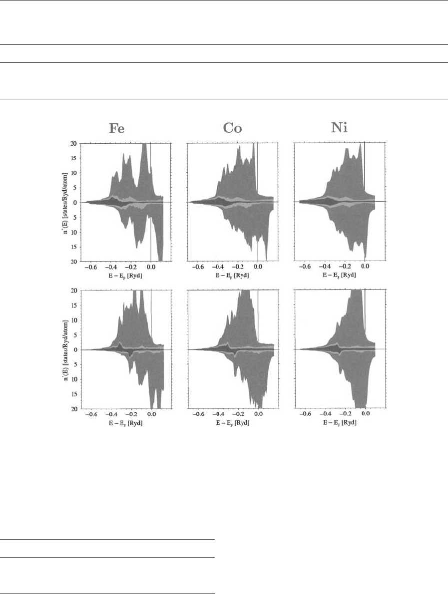
ferromagnetism. Fig. 3 shows the densities of states for
Fe, Co, and Ni computed by means of a spin-density
functional calculation. The results show the dominat-
ing contribution of the 3d electrons. The density of
states is exchange split and otherwise very complicated.
The spin-up and spin-down densities of states remain,
however, very similar which confirms the validity of a
Stoner model.
The corresponding spin moments in comparison to
experimental results for spin moments and total mo-
ments are given in Table 2. The exchange integral I is
an interatomic property that depends on the type of
Figure 2
Spin–split band structure and density of states for free electrons; majority and minority bands are shifted downwards
and upwards, respectively.
1225
Thin Film Magnetism: Band Calculations
atom but is independent on structure and environ-
ment. According to Gunnarsson (1976) and Janak
(1977) the exchange integrals of the transition metals
have the following trend
I
3d
4I
4d
4I
5d
ð18Þ
The density of states at the Fermi level is reciprocal
with the bandwidth
nðE
F
ÞBW ð19Þ
Table 1
Density of states n(E
F
) (in eV
1
) at the Fermi energy calculated for nonmagnetic systems, Stoner parameter I (in eV),
and In(E
F
) (Gunnarsson 1976, Janak 1977).
Metal Na Al Cr Mn Fe Co Ni Cu Pd Pt
n(E
F
) 0.23 0.21 0.35 0.77 1.54 1.72 2.02 0.14 1.14 0.79
I 1.82 1.22 0.76 0.82 0.93 0.99 1.01 0.73 0.68 0.63
In(E
F
) 0.41 0.25 0.27 0.63 1.43 1.70 2.04 0.11 0.78 0.50
Figure 3
Spin–polarized density of states of Fe, Co, and Ni in a bulk phase (upper panel) and for the corresponding atom in a
(100) surface (lower panel). The shades denote the contribution of s (darkest), p (lightest), and d electrons (medium
shade).
Table 2
Magnetic moments (m
B
atom) for Fe, Co, and Ni
calculated in the LSDA approximation (Moruzzi et al.
1978) in comparison to experimental values for the spin
moment and the total moment.
Metal M
LSDA
M
spin
M
Fe 2.15 2.12 2.22
Co 1.56 1.57 1.71
Ni 0.59 0.55 0.61
1226
Thin Film Magnetism: Band Calculations
The stronger the electrons are localized, the smaller
the bandwidth, the larger is the density of states. The
bandwidth is zero in the atomic limit. The Stoner
criterion is always fulfilled. The magnetic moment is
determined by Hunds rule. If we restrict our further
considerations to the tightly bound d electrons of
transition metals, the bandwidth can be expressed by
means of a tight-binding model
W
d
¼ 2
ffiffiffiffiffiffiffiffi
N
nn
p
h
d
ðR
nn
Þð20Þ
According to this approximation the bandwidth
depends on the overlap integral or hopping matrix
element h
d
of the d electrons and the number of
nearest neighbor atoms or coordination number N
nn
.
The overlap integral h
d
is element specific, is deter-
mined by the overlap of the d wavefunctinos, and, of
course, is reciprocal with the nearest neighbor dis-
tance R
nn
. The trend in the transition metals follows
the relation
h
3d
oh
4d
oh
5d
) W
3d
oW
4d
oW
5d
) n
3d
4n
4d
4n
5d
ð21Þ
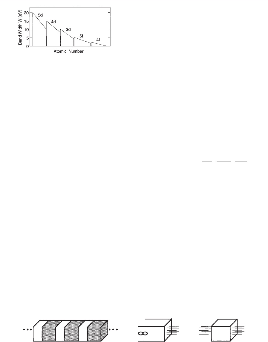
In Fig. 4 the general trend of the bandwidths of
transition metals, lanthanides, and actinides is shown
(Blu
¨
gel 1995).
3. Magnetism in Reduced Dimensions
Understanding magnetism in low-dimensional sys-
tems looks rather straightforward. The simplest low-
dimensional system is the isolated atom, for which it
is known that nearly all transition metal atoms have
magnetic moments. The magnetism is well described
by Hund’s rules. On the other hand, it is known that
among the transition metals only five remain mag-
netic in their bulk crystalline phase: Co and Ni are
ferromagnetic, Cr is antiferromagnetic, and Mn and
Fe are ferromagnetic or antiferromagnetic depending
on their crystal structure. Low-dimensional transition
metals, that is interfaces, surfaces, ultrathin films, and
monolayers, should behave between these two ex-
tremes. As a consequence an enormous number of ab
initio calculations have been performed in this field in
the 1990s (Blu
¨
gel 1995, Weinert and Blu
¨
gel 1993,
Asada et al. 1999).
Following the considerations above a reduction of
the coordination number N
nn
would cause band nar-
rowing and would enhance the tendency towards
magnetism due to the Stoner criterion.
The reduction of the coordination number, how-
ever, can be achieved by a reduction in the dimension
of the system under consideration. An fcc crystal has
a coordination number of N
fcc
¼12, an atom of the
(100) surface of the same crystal is surrounded by
eight atoms N
(100)
¼8, and the monolayer atoms have
only four nearest neighbors, N
ML
¼4. Keeping the
nearest neighbor distance and the overlap integrals
fixed, the density of states and the bandwidths would
obey the following relation
n
fcc
: n
ð100Þ
: n
ML
¼
1
W
fcc
:
1
W
ð100Þ
:
1
W
ML
¼1 : 1:22 : 1:73 ð22Þ
The reduction in the coordination number is hence
the origin of the enhancement of magnetism at in-
terfaces, surfaces, and for monolayers or ultrathin
films. As a consequence these systems offer the op-
portunity to find totally new magnetic materials.
For the ab initio description of surfaces and ultra-
thin magnetic films, different geometries are used. In
a superlattice the metallic films are separated by vac-
uum layers but otherwise are periodically repeated in
all spatial directions. Surface geometry consists of
semi-infinite metal and vacuum parts whereas slab
geometry is a freestanding metal film embedded in
vacuum layers (Fig. 5).
Figure 3 shows the local densities of states for Fe,
Co, and Ni (100) surfaces computed by means of a
spin-density functional calculation. The comparison
of bulk (upper panel) and surface (lower panel) den-
sity of states illustrates the effect of band narrowing
clearly. Enhancement of the magnetic moments at the
surface in comparison to the bulk material is given in
Table 3.
Figure 5
Superlattice, semi-infinite surface system, and slab.
Figure 4
Trend in the bandwidth of the transition metal,
lanthanides and actinides as a function of the atomic
number.
1227
Thin Film Magnetism: Band Calculations
The magnetic moment of the Ni (110) surface is
higher than for the (100) surface. The results for Fe
are opposite. This behavior is related to the surface
orientation, which determines the coordination num-
ber and the next nearest neighbor distance between
the surface atoms. The largest magnetic moment will
be obtained for the most open surface. Ni is an fcc
system: the low-index surface (110) is the most open,
(100) is intermediate, and (111) is the closed packed
surface. The trend for bcc systems is opposite. Fe is a
bcc system.
It is implied in the discussion so far that the co-
ordination number is the crucial feature in interface
magnetism but this is not the whole truth. The fol-
lowing features have to be taken into account to
achieve a quantitative description. First of all, the
point symmetry of a surface is lower in comparison to
the bulk system. Symmetry reduction causes level
splitting, changes the shape of the density of states,
and is unfavourable for magnetism. Second, the re-
laxation of the charge density at the surface changes
hybridization at the surface and causes a redistribu-
tion of spectral density. As a consequence, a relative
shift of the sp band against the d band and/or a
change of spectral weight is obtained (see Fig. 3). The
number of d states is changed. As a result magnetism
can be enhanced or reduced (see Ni). The neglect of
these interplaying effects have caused confusion in
the past in the field of surface and interface magnet-
ism. But they are readily included in self-consistent
electronic structure calculations.
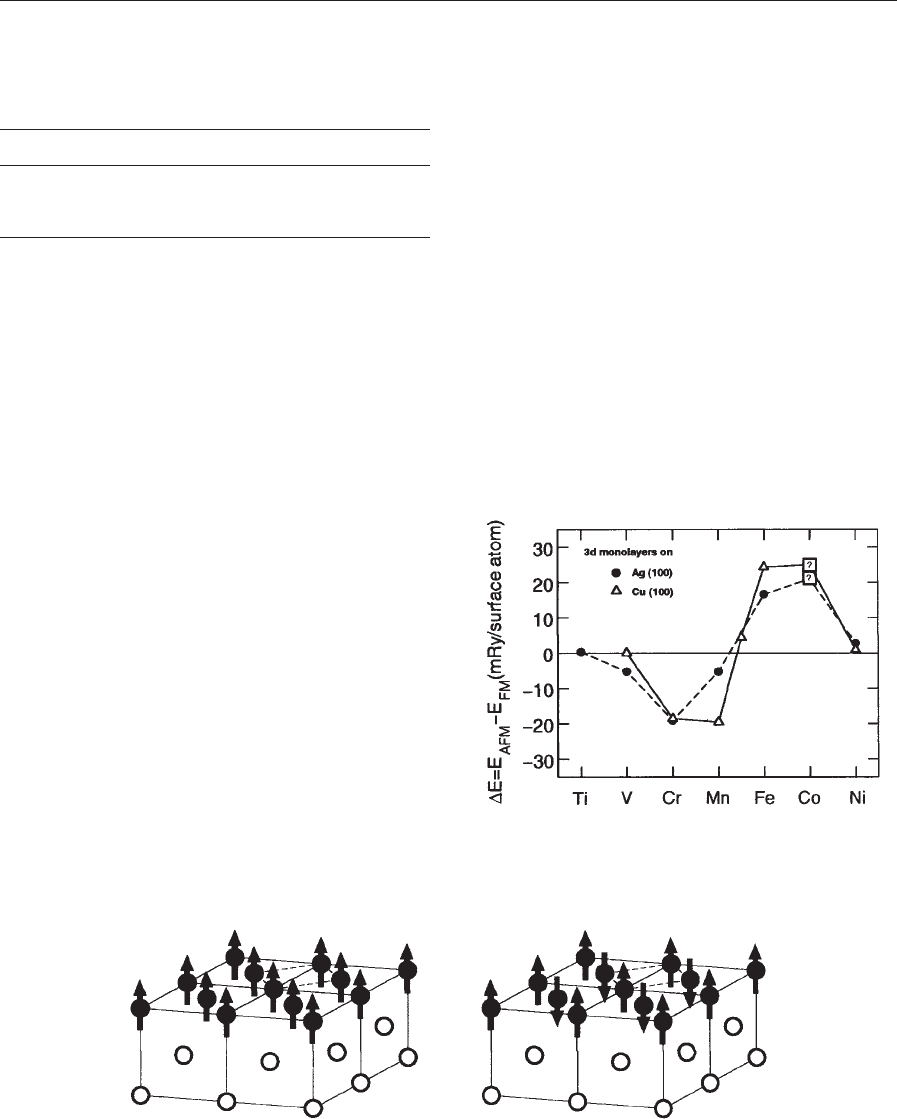
4. Ground State Magnetic Order
Based on the calculation of the total energy (Eqn.
(10)), ab initio electronic structure calculations pro-
vide a perfect tool for investigating the magnetic or-
der. Comparison of the total energy for different
magnetic configurations DE allows a systematic
search of the magnetic order in the ground state.
As an example, the magnetic order of 3d transition
metal monolayers on top of a Cu substrate was in-
vestigated (Blu
¨
gel 1995). The total energy of a ferro-
magnetic configuration E
F
is compared to the total
energy of an antiferromagnetic configuration E
AF
(Fig. 6). The energy differences
DE ¼ E
AF
E
F
ð23Þ
are shown in Fig. 7. Fe, Co, and Ni layers prefer
ferromagnetic, whereas V, Cr, and Mn prefer an an-
tiferromagnetic order.
This is an arbitrary example chosen to demonstrate
the method. Similar calculations can be applied to
various other problems concerning the magnetic con-
figuration, for example surface steps and edges or
Table 3
Magnetic moments (m
B
atom) for Fe, Co, and Ni
calculated in LSDA approximation (Blu
¨
gel 1995) for
(100) and (110) surfaces in comparison to bulk moments
(Moruzzi et al. 1978).
Metal M
LSDA
M
(100)
M
(110)
Fe 2.15 2.88 2.43
Co 1.56 1.85 —
Ni 0.59 0.68 0.74
Figure 6
Schematic representation of ferromagnetic and c (2 2) antiferromagnetic order of a monolayer film grown as an
overlayer on a fcc (001) substrate.
Figure 7
Total energy difference per atom for a ferromagnetic
and antiferromagnetic phase for 3d monolayers on a n,
Cu (100) and *, Ag (100) substrate.
1228
Thin Film Magnetism: Band Calculations

surface alloys (Freyss et al. 1996) also including non-
collinear magnetic order (Sandratskii 1998)
The capability of this method was nicely confirmed
by experimental observation of the local magnetic
order in surface alloys by means of spin-polarized
scanning tunneling spectroscopy (Heinze et al. 2000).
See also: Monolayer Films: Magnetism; Density
Functional Theory: Magnetism; Itinerant Electron
Systems: Magnetism (Ferromagnetism); Multilayers:
Interlayer Coupling
Bibliography
Asada T, Bihlmayer G, Handschuh S, Heinze S, Kurz Ph,
Blu
¨
gel S 1999 J. Phys.: Condens. Matter. 11, 9347
Barth U V, Hedin L 1972 J. Phys. C 5, 1629
Blu
¨
gel S 1995 Habilitation. RWTA, Aachen
Born M, Oppenheimer J R 1927 Ann. Phys. 84, 457
Freyss M, Stoeffler D, Dreysse
´
1996 Phys. Rev. B 54, 12677
Gunnarsson O 1976 J. Phys. F: Met. Phys. 6, 587
Gunnarsson O, Lundqvist B I 1976 Phys. Rev. B 13, 4274
Gunnarsson O, Lundqvist B I, Lundqvist S 1972 Solid State
Commun. 11, 149
Hedin L, Lundqvist B 1971 J. Phys. C 4, 2064
Heinze S, Bode M, Kubetzka A, Pietzsch O, Nie X, Bluegel S,
Wiesendanger R 2000 Science 288, 1805
Hohenberg P, Kohn W 1964 Phys. Rev. 136, 864
Janak J F 1977 Phys. Rev. B 16, 255
Kohn W, Sham L J 1965 Phys. Rev. 140, 1133
Moruzzi V R, Janak J F, Williams A R 1978 Calculated Elec-
tronic Properties of Metals. Pergamon Press, New York
Rajagopal A K 1980 Adv. Chem. Phys. 41,59
Sandratskii L M 1998 Adv. Phys. 47,91
Sham L J, Kohn W 1966 Phys. Rev. 145, 561
Stoner E C 1938 Proc. R. Soc. London 165, 372
Vosko S H, Perdew J P 1975 Can. J. Phys. 53, 1385
Vosko S H, Wilk L, Nussair N 1980 Can. J. Phys. 58, 1200
Weinert M, Blu
¨
gel S 1993 In: Bennet L H, Watson R E (eds.)
Magnetic Multilayers. World Scientific, Singapore
I. Mertig
Technische Universita
¨
t Dresden, Germany
Thin Film Magnetism: PEEM Studies
A Photoemission Electron Microscope (PEEM) im-
ages photo and/or secondary electrons, which are
generated at the surface of a solid sample by the ab-
sorption of x-ray or ultraviolet (UV) radiation. De-
pending on the wavelength of the radiation different
contrast mechanisms are present, providing informa-
tion on the elemental and chemical composition, and
the electronic and magnetic properties of the mate-
rial. Magnetic domain imaging is a major application
of the PEEM technique, exploiting strong x-ray mag-
netic dichroism effects, e.g., at the L edges of the
magnetic 3d transition metals (e.g., Cr, Mn, Fe, Co,
Ni). For a comparison with other domain imaging
techniques see Kerr Microscopy, Magnetic Materials:
Transmission Electron Microscopy, Magnetic Force
Microscopy, and references therein.
The PEEM technique stands out against other
magnetic imaging techniques through its surface sen-
sitivity and element specificity, making PEEM an
ideal tool for the investigation of ultra-thin magnetic
films, multi-layers, and alloys. In contrast to other
high resolution, magnetic imaging techniques, PEEM
is also sensitive to antiferromagnetic order. The spa-
tial resolution of PEEM for magnetic domain imag-
ing is typically about 50–100 nm, which positions
PEEM between Transmission Electron Microscopy
and optical techniques, such as Kerr Microscopy.
1. Photoemission Electron Microscopy
PEEM was first used in the 1930s and has since then
matured into an established surface science technique
(Sto
¨
hr et al. 1993, Tonner et al. 1995, Sto
¨
hr et al.
1998, Anders et al. 1999). PEEM is closely related to
the Low Energy Electron Microscope (LEEM) and
the Spin-polarized Low-energy Electron Microscope
(SPLEEM), which were pioneered by Bauer (1994)
and Duden and Bauer (1998). All three techniques
utilize low-energy electrons to form an image repre-
senting physical properties of the sample surface.
LEEM and SPLEEM image diffracted low-energy
electrons and thereby provide information on the
local crystallographic (LEEM) and magnetic
(SPLEEM) structure of the surface of a crystalline
sample. PEEM, in contrast, images electrons gener-
ated by photoionization and therefore is not limited
to the study of crystalline samples (see also Photo-
emission: Spin-polarized and Angle-resolved). As light
sources, UV gas discharge lamps, UV lasers, and
synchrotron radiation sources have been used. Syn-
chrotron radiation offers the important advantage of
tunability of the wavelength of the illumination,
thereby allowing a selection between various mech-
anisms of contrast. X-ray PEEM thus combines as-
pects of spectroscopic and microscopic methods and
is called a spectromicroscopy technique.
A typical PEEM setup using synchrotron radiation
from a bending magnet is shown in Fig. 1 (Anders
et al. 1999, Scholl et al. 2002). X rays pass through a
moveable aperture, which selects the polarization of
the radiation. A spherical grating monochromator
and an exit slit monochromatize and focus the beam
onto the sample. A schematic kinetic energy spectrum
of the emitted electrons after x-ray absorption is
shown in Fig. 2(a). Photoemission lines appear at
high kinetic energy, followed by a broad tail of sec-
ondary electrons which peak at low energies close
to the work function cut-off. PEEM microscopes
usually accept the total electron yield without prior
1229
Thin Film Magnetism: PEEM Studie s