Bhushan B. Nanotribology and Nanomechanics: An Introduction
Подождите немного. Документ загружается.

212 Markus Morgenstern et al.
relax downward [134]. This corresponds to the situation in Fig. 5.19a, where the tip
is relatively far away and an inward relaxation of the two arsenic atoms is observed.
The considerablylarger attractive force in Fig. 5.19b, however, pulls the two arsenic
atoms toward the tip. All other arsenic atoms are also pulled, but they are less dis-
placed, becausethey have three bonds to the bulk,while the two arsenic atoms in the
neighborhood of an indium vacancy have only two bonds. This direct experimental
proof of the presence of tip-induced relaxations is also relevant for STM measure-
ments, because the tip–sample distances are similar during atomic-resolution imag-
ing. Moreover, the result demonstrates that FM-AFM can probe elastic properties
on an atomic level.
Imaging of Weakly Interacting van der Waals Surfaces
For weakly interacting van der Waals surfaces, much smaller atomic corrugation
amplitudes are expected compared to strongly interacting surfaces of semiconduc-
tors. A typical example is graphite, a layered material, where the carbon atoms are
covalently bonded and arranged in a honeycomb structure within the (0001) plane.
Individual graphene layers stick together by van der Waals forces. Due to the ABA
stacking, three distinctive sites exist on the (0001) surface: carbon atoms with (A-
type)andwithout(B-type)neighborin thenextgraphitelayerand the hollowsite (H-
site) in the hexagon center. In static contact force microscopy as well as in STM the
contrast exhibits usually a trigonal symmetry with a periodicity of 246pm, where
A-andB-site carbon atoms could not be distinguished. However, in high-resolution
FM-AFM images acquired at low temperatures, a large maximum and two different
minima have been resolved, as demonstrated by the profiles along the three equiv-
alent [1-100] directions in Fig. 5.20a. A simulation using the Lennard–Jones po-
tential, given by the short-range interatomic van der Waals force, reproduced these
three features very well (dotted line). Therefore, the large maximum could be as-
signed to the H-site, while the two different minima represent A-andB-type carbon
atoms [132].
Compared to graphite, the carbon atoms in a single-walled carbon nanotube
(SWNT), which consists of a single rolled up graphene layer, are indistinguish-
able. For the first time Ashino et al. [133] successfully imaged the curved surface
of a SWNT with atomic resolution. Note that for geometric reasons, atomic resolu-
tion is only achieved on the top (see Fig. 5.20b). Indeed, as shown in Fig. 5.20b, all
profilesbetween two hollowsites across twoneighboring carbonatoms are symmet-
ric [135]. Particularly, curve1 and 2 exhibittwo minima of equal depth, as predicted
by theory (cf., dotted line). The assumption used in the simulation (dotted lines in
the profiles of Fig. 5.20) that interatomic van der Waals forces are responsible for
the atomic-scale contrast has been supported by a quantitative evaluation of force
spectroscopy data obtained on SWNTs [133].
Interestingly, the image contrast on graphite and SWNTs is inverted with re-
spect to the arrangement of the atoms, i.e., the minima correspond to the position
of the carbon atoms. This can be related to the small carbon–carbon distance of
only 142pm, which is in fact the smallest interatomic distance that has been re-
5 Low-Temperature Scanning Probe Microscopy 213
426 pm
426 pm
900 pm
Corrugation a.u.
1
2
H
B
H
A
1
3
2
3
2
1
HH
CC
2
Xe Xe
1
Xe Xe Xe
a)
3
2
1
Corrugation a.u.
b)
Corrugation a.u.
c)
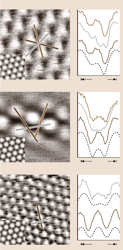
Fig. 5.20. FM-AFM images
of graphite(0001) (a) a single-
walled carbon nanotube
(SWNT) (b) and Xe(111) (c)
recorded at 22 K. On the right
side, line sections taken from
the experimental data (solid
lines) are compared to simu-
lations (dotted lines). A-and
B-type carbon atoms, as well
as the hollow site (H-site) on
graphite can be distinguished,
but are imaged with inverted
contrast, i.e., the carbon sites
are displayed as minima.
Such an inversion does not
occur on Xe(111)
solved with FM-AFM so far. The van der Waals radius of the front tip atom, (e.g.,
210 pm for silicon) has a radius that is significantly larger than the intercarbon dis-
tance. Therefore, next-nearest-neighborinteractions become important and result in
a contrast inversion [135].
While experiments on graphite and SWNTs basically take advantage of the in-
creased stability and signal-to-noise ratio at low temperatures, solid xenon (melting
temperature T
m
= 161K) can only be observed at sufficient low temperatures [8]. In
214 Markus Morgenstern et al.
addition,xenonis a pure vander Waals crystaland, since it is an insulator, FM-AFM
is the only real-space method available today that allows the study of solid xenon
on the atomic scale.
Allers et al. [8] adsorbed a well-ordered xenon film on cold graphite(0001)
(T < 55K) and studied it subsequently at 22 K by FM-AFM (see Fig. 5.20c). The
sixfold symmetry and the distance between the protrusions corresponds well with
the nearest-neighbordistance in the close-packed (111) plane of bulk xenon, which
crystallizes in a face-centered cubic structure. A comparison between experiment
and simulation confirmed that the protrusions correspond to the position of the
xenon atoms [132]. However, the simulated corrugation amplitudes do not fit as
well as for graphite (see sections in Fig. 5.20c). A possible reasonis that tip-induced
relaxations, which were not considered in the simulations, are more important for
this pure van der Waals crystal xenon than they are for graphite, because in-plane
graphite exhibits strong covalent bonds. Nevertheless, the results demonstrated for
the first time that a weakly bonded van der Waals crystal could be imaged nonde-
structively on the atomic scale. Note that on Xe(111) no contrast inversion exists,
presumably because the separation between Xe sites is about 450pm, i.e., twice as
large as the van der Waals radius of a silicon atom at the tip end.
Atomic Resolution Using Small Oscillation Amplitudes
All the examplesabove described used spring constants and amplitudeson the order
of 40 N/m and 10nm, respectively, to obtain atomic resolution. However, Giessibl
et al. [137] pointed out that the optimal amplitude should be on the order of the
characteristic decay length λ of the relevant tip–sample interaction. For short-range
interactions, which are responsible for the atomic-scale contrast, λ is on the order of
0.1nm. On the other hand, stable imaging without a jump-to-contact is only possi-
ble as long as the restoring force c
z
A at the lower turnaround point of each cycle is
larger than the maximal attractive tip–sample force. Therefore, reducing the desired
amplitude by a factor of 100 requires a 100 times larger spring constant. Indeed,
Hembacher et al. [136] could demonstrate atomic resolution with small amplitudes
(about 0.25nm) and large spring constants (about 1800N/m) utilizing a qPlus sen-
sor [138]. Figure 5.21 shows a constant-height image of graphite recorded at 4.9K
within the repulsive regime. Note that compared to Fig. 5.20a,b the contrast is in-
verted,i.e., the carbon atoms appearas maxima.This is expected,because the imag-
ing interaction is switched from attractive to repulsive regime [131,135].
5.5.2 Force Spectroscopy
A wealth of information about the nature of the tip–sample interaction can be ob-
tained by measuring its distance dependence. This is usually done by recording
the measured quantity (deflection, frequency shift, amplitude change, phase shift)
and applying an appropriate voltage ramp to the z-electrode of the scanner piezo,
while the z-feedback is switched off. According to (5.2), low temperatures and high
Q-factors (vacuum) considerably increase the force resolution. In the static mode,
5 Low-Temperature Scanning Probe Microscopy 215
1.49
0.86
Fig. 5.21. Constant-
height FM-AFM image of
graphite(0001) recorded at
4.9K using asmall am-
plitude (A = 0.25 nm) and
a large spring constant
(c
z
= 1800 N/m). As in
Fig. 5.20a, A-andB-site
carbon atoms can be dis-
tinguished. However, they
appear as maxima, because
imaging has been performed
in the repulsive regime
(courtesy of F. J. Giessibl;
cf. [136])
long-rangeforces and contact forces can be examined. Force measurements at small
tip–sample distances are inhibited by the jump-to-contact phenomenon: If the force
gradient ∂F
ts
/∂z becomes larger than the spring constant c
z
, the cantilever cannot
resist the attractive tip–sample forces and the tip snaps onto the surface. Sufficiently
large spring constants prevent this effect, but reduce the force resolution. In the dy-
namic modes, the jump-to-contact can be avoided due to the additional restoring
force (c
z
A) at the lower turnaround point. The highest sensitivity can be achieved
in vacuum by using the FM technique, i.e., by recording Δ f(z)-curves. An alterna-
tive FM spectroscopy method, the recording of Δ f(A)-curves, has been suggested
by Hölscher et al. [139]. Note that, if the amplitude is much larger than the charac-
teristic decay length of the tip–sample force, the frequency shift cannot simply be
convertedinto force gradientsby using ∂F
ts
/∂z= 2c
z
·Δ f/ f
0
[140].Severalmethods
have been published to convert Δ f(z) data into the tip–sample potential V
ts
(z)and
tip–sample force F
ts
(z) (see, e.g., [141–144]).
Measurement of Interatomic Forces at Specific Atomic Sites
FM force spectroscopy has been successfully used to measure and determine quan-
titatively the short-range chemical force between the foremost tip atom and specific
surface atoms [109, 145, 146]. Figure 5.22 displays an example for the quantita-
tive determination of the short-range force. Figure 5.22a shows two Δ f(z)-curves
measured with a silicon tip above a corner hole and above an adatom. Their po-
sition is indicated by arrows in the inset, which displays the atomically resolved
Si(111)-(7×7) surface. The two curves differ from each other only for small tip0-
sample distances, because the long-range forces do not contribute to the atomic-
scale contrast. The low, thermally induced lateral drift and the high stability at low
216 Markus Morgenstern et al.
Δf (Hz)
z-displacement (nm)
0
2.0
0
–20
–40
–60
–80
0.5 1.0 1.5
Force (nN)
z-displacement (nm)
0
1.2
0
–1
–2
–3
0.4 0.6 0.8 1.0
3 nm
a)
b)
Short-range
force F
sr
Total
force F
tot
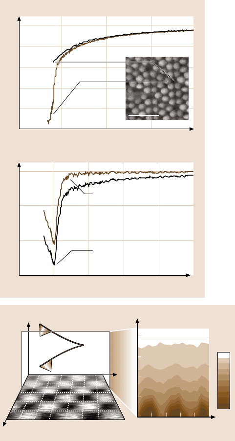
Fig. 5.22. FM force spec-
troscopy on specific atomic
sites at 7.2K. In (a), an
FM-SFM image of the
Si(111)-(7×7) surface is
displayed together with two
Δ f(z)-curves, which have
been recorded at the posi-
tions indicated by the arrows,
i.e., above the corner hole
(brown) and above an adatom
(black). In (b), the total force
above an adatom (brown line)
has been recovered from the
Δ f(z)-curve. After subtrac-
tion of the long-range part,
the short-range force can be
determined (black line) (cour-
tesy of H.J. Hug; cf. [145])
z
x
0
0.4
0.3
0.2
0.1
0.2 0.4 0.6 0.8 1
a)
x
y
z
b)
–0.2
nN
–1.0
nN
Fig. 5.23. Principle of the 3-D force field spectroscopy method (a) and a 2-D cut through
the 3-D force field F
ts
(x,y,z) recorded at 14 K (b). At all 32×32 image points of the 1nm
× 1 nm scan area on NiO(001), a Δ f(z)-curve has been recorded. The Δ f(x, y,z) data set
obtained is then converted into the 3-D tip–sample force field F
ts
(x, y, z). The shaded slice
F
ts
(x, y = const, z)in(a) corresponds to a cut along the [100]-direction and demonstrates
that atomic resolution has been obtained, because the distance between the protrusions
corresponds well to the lattice constant of nickel oxide
5 Low-Temperature Scanning Probe Microscopy 217
temperatures were required to precisely address the two specific sites. To extract the
short-range force, the long-range van der Waals and/or electrostatic forces can be
subtracted from the total force. The grey curve in Fig. 5.22b has been reconstructed
from the Δ f(z)-curve recorded above an adatom and represents the total force. After
removing the long-range contribution from the data, the much steeper black line is
obtained, which corresponds to the short-range force between the adatom and the
atom at the tip apex. The measuredmaximum attractive force(−2.1nN) agrees well
with first-principles calculations (−2.25nN).
Three-Dimensional Force Field Spectroscopy
Further progress with the FM technique has been made by Hölscher et al. [147].
They acquired a complete 3-D force field on NiO(001) with atomic resolution (3-D
force field spectroscopy). In Fig. 5.23a, the atomically resolved FM-AFM image of
NiO(001) is shown together with the coordinate system used and the tip to illustrate
the measurement principle. NiO(001) crystallizes in the rock-salt structure. The dis-
tance between the protrusions corresponds to the lattice constant of 417pm, i.e.,
only one type of atom (most likely the oxygen)is imaged as a protrusion. In an area
of 1nm×1nm,32×32 individual Δ f(z)-curves have been recorded at every (x, y)
image point and converted into F
ts
(z)-curves.The Δ f(x, y,z) data set is thereby con-
verted into the 3-D force field F
ts
(x, y, z). Figure 5.23b, where a specific x–z-plane
is displayed, demonstrates that atomic resolution is achieved. It represents a 2-D
cut F
ts
(x, y = const,z) along the [100]-direction (corresponding to the shaded slice
marked in Fig. 5.23a). Since a large number of curves have been recorded, Langkat
et al. [146] could evaluate the whole data set by standard statistical means to extract
the long- and short-range forces. A possible future application of 3-D force field
spectroscopy could be to map the short-range forces of complex molecules with
functionalized tips in order to resolve locally their chemical reactivity. A first step
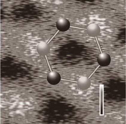
in this direction has been accomplished on SWNTs. Its structural unit, a hexagonal
carbon ring, is common to all aromatic molecules.Like the constant frequency-shift
image of an SWNT shown in Fig. 5.20b the force map shows clear differences be-
tween hollow sites and carbon sites [133]. Analyzing site-specific individual force
curves extracted from the 3-D data revealed a maximum attractive force of about
−0.106nN above H-sites and about −0.075nN above carbon sites. Since the at-
traction is one order of magnitude weaker than on Si(111)-(7×7) (cf., Fig. 5.22b),
it has been inferred that the short-range interatomic van der Waals force and not
a chemical force is responsible for atomic-scale contrast formation on such nonre-
active surfaces.
Noncontact Friction
Another approach to achieve small tip–sample distances in combination with high
force sensitivity is to use soft springs in a perpendicular configuration. The much
higher cantilever stiffness along the cantilever axis prevents the jump-to-contact,
but the lateral resolution is limited by the magnitude of the oscillation amplitude.
However, with such a set-up at low temperatures, Stipe et al. [148] measured the
218 Markus Morgenstern et al.
a) b) c)
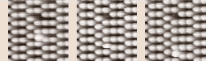
Fig. 5.24. Consecutively recorded FM-AFM images showing the tip-induced manipulation
of a Ge adatom on Ge(111)-c(2×8) at 80 K. Scanning was performed from bottom to top
(courtesy of N. Oyabu; cf. [150])
distance dependence of the very small force due to noncontact friction between
the tip and sample in vacuum. The effect was attributed to electric charges, which
are moved parallel to the surface by the oscillating tip. Since the topography was
not recorded in situ, the influence of contaminants or surface steps remained un-
known.
5.5.3 Atomic Manipulation
Nowadays, atomic-scale manipulation is routinely performed using an STM tip (see
Sect. 5.4.1). In most of these experiments an adsorbate is dragged with the tip due
to an attractive force between the foremost tip apex atoms and the adsorbate. By
adjusting a large or a small tip–surface distance via the tunneling resistance, it is
possible to switch between imaging and manipulation. Recently, it has been demon-
strated that controlled manipulation of individual atoms is also possible in the dy-
namic mode of atomic force microscopy, i.e., FM-AFM. Vertical manipulation was
demonstrated by pressing the tip in a controlled manner into the Si(111)-(7×7) sur-
face [149]. The strong repulsion leads to the removal of the selected silicon atom.
The process could be traced by recording the frequency shift and the damping sig-
nal during the approach. For lateral manipulation a rubbing technique has been uti-
lized [150], where the slow scan axis is halted above a selected atom, while the
tip–surface distance is gradually reduced until the selected atom hops to a new sta-
ble position. Figure 5.24 shows a Ge adatom on Ge(111)-c(2×8) that was moved
during scanning in two steps from its original position (a) to its final position (c). In
fact, manipulation by FM-AFM is reproducible and fast enough to write nanostruc-
tures in a bottom-up process with single atoms [151].
5.5.4 Electrostatic Force Microscopy
Electrostatic forces are readily detectable by a force microscope, because the tip
and sample can be regarded as two electrodes of a capacitor. If they are electrically
connected via their back sides and have different work functions, electrons will flow
5 Low-Temperature Scanning Probe Microscopy 219
between the tip and sample until their Fermi levels are equalized. As a result, an
electric field and, consequently, an attractive electrostatic force exists between them
at zero bias. This contact potential difference can be balanced by applying an ap-
propriate bias voltage. It has been demonstrated that individual doping atoms in
semiconducting materials can be detected by electrostatic interactions due to the
local variation of the surface potential around them [152,153].
Detection of Edge Channels in the Quantum Hall Regime
At low temperatures, electrostatic force microscopy has been used to measure the
electrostatic potentialin the quantum Hall regimeof a two-dimensional electron gas
(2-DEG) buried in epitaxially grown GaAs/AlGaAs heterostructures [154–157]. In
the 2-DEG, electrons can move freely in the x–y-plane, but they cannot move in
z-direction. Electrical transport properties of a 2-DEG are very different compared
to normal metallic conduction. Particularly, the Hall resistance R
H
= h/ne
2
(where
h represents Planck’s constant, e is the electron charge, and n = 1, 2,...) is quan-
tized in the quantum Hall regime, i.e., at sufficiently low temperatures (T < 4K)
and high magnetic fields (up to 20 T). Under these conditions, theoretical calcu-
lations predict the existence of edge channels in a Hall bar. A Hall bar is a strip
conductor that is contacted in a specific way to allow longitudinal and transversal
transport measurements in a perpendicularmagnetic field. The current is not evenly
distributed over the cross section of the bar, but passes mainly along rather thin
paths close to the edges. This prediction has been verified by measuring profiles
of the electrostatic potential across a Hall bar in different perpendicular external
magnetic fields [154–156].
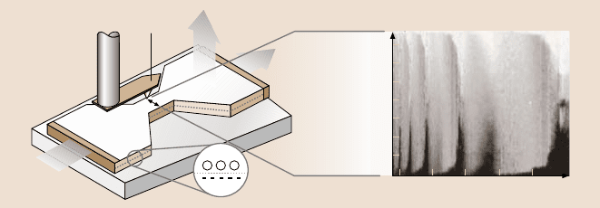
Figure5.25a shows theexperimentalset-up usedto observe these edge channels
on top of a Hall bar with a force microscope. The tip is positioned above the surface
Tip position (μm)
212
14
12
10
8
6
4
2
0
46810
B (T)
SFM
tip
B
I
+
+
+
AlGaAs
GaAs 2DEG
a) b)
Fig. 5.25. Configuration of the Hall bar within a low temperature (T < 1 K) force micro-
scope (a) and profiles (y-axis) at different magnetic field (x-axis) of the electrostatic potential
across a 14-µm-wide Hall bar in the quantum Hall regime (b). The external magnetic field
is oriented perpendicular to the 2-DEG, which is buried below the surface. Bright and dark
regions reflect the characteristic changes of the electrostatic potential across the Hall bar at
different magnetic fields and can be explained by the existence of the theoretically predicted
edge channels (courtesy of E. Ahlswede; cf. [156])
220 Markus Morgenstern et al.
of a Hall bar under which the 2-DEG is buried. The direction of the magnetic field
is oriented perpendicular to the 2-DEG. Note that, although the 2-DEG is located
several tens of nanometers below the surface, its influence on the electrostatic sur-
face potential can be detected. In Fig. 5.25b, the results of scans perpendicular to
the Hall bar are plotted against the magnitude of the external magnetic field. The
value of the electrostatic potential is grey-coded in arbitrary units. In certain field
ranges, the potential changes linearly across the Hall bar, while in other field ranges
the potential drop is confined to the edges of the Hall bar. The predicted edge chan-
nels can explain this behavior. The periodicity of the phenomenon is related to the
filling factor ν, i.e., the number of Landau levels that are filled with electrons (see
also Sect. 5.4.4). Its value depends on 1/B and is proportional to the electron con-
centration n
e
in the 2-DEG (ν = n
e
h/eB,whereh represents Planck’s constant and
e the electron charge).
5.5.5 Magnetic Force Microscopy
To detect magnetostatic tip–sample interactions with magnetic force microscopy
(MFM), a ferromagnetic probe has to be used. Such probes are readily prepared by
evaporating a thin magnetic layer, e.g., 10nm iron, onto the tip. Due to the in-plane
shape anisotropy of thin films, the magnetization of such tips lies predominantly
along the tip axis, i.e., perpendicular to the surface.Since magnetostatic interactions
are long-range, they can be separated from the topography by scanning in a certain
constant height (typically around 20nm) above the surface, where the z-component
of the sample stray field is probed (see Fig. 5.26a). Therefore, MFM is always op-
erated in noncontact mode. The signal from the cantilever is directly recorded while
the z-feedback is switched off. MFM can be operatedin the static mode or in the dy-
namic modes (AM-MFM at ambient pressures and FM-MFM in vacuum). A lateral
resolution below 50nm can be routinely obtained.
Observation of Domain Patterns
MFM is widely used to visualize domain patterns of ferromagneticmaterials. At low
temperatures, Moloni et al. [158] observed the domain structure of magnetite below
its Verwey transition temperature (T
V
= 122K), but most of the work concentrated
on thin films of La
1−x
Ca
x
MnO
3
[159–161]. Below T
V
, the conductivity decreases
bytwo ordersof magnitudeand a smallstructuraldistortionis observed.The domain
structure of this mixed-valence manganite is of great interest, because its resistivity
strongly depends on the external magnetic field, i.e., it exhibits a large colossal-
magnetoresistive effect. To investigate the field dependence of the domain patterns
under ambient conditions, electromagnets have to be used. They can cause severe
thermal drift problems due to Joule heating of the coils by large currents. Fields
on the order of 100mT can be achieved. In contrast, much larger fields (more than
10T) can be rather easily produced by implementing a superconducting magnet in
low-temperature set-ups. With such a design, Liebmann et al. [161] recorded the
5 Low-Temperature Scanning Probe Microscopy 221
400 mT
800 mT
0 mT
205 mT
360 mT
a) b) c)
d)
e)
f)
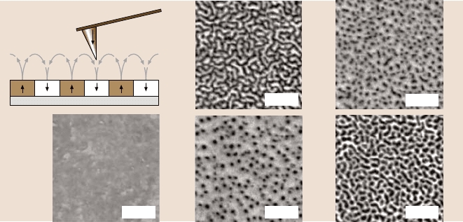
Fig. 5.26. Principle of MFM operation (a) and field-dependent domain structure of a ferro-
magnetic thin film (b)–(f) recorded at 5.2 K with FM-MFM. All images were recorded on
the same 4 µm×4 µm scan area. The La
0.7
Ca
0.3
MnO
3
/LaAlO
3
system exhibits a substrate-
induced out-of-plane anisotropy. Bright and dark areas are visible and correspond to attrac-
tive and repulsive magnetostatic interactions, respectively. The series shows how the domain
pattern evolves along the major hysteresis loop from, i.e., zero field to saturation at 600 mT
and back to zero field
domain structure along the major hysteresis loop of La
0.7
Ca
0.3
MnO
3
epitaxially
grownonLaAlO
3
(see Fig. 5.26b–f). The film geometry (the thickness is 100nm)
favors an in-plane magnetization,but the lattice mismatch with the substrate induces
an out-of-planeanisotropy. Thereby, an irregular pattern of strip domains appears at
zero field. If the external magnetic field is increased, the domains with antiparallel
orientation shrink and finally disappear in saturation (see Fig. 5.26b,c). The residual
contrast in saturation (d) reflects topographic features. If the field is decreased after
saturation(see Fig.5.26e,f),cylindricaldomainsfirst nucleateand then start to grow.
At zero field, the maze-type domain pattern has evolved again. Such data sets can
be used to analyze domain nucleation and the domain growth mode. Moreover, due
to the negligible drift, domain structure and surface morphology can be directly
compared, because every MFM can be used as a regular topography-imaging force
microscope.
Detection of Individual Vortices in Superconductors
Numerous low-temperature MFM experiments have been performed on supercon-
ductors [162–169]. Some basic features of superconductors have been mentioned
already in Sect. 5.4.5. The main difference of STM/STS compared to MFM is its
high sensitivity to the electronicproperties of the surface. Therefore, careful sample
preparation is a prerequisite. This is not so important for MFM experiments, since
the tip is scanned at a certain distance above the surface.