Berg J.M., Tymoczko J.L., Stryer L. Biochemistry
Подождите немного. Документ загружается.

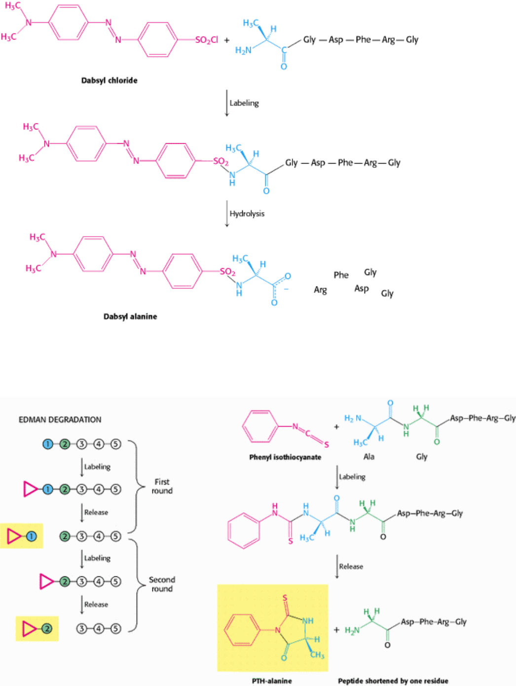
Figure 4.20. Determination of the Amino-Terminal Residue of a Peptide. Dabsyl chloride labels the peptide, which
is then hydrolyzed with the use of hydrochloric acid. The dabsyl-amino acid (dabsyl-alanine in this example) is
identified by its chromatographic characteristics.
I. The Molecular Design of Life 4. Exploring Proteins 4.2. Amino Acid Sequences Can Be Determined by Automated Edman Degradation
Figure 4.21. The Edman Degradation. The labeled amino-terminal residue (PTH-alanine in the first round) can be
released without hydrolyzing the rest of the peptide. Hence, the amino-terminal residue of the shortened peptide (Gly-
Asp-Phe-Arg-Gly) can be determined in the second round. Three more rounds of the Edman degradation reveal the
complete sequence of the original peptide.
I. The Molecular Design of Life 4. Exploring Proteins 4.2. Amino Acid Sequences Can Be Determined by Automated Edman Degradation
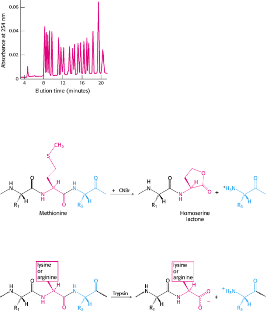
Figure 4.22. Separation of PTH-Amino Acids. PTH-amino acids can be rapidly separated by high-pressure liquid
chromatography (HPLC). In this HPLC profile, a mixture of PTH-amino acids is clearly resolved into its components.
An unknown amino acid can be identified by its elution position relative to the known ones.
I. The Molecular Design of Life 4. Exploring Proteins 4.2. Amino Acid Sequences Can Be Determined by Automated Edman Degradation
Figure 4.23. Cleavage by Cyanogen Bromide. Cyanogen bromide cleaves polypeptides on the carboxyl side of
methionine residues.
I. The Molecular Design of Life 4. Exploring Proteins 4.2. Amino Acid Sequences Can Be Determined by Automated Edman Degradation
Figure 4.24. Cleavage by Trypsin. Trypsin hydrolyzes polypeptides on the carboxyl side of arginine and lysine
residues.
I. The Molecular Design of Life 4. Exploring Proteins 4.2. Amino Acid Sequences Can Be Determined by Automated Edman Degradation
Table 4.3. Specific cleavage of polypeptides
Reagent Cleavage site
Chemical cleavage
Cyanogen bromide Carboxyl side of methionine residues
O-Iodosobenzoate Carboxyl side of tryptophan residues
Hydroxylamine Asparagine-glycine bonds
2-Nitro-5-thiocyanobenzoate Amino side of cysteine residues
Enzymatic cleavage
Trypsin Carboxyl side of lysine and arginine residues
Clostripain Carboxyl side of arginine residues
Staphylococcal protease Carboxyl side of aspartate and glutamate residues (glutamate only under certain
conditions)
Thrombin Carboxyl side of arginine
Chymotrypsin Carboxyl side of tyrosine, tryptophan, phenylalanine, leucine, and methionine
Carboxypeptidase A Amino side of C-terminal amino acid (not arginine, lysine, or proline)
I. The Molecular Design of Life 4. Exploring Proteins 4.2. Amino Acid Sequences Can Be Determined by Automated Edman Degradation
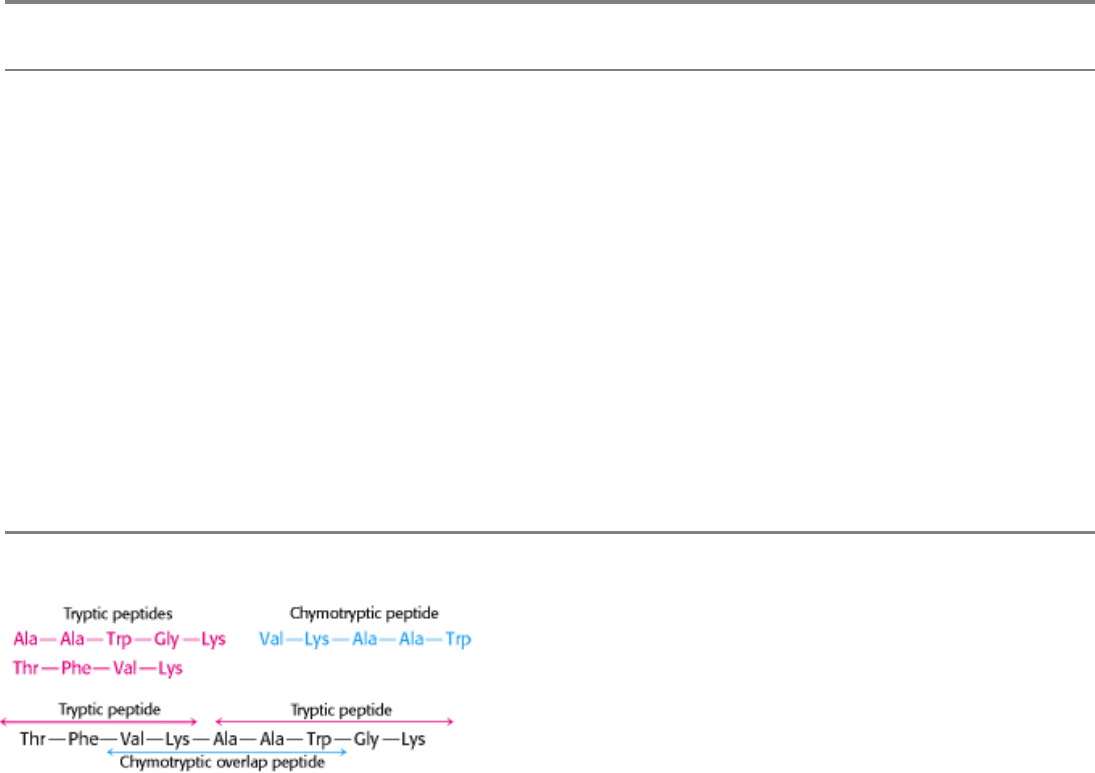
Figure 4.25. Overlap Peptides. The peptide obtained by chymotryptic digestion overlaps two tryptic peptides,
establishing their order.
I. The Molecular Design of Life 4. Exploring Proteins 4.2. Amino Acid Sequences Can Be Determined by Automated Edman Degradation
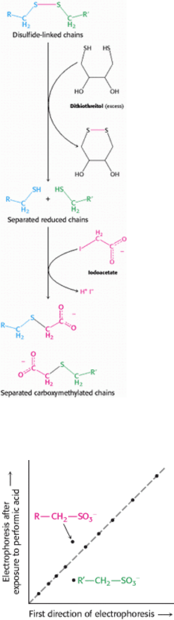
Figure 4.26. Disulfide-Bond Reduction. Polypeptides linked by disulfide bonds can be separated by reduction with
dithiothreitol followed by alkylation to prevent reformation.
I. The Molecular Design of Life 4. Exploring Proteins 4.2. Amino Acid Sequences Can Be Determined by Automated Edman Degradation
Figure 4.27. Diagonal Electrophoresis. Peptides joined together by disulfide bonds can be detected by diagonal
electrophoresis. The mixture of peptides is subjected to electrophoresis in a single lane in one direction (horizontal) and
then treated with performic acid, which cleaves and oxidizes the disulfide bonds. The sample is then subjected to
electrophoresis in the perpendicular direction (vertical).

I. The Molecular Design of Life 4. Exploring Proteins 4.2. Amino Acid Sequences Can Be Determined by Automated Edman Degradation
Figure 4.28. Repeating Motifs in a Protein Chain.
Calmodulin, a calcium sensor, contains four similar units in a single
polypeptide chain shown in red, yellow, blue, and orange. Each unit binds a calcium ion (shown in green).
I. The Molecular Design of Life 4. Exploring Proteins 4.2. Amino Acid Sequences Can Be Determined by Automated Edman Degradation
Figure 4.29. DNA Sequence Yields the Amino Acid Sequence. The complete nucleotide sequence of HIV-1 (human
immunodeficiency virus), the cause of AIDS (acquired immune deficiency syndrome), was determined within a year
after the isolation of the virus. A part of the DNA sequence specified by the RNA genome of the virus is shown here
with the corresponding amino acid sequence (deduced from a knowledge of the genetic code).
I. The Molecular Design of Life 4. Exploring Proteins
4.3. Immunology Provides Important Techniques with Which to Investigate Proteins
Immunological methods have become important tools used to purify a protein, locate it in the cell, or quantify how much
of the protein is present. These methods are predicated on the exquisite specificity of antibodies for their target proteins.
Labeled antibodies provide a means to tag a specific protein so that it can be isolated, quantified, or visualized.
4.3.1. Antibodies to Specific Proteins Can Be Generated
Immunological techniques begin with the generation of antibodies to a particular protein. An antibody (also called an
immunoglobulin, Ig) is a protein synthesized by an animal in response to the presence of a foreign substance, called an
antigen, and normally functions to protect the animal from infection (Chapter 33). Antibodies have specific and high
affinity for the antigens that elicited their synthesis. Proteins, polysaccharides, and nucleic acids can be effective
antigens. An antibody recognizes a specific group or cluster of amino acids on a large molecule called an antigenic
determinant, or epitope (Figures 4.30 and 4.31). Small foreign molecules, such as synthetic peptides, also can elicit
antibodies, provided that the small molecule contains a recognized epitope and is attached to a macromolecular carrier.
The small foreign molecule itself is called a hapten. Animals have a very large repertoire of antibody-producing cells,
each producing an antibody of a single specificity. An antigen acts by stimulating the proliferation of the small number
of cells that were already forming an antibody capable of recognizing the antigen (Chapter 33).
Immunological techniques depend on our being able to generate antibodies to a specific antigen. To obtain antibodies
that recognize a particular protein, a biochemist injects the protein into a rabbit twice, 3 weeks apart. The injected protein
stimulates the reproduction of cells producing antibodies that recognize the foreign substance. Blood is drawn from the
immunized rabbit several weeks later and centrifuged to separate blood cells from the supernatant, or serum. The serum,
called an antiserum, contains antibodies to all antigens to which the rabbit has been exposed. Only some of them will be

antibodies to the injected protein. Moreover, antibodies of a given specificity are not a single molecular species. For
instance, 2,4- dinitrophenol (DNP) has been used as a hapten to generate antibodies to DNP. Analyses of anti-DNP
antibodies revealed a wide range of binding affinities the dissociation constants ranged from about 0.1 nM to 1 µ M.
Correspondingly, a large number of bands were evident when anti-DNP antibody was subjected to isoelectric focusing.
These results indicate that cells are producing many different antibodies, each recognizing a different surface feature of
the same antigen. The antibodies are heterogeneous, or polyclonal (Figure 4.32). This heterogeneity is a barrier, which
can complicate the use of these antibodies.
4.3.2. Monoclonal Antibodies with Virtually Any Desired Specificity Can Be Readily
Prepared
The discovery of a means of producing monoclonal antibodies of virtually any desired specificity was a major
breakthrough that intensified the power of immunological approaches. Just as working with impure proteins makes it
difficult to interpret data and understand function, so too does working with an impure mixture of antibodies. The ideal
would be to isolate a clone of cells that produce only a single antibody. The problem is that antibody-producing cells
isolated from an organism die in a short time.
Immortal cell lines that produce monoclonal antibodies do exist. These cell lines are derived from a type of cancer,
multiple myeloma, a malignant disorder of antibody-producing cells. In this cancer, a single transformed plasma cell
divides uncontrollably, generating a very large number of cells of a single kind. They are a clone because they are
descended from the same cell and have identical properties. The identical cells of the myeloma secrete large amounts of
normal immunoglobulin of a single kind generation after generation. A myeloma can be transplanted from one mouse to
another, where it continues to proliferate. These antibodies were useful for elucidating antibody structure, but nothing is
known about their specificity and so they are useless for the immunological methods described in the next pages.
Cesar Milstein and Georges Köhler discovered that large amounts of homogeneous antibody of nearly any desired
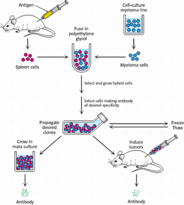
specificity could be obtained by fusing a short-lived antibody-producing cell with an immortal myeloma cell. An antigen
is injected into a mouse, and its spleen is removed several weeks later (Figure 4.33). A mixture of plasma cells from this
spleen is fused in vitro with myeloma cells. Each of the resulting hybrid cells, called hybridoma cells, indefinitely
produces homogeneous antibody specified by the parent cell from the spleen. Hybridoma cells can then be screened, by
using some sort of assay for the antigen-antibody interaction, to determine which ones produce antibody having the
desired specificity. Collections of cells shown to produce the desired antibody are subdivided and reassayed. This
process is repeated until a pure cell line, a clone producing a single antibody, is isolated. These positive cells can be
grown in culture medium or injected into mice to induce myelomas. Alternatively, the cells can be frozen and stored for
long periods.
The hybridoma method of producing monoclonal antibodies has opened new vistas in biology and medicine. Large
amounts of homogeneous antibodies with tailor-made specificities can be readily prepared. They are sources of insight
into relations between antibody structure and specificity. Moreover, monoclonal antibodies can serve as precise
analytical and preparative reagents. For example, a pure antibody can be obtained against an antigen that has not yet
been isolated (Section 4.4). Proteins that guide development have been identified with the use of monoclonal antibodies
as tags (Figure 4.34). Monoclonal antibodies attached to solid supports can be used as affinity columns to purify scarce
proteins. This method has been used to purify interferon (an antiviral protein) 5000-fold from a crude mixture. Clinical
laboratories are using monoclonal antibodies in many assays. For example, the detection in blood of isozymes that are
normally localized in the heart points to a myocardial infarction (heart attack). Blood transfusions have been made safer
by antibody screening of donor blood for viruses that cause AIDS (acquired immune deficiency syndrome), hepatitis,
and other infectious diseases. Monoclonal antibodies are also being evaluated for use as therapeutic agents, as in the
treatment of cancer. Furthermore, the vast repertoire of antibody specificity can be tapped to generate catalytic
antibodies having novel features not found in naturally occurring enzymes.
4.3.3. Proteins Can Be Detected and Quantitated by Using an Enzyme-Linked
Immunosorbent Assay

Antibodies can be used as exquisitely specific analytic reagents to quantify the amount of a protein or other antigen. The
technique is the enzyme-linked immunosorbent assay (ELISA). In this method, an enzyme, which reacts with a colorless
substrate to produce a colored product, is covalently linked to a specific antibody that recognizes a target antigen. If the
antigen is present, the antibody-enzyme complex will bind to it, and the enzyme component of the antibody-enzyme
complex will catalyze the reaction generating the colored product. Thus, the presence of the colored product indicates the
presence of the antigen. Such an enzyme-linked immunosorbent assay, which is rapid and convenient, can detect less
than a nanogram (10
-9
g) of a protein. ELISA can be performed with either polyclonal or monoclonal antibodies, but the
use of monoclonal antibodies yields more reliable results.
We will consider two among the several types of ELISA. The indirect ELISA is used to detect the presence of
antibody and is the basis of the test for HIV infection. In that test, viral core proteins (the antigen) are absorbed to
the bottom of a well. Antibodies from a patient are then added to the coated well and allowed to bind to the antigen.
Finally, enzyme-linked antibodies to human antibodies (for instance, goat antibodies that recognize human antibodies)
are allowed to react in the well and unbound antibodies are removed by washing. Substrate is then applied. An enzyme
reaction suggests that the enzyme-linked antibodies were bound to human antibodies, which in turn implies that the
patient had antibodies to the viral antigen (Figure 4.35).
The sandwich ELISA allows both the detection and the quantitation of antigen. Antibody to a particular antigen is first
absorbed to the bottom of a well. Next, the antigen (or blood or urine containing the antigen) is added to the well and
binds to the antibody. Finally, a second, different antibody to the antigen is added. This antibody is enzyme linked and is
processed as described for indirect ELISA. In this case, the extent of reaction is directly proportional to the amount of
antigen present. Consequently, it permits the measurement of small quantities of antigen (see Figure 4.35).
4.3.4. Western Blotting Permits the Detection of Proteins Separated by Gel
Electrophoresis
Often it is necessary to detect small quantities of a particular protein in the presence of many other proteins, such as a
viral protein in the blood. Very small quantities of a protein of interest in a cell or in body fluid can be detected by an
immunoassay technique called Western blotting (Figure 4.36). A sample is subjected to electrophoresis on an SDS-
polyacrylamide gel. Blotting (or more typically electroblotting) transfers the resolved proteins on the gel to the surface of
a polymer sheet to make them more accessible for reaction. An antibody that is specific for the protein of interest is
added to the sheet and reacts with the antigen. The antibody-antigen complex on the sheet then can be detected by
rinsing the sheet with a second antibody specific for the first (e.g., goat antibody that recognizes mouse antibody). A
radioactive label on the second antibody produces a dark band on x-ray film (an autoradiogram). Alternatively, an
enzyme on the second antibody generates a colored product, as in the ELISA method. Western blotting makes it possible
to find a protein in a complex mixture, the proverbial needle in a haystack. It is the basis for the test for infection by
hepatitis C, where it is used to detect a core protein of the virus. This technique is also very useful in the cloning of
genes.
4.3.5. Fluorescent Markers Make Possible the Visualization of Proteins in the Cell
Biochemistry is often performed in test tubes or polyacrylamide gels. However, most proteins function in the context of
a cell. Fluorescent markers provide a powerful means of examining proteins in their biological context. For instance,
cells can be stained with fluorescence-labeled antibodies or other fluorescent proteins and examined by fluorescence
microscopy to reveal the location of a protein of interest. Arrays of parallel bundles are evident in cells stained with
antibody specific for actin, a protein that polymerizes into filaments (Figure 4.37). Actin filaments are constituents of the
cytoskeleton, the internal scaffolding of cells that controls their shape and movement. By tracking protein location,
fluorescent markers also provide clues to protein function. For instance, the glucocorticoid receptor protein is a
transcription factor that controls gene expression in response to the steroid hormone cortisone. The receptor was linked
to green fluorescent protein (GPF), a naturally fluorescent protein isolated from the jellyfish Aequorea victoria (Section
3.6.5). Fluorescence microscopy revealed that, in the absence of the hormone, the receptor is located in the cytoplasm
(Figure 4.38A). On addition of the steroid, the receptor is translocated to the nucleus, where it binds to DNA (Figure
4.38B).
The highest resolution of fluorescence microscopy is about 0.2 µ m (200 nm, or 2000 Å), the wavelength of visible light.
Finer spatial resolution can be achieved by electron microscopy by using antibodies tagged with electron-dense markers.
For example, ferritin conjugated to an antibody can be readily visualized by electron microscopy because it contains an
electron-dense core rich in iron. Clusters of gold also can be conjugated to antibodies to make them highly visible under
the electron microscope. Immunoelectron microscopy can define the position of antigens to a resolution of 10 nm (100
Å) or finer (Figure 4.39).
I. The Molecular Design of Life 4. Exploring Proteins 4.3. Immunology Provides Important Techniques with Which to Investigate Proteins
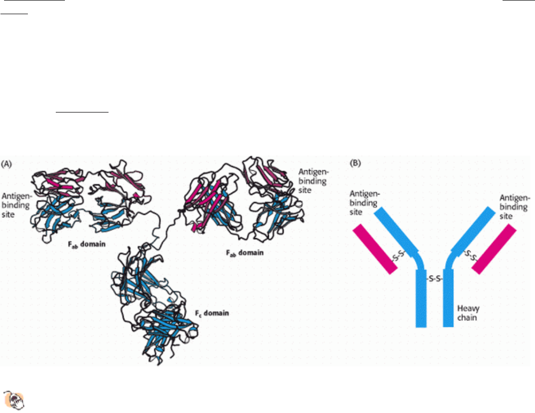
Figure 4.30. Antibody Structure.
(A) IgG antibodies consist of four chains, two heavy chains (blue) and two light
chains (red), linked by disulfide bonds. The heavy and light chains come together to form Fab domains, which
have the antigen-binding sites at the ends. The two heavy chains form the Fc domain. The Fab domains are linked
to the Fc domain by flexible linkers. (B) A more schematic representation of an IgG molecule.
I. The Molecular Design of Life 4. Exploring Proteins 4.3. Immunology Provides Important Techniques with Which to Investigate Proteins
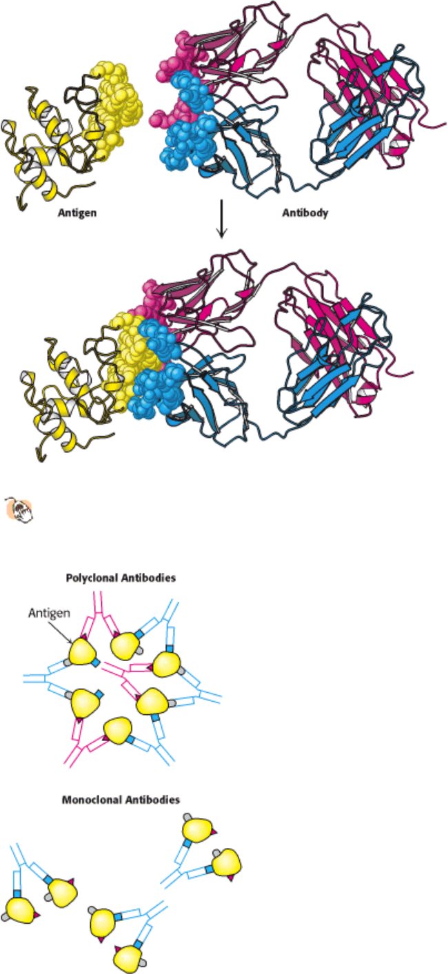
Figure 4.31. Antigen-Antibody Interactions.
A protein antigen, in this case lysozyme, binds to the end of an Fab
domain from an antibody. The end of the antibody and the antigen have complementary shapes, allowing a large
amount of surface to be buried on binding.
I. The Molecular Design of Life 4. Exploring Proteins 4.3. Immunology Provides Important Techniques with Which to Investigate Proteins
Figure 4.32. Polyclonal and Monoclonal Antibodies. Most antigens have several epitopes. Polyclonal antibodies are
heterogeneous mixtures of antibodies, each specific for one of the various epitopes on an antigen. Monoclonal antibodies
are all identical, produced by clones of a single antibody-producing cell. They recognize one specific epitope. [After R.
A. Goldsby, T. J. Kindt, B. A. Osborne, Kuby Immunology, 4th ed. (W. H. Freeman and Company, 2000), p. 154.]
I. The Molecular Design of Life 4. Exploring Proteins 4.3. Immunology Provides Important Techniques with Which to Investigate Proteins
Figure 4.33. Preparation of Monoclonal Antibodies. Hybridoma cells are formed by fusion of antibody-producing
cells and myeloma cells. The hybrid cells are allowed to proliferate by growing them in selective medium. They are then
screened to determine which ones produce antibody of the desired specificity. [After C. Milstein. Monoclonal antibodies.
Copyright © 1980 by Scientific American, Inc. All rights reserved.]
I. The Molecular Design of Life 4. Exploring Proteins 4.3. Immunology Provides Important Techniques with Which to Investigate Proteins