Журнал - Проблемы криобиологии 2009 №3
Подождите немного. Документ загружается.

361
PROBLEMS
OF CRYOBIOLOGY
Vol. 19, 2009, ¹3
ÏÐÎÁËÅÌÛ
ÊÐÈÎÁÈÎËÎÃÈÈ
Ò. 19, 2009, ¹3
Chen K., Wei Y., Sharp G.C., Braley-Mullen H. Induction of
experimental autoimmune thyroiditis in IL-12
-/-
mice // J.
Immunol.– 2001.– Vol. 167, N3.– P. 1720–1727.
Fistfalen M.E., De Groot Z.J. Molecular Endocrinology. Basic
Concepts and Clinical Correlation / Ed. B.D. Weintraub.– New
York, 1995.– P. 319–370.
Goltsev A.N., Babenko N.N., Dubrava T.G. et al. Modification
of the state of bone marrow hematopoietic cells after
cryopreservation // International Journal of Refrigeration.–
2006.– Vol. 29, N3.– Р. 358–367.
Goltsev A.N., Grischenko V.I., Sirous M.A. et al. Cryopreser-
vation: an optimizing factor for therapeutic potential of feto-
placental complex products // Biopreservation and
Biobanking.– 2009.– Vol. 7, N1.– P. 29–38.
Kvanstrom M., Jenmalm M.C., Ekerfelt C. Effect of cryo-
preservation on expression of Th1 and Th2 cytokines in blood
mononuclear cells from patients with different cytokine
profiles, analysed with three common assays: an overall
decrease of interleukin-4 // Cryobiology.– 2004.– Vol. 49, N2.–
P. 157–168.
Morrison S., Hemmati H., Wandyez A., Weissmon I. The
purification characterization of fetal liver hematopoietic stem
cells // Proc. Nat. Acad. Sci. USA.– 1995.– Vol. 92, N22.–
P. 10302–10306.
Parish N.M., Cooke A. Mechanisms of autoimmune thyroid
disease // Drug Discovery Today: Disease Mechanisms.–
2004.– Vol. 1, N3.– P. 337–344.
Ringden O., Uzunel M., Rasmusson I. et al. Mesenchymal
stem cells for treatment of therapy-resistant graft-versus-
host disease // Transplantation.– 2006.– Vol. 81, N10.–
P. 1390–1397.
Sanchez А., Pagan R., Alvarez A.M. et al. Transforming
growth factor-β (TGF-β) and EGF promote cord-like structures
that indicate terminal differentiation of fetal hepatocytes in
primary culture// Exp. Cell Res.– 1998.– Vol. 242, N1.– P. 27–
37.
Taylor M.J., Duffy T.J., Hunt C.J. et al. Transplantation and in
vitro perfusion of rat islets of Langerhans after slow cooling
and warming in the presence of either glycerol or dimethil
sulfoxide // Cryobiology.– 1983.– Vol. 2, N2.– P. 185–204.
Tyndall A., Passwerg J., Gratwohl A. Haemopoietic stem cell
transplantation in the treatment of severe autoimmune
diseases 2000 // Ann. Rheum. Dis.– 2001.– Vol. 60, N7.–
Р. 702–707.
Witebsky E., Rose N.R., Terplan K., Rain J.R. Chronic
thyroiditis and autoimmunization // J.A.M.A.– 1957.– Vol.164,
N13.– P. 1439–1447.
Поступила 19.05.2009
Рецензент Е.А. Гордиенко
26.
27.
28.
29.
30.
31.
32.
33.
34.
35.
36.
37.
Chen K., Wei Y., Sharp G.C., Braley-Mullen H. Induction of
experimental autoimmune thyroiditis in IL-12
-/-
mice // J.
Immunol.– 2001.– Vol. 167, N3.– P. 1720–1727.
Fistfalen M.E., De Groot Z.J. Molecular Endocrinology. Basic
Concepts and Clinical Correlation / Ed. B.D. Weintraub.– New
York, 1995.– P. 319–370.
Goltsev A.N., Babenko N.N., Dubrava T.G. et al. Modification
of the state of bone marrow hematopoietic cells after
cryopreservation // International Journal of Refrigeration.–
2006.– Vol. 29, N3.– Р. 358–367.
Goltsev A.N., Grischenko V.I., Sirous M.A. et al. Cryopreser-
vation: an optimizing factor for therapeutic potential of feto-
placental complex products // Biopreservation and
Biobanking.– 2009.– Vol. 7, N1.– P. 29–38.
Kvanstrom M., Jenmalm M.C., Ekerfelt C. Effect of cryo-
preservation on expression of Th1 and Th2 cytokines in blood
mononuclear cells from patients with different cytokine
profiles, analysed with three common assays: an overall
decrease of interleukin-4 // Cryobiology.– 2004.– Vol. 49, N2.–
P. 157–168.
Morrison S., Hemmati H., Wandyez A., Weissmon I. The
purification characterization of fetal liver hematopoietic stem
cells // Proc. Nat. Acad. Sci. USA.– 1995.– Vol. 92, N22.–
P. 10302–10306.
Parish N.M., Cooke A. Mechanisms of autoimmune thyroid
disease // Drug Discovery Today: Disease Mechanisms.–
2004.– Vol. 1, N3.– P. 337–344.
Ringden O., Uzunel M., Rasmusson I. et al. Mesenchymal
stem cells for treatment of therapy-resistant graft-versus-
host disease // Transplantation.– 2006.– Vol. 81, N10.–
P. 1390–1397.
Sanchez А., Pagan R., Alvarez A.M. et al. Transforming
growth factor-β (TGF-β) and EGF promote cord-like structures
that indicate terminal differentiation of fetal hepatocytes in
primary culture// Exp. Cell Res.– 1998.– Vol. 242, N1.– P. 27–
37.
Taylor M.J., Duffy T.J., Hunt C.J. et al. Transplantation and in
vitro perfusion of rat islets of Langerhans after slow cooling
and warming in the presence of either glycerol or dimethil
sulfoxide // Cryobiology.– 1983.– Vol. 2, N2.– P. 185–204.
Tyndall A., Passwerg J., Gratwohl A. Haemopoietic stem cell
transplantation in the treatment of severe autoimmune
diseases 2000 // Ann. Rheum. Dis.– 2001.– Vol. 60, N7.–
Р. 702–707.
Witebsky E., Rose N.R., Terplan K., Rain J.R. Chronic
thyroiditis and autoimmunization // J.A.M.A.– 1957.– Vol.164,
N13.– P. 1439–1447.
Accepted in 19.05.2009
26.
27.
28.
29.
30.
31.
32.
33.
34.
35.
36.
37.

362
PROBLEMS
OF CRYOBIOLOGY
Vol. 19, 2009, ¹3
ÏÐÎÁËÅÌÛ
ÊÐÈÎÁÈÎËÎÃÈÈ
Ò. 19, 2009, ¹3
* Автор, которому необходимо направлять корреспонденцию:
ул. Переяславская, 23, г. Харьков, Украина 61015; тел.:+38
(057) 373-41-35, факс: +38 (057) 373-30-84, электронная почта:
cryo@online.kharkov.ua
* To whom correspondence should be addressed: 23, Pereyaslavskaya
str., Kharkov, Ukraine 61015; tel.:+380 57 373 4135, fax: +380 57
373 3084, e-mail: cryo@online.kharkov.ua
Institute for Problems of Cryobiology and Cryomedicine of the Na-
tional Academy of Sciences of Ukraine, Kharkov, Ukraine
Институт проблем криобиологии и криомедицины
НАН Украины, г. Харьков
УДК 616.72-018.3:615.361.018.5.013.8
А.К. ГУЛЕВСКИЙ*, В.И. ГРИЩЕНКО, Е.Г. ИВАНОВ
Стимуляция репаративной регенерации суставного хряща
под влиянием низкомолекулярной фракции
кордовой крови (до 5 кДа)
UDC 616.72-018.3:615.361.018.5.013.8
A.K. GULEVSKY*, V.I. GRISCHENKO, E.G. IVANOV
Stimulation of Reparative Regeneration of Articular Cartilage
Under the Effect of Cord Blood Low Molecular
Fraction (below 5 kDa)
Изучали особенности репаративной регенерации суставного хряща у крыс под влиянием низкомолекулярной фракции
(до 5 кДа), выделенной из криогемолизата кордовой крови, и препарата сравнения “Актовегина”. Установлены особенности
биохимического состава хрящевой ткани в условиях эксперимента. Рентгенологически выявлено положительное морфоло-
гическое изменение состояния хрящевой ткани. Проведенное исследование показало нормализацию двигательной активности
крыс к 28 суткам эксперимента.
Ключевые слова: репаративная регенерация, хрящ, жидкий азот, кордовая кровь, “Актовегин”, оксипролин, тирозин,
гексозамин, гексуроновые кислоты, гиалуроновая кислота, хондроитинсульфаты, гепарин.
Вивчали особливості репаративної регенерації суглобного хряща у щурів під впливом низькомолекулярної фракції (до 5
кДа), виділеної з кріогемолізату кордової крові, та препарату порівняння “Актовегіну”. Встановлені особливості біохімічного
складу хрящьової тканини в умовах експерименту. Рентгенологічно виявлена позитивна морфологічна зміна стану хрящьової
тканини. Проведене дослідження показало нормалізацію рухової активності щурів на 28 добу експерименту.
Ключові слова: репаративна регенерація, хрящ, рідкий азот, кордова кров, “Актовегін”, оксипролін, тирозин, гексозамін,
гексуронові кислоти, гіалуронова кислота, хондроїтінсульфати, гепарин.
There were studied the features of reparative regeneration of articular cartilage in rats under the effect of low molecular fraction
(below 5 kDa), isolated from cord blood cryohemolysate and the reference substance “Actovegin”. The peculiarities of biochemical
composition of cartilage tissue under experimental conditions were established. Positive morphological change in cartilage tissue state
was radiologically shown. The research performed demonstrated the normalisation of locomotor activity in rats to the 28
th
day of
research.
Key-words: reparative regeneration, cartilage, liquid nitrogen, Actovegin, oxyproline, tyrosine, hexosamine, hexuronic acids, hyaluronic
acid, chondroitin sulfates, heparin.
Одной из актуальных задач современной трав-
матологии является эффективное лечение повреж-
дений суставного хряща различной этиологии.
Среди лекарственных препаратов, способствую-
щих репаративной регенерации хряща, наиболее
эффективны те, в состав которых включены гиа-
луроновая кислота, хондроитинсульфат и глюкоза-
мин (“Артрон”, “Хондроксид”, “Терафлекс”). Их
действие связано с активацией ферментных систем
хондроцитов, ответственных за синтез элементов
матрикса хряща по типу обратной связи [4–6]. Кро-
ме того, существуют препараты, активирующие
энергетический обмен в хондроцитах за счет низ-
комолекулярной фракции (до 5 кДа) из крови телят
(“Солкосерил” и “Актовегин”). В результате наших
исследований [2] сделано предположение, что
препарат на основе низкомолекулярной фракции (до
One of the actual tasks in current traumatology is
an efficient treatment of articular cartilage damages
of different etiology. Among the drugs, contributing to
the cartilage reparation, the most efficient are those,
comprising hyaluronic acid, chondroitin sulfate and
glucosamine (“Arthron”, “Chondroxide”, “Theraflex”).
Their effect is associated to the activation of chondro-
cyte enzyme systems, responsible for the feedback
synthesis of cartilage matrix elements [4–6]. In
addition, there are the preparations, activating an ener-
getic metabolism in chondrocytes due to a low mole-
cular fraction (below 5 kDa) from calf blood (“Solco-
seryl” and “Actovegin”). As a result of our research
[2], the preparation, based on a low molecular fraction
(below 5 kDa) of bovine blood, isolated from cattle
cord blood cryohemolysate, was suggested as capable
to stimulate the cartilage tissue regeneration.
363
PROBLEMS
OF CRYOBIOLOGY
Vol. 19, 2009, ¹3
ÏÐÎÁËÅÌÛ
ÊÐÈÎÁÈÎËÎÃÈÈ
Ò. 19, 2009, ¹3
5 кДа) крови коров, выделенной из криогемолизата
кордовой крови крупного рогатого скота, может
стимулировать регенерацию хрящевой ткани.
Цель работы – изучение особенностей регене-
рации суставного хряща крыс после механической
травмы, применения низкомолекулярной фракции
из кордовой крови коров и “Актовегина”.
Материалы и методы
Низкомолекулярную фракцию кордовой крови
(ФКК) коров выделили по методу [2]. Исследова-
ние выполняли на 40 крысах-самцах линии Wistar
массой 290–310 г.
Животных распределили на 4 группы по 10 осо-
бей в каждой: 1 – здоровые животные (норма); 2 –
животные с травмой хряща, не получавшие лечения
(контроль); 3 – экспериментальные животные с
травмой хряща, которым в течение всего срока
исследования внутримышечно вводили ФКК в дозе
1,17 мг на 100 г массы тела (группа ФКК); 4 – экс-
периментальные животные с травмой хряща, кото-
рым в течение всего срока исследования внутри-
мышечно вводили “Актовегин” (Nycomed, Авст-
рия) в дозе 1,17 мг на 100 г массы тела (группа
“Актовегин”).
Механический дефект хряща был осуществлен
[3, 7] электрической бормашиной с наконечником
в форме усеченного конуса (длина рабочей поверх-
ности наконечника составляет 4 мм, диаметр вни-
зу – 0,7 мм, вверху – 0,5 мм) высверливанием
отверстия в хряще (дистальный отдел бедренной
кости от межмыщелковой зоны вглубь метадиа-
физа) длиной 4 мм и диаметром до 0,7 мм.
На 7, 14, 21 и 28-е сутки с момента моделиро-
вания механического повреждения хряща колен-
ного сустава оценивали состояние двигательной
активности крыс по методу [1], а также определяли
содержание биохимических компонентов матрикса
в регенерате хряща: тирозина [6], гексозамина [8],
оксипролина [12], гексуроновых кислот [9], гиалу-
роновых кислот, гепарина и хондроитинсульфатов
(гликозаминогликаны) [6]. Сразу после моделиро-
вания механической травмы хряща и на 28-е сутки
наблюдения с помощью аппарата РУМ-4 (Россия)
проведено рентгенологическое исследование пов-
режденного участка.
Эксперименты проведены в соответствии с
“Общими принципами экспериментов на живот-
ных”, одобренными II Национальным конгрессом
по биоэтике (20.09.04, Киев, Украина) и согласован-
ными с положениями “Европейской конвенции о
защите позвоночных животных, используемых для
экспериментальных и других научных целей”
(Страсбург, 1985).
The research aim is to study the regeneration
peculiarities of articular cartilage in rats after mecha-
nical trauma, application of low molecular fraction from
bovine cord blood and “Actovegin”.
Materials and methods
The low molecular bovine cord blood fraction
(CBF) was isolated according to the method [2]. The
research was done in 40 Wistar male rats of 290–310 g.
Animals were divided into 4 groups by 10 indivi-
duals in each: 1 – healthy animals (the norm); 2 – the
animals with cartilage trauma, non-treated (the cont-
rol); 3 – experimental animals with cartilage trauma
with CBF intramuscular injection in the dose of 1.17 mg
per 100 g of body weight within the whole research
period (CBF group); 4 – those with cartilage trauma
with intramuscular injection of “Actovegin” (Nycomed,
Austria) in the dose of 1.17 mg per 100 g of body
weight within the whole research period (“Actovegin”
group).
Mechanical cartilage defect was done [3, 7] using
an electric drill with a handpiece in the form of flatte-
ned cone (4 mm length of handpiece operative surface,
0.7 and 0.5 mm of upper and lower diameters, corres-
pondingly) by drilling the hole in the cartilage (femur
distal part from an intercondylar area into the depth of
methadiaphys) of 4 mm length and up to 0.7 mm dia-
meter.
To the 7
th
, 14
th
, 21
st
and 28
th
days from the moment
of modelling of knee joint cartilage mechanical damage
there was assessed the state of locomotor activation
in rats by the method [1], as well as the content of
biochemical component of matrix in cartilage regene-
rate: tyrosine [6], hexosamine [8], oxyproline [12],
hexuronic acids [9], hyaluronic acids, heparin and
chondroitin sulfate (glycosaminglycans) [6] was deter-
mined. Right after modelling the cartilage mechanical
damage and to the 28
th
observation day using the
RUM-4 device (Russia) there was performed an X-ray
study of a damaged site.
Experiments were done according to the “General
ethical principles of experiments in animals”, approved
by the 2
nd
National Congress on Bioethics (Kiev, 2004)
and agreed with the statements of the “European
Convention for the Protection of Vertebrate Animals
Used for Experimental and Other Scientific Purposes”
(Strasbourg, 1985).
The data were statistically processed with the
Microsoft Excel 2003 software using the Mann-
Whitney U-test.
Results and discussion
Cord blood and the one of animals of early onto-
genesis has quite a high biological potential, stipulated

0
2
4
6
8
10
12
14
Контроль ФКК "Актовегин"
*
*
364
PROBLEMS
OF CRYOBIOLOGY
Vol. 19, 2009, ¹3
ÏÐÎÁËÅÌÛ
ÊÐÈÎÁÈÎËÎÃÈÈ
Ò. 19, 2009, ¹3
Данные статистически обрабатывали с по-
мощью программы Microsoft Excel 2003 с использо-
ванием U-критерия Манна-Уитни.
Результаты и обсуждение
Кордовая кровь и кровь животных раннего
онтогенеза обладает чрезвычайно высоким биоло-
гическим потенциалом, который обусловлен сба-
лансированностью специфичных компонентов,
поддерживающих и активирующих клеточный
метаболизм. Особый интерес в этом отношении
представляет низкомолекулярная фракция (до
5 кДа) кордовой крови крупного рогатого скота,
которая способна стимулировать клеточный мета-
болизм, что в частности было установлено для
“Солкосерила” и “Актовегина”, полученных из
онтогенетически близкого источника – крови мо-
лочных телят[2].
Установлено, что ФКК стимулирует образова-
ние хрящевой ткани после механической травмы,
однако рентгенологический анализ показал, что
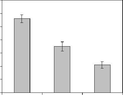
даже на 28-е сутки дефект хряща сохраняется. В
группе животных с введением ФКК площадь
повреждения на 28-е сутки составляла 7 мм
2
, что
достоверно меньше, чем в контроле и больше , чем
в группе с введением “Актовегина” (рис. 1).
Интенсивность образования хрящевой ткани
определяется биосинтезом в хондроцитах матрик-
са гексозамина, гексуроновых кислот, тирозина,
оксипролина, гиалуроновой кислоты, гепарина,
хондроитинсульфатов и активностью фиброблас-
тов, синтезирующих соединительнотканные эле-
менты регенерата хряща [6, 10, 11].
Данные о содержании гликозаминогликановых
и протеогликановых компонентов в ткани хряща
на 21-е сутки после нанесения травмы представ-
лены в таблице.
Под влиянием ФКК и “Актовегина” в регене-
рате хряща достоверно увеличивается содержание
всех исследуемых биохимических компонентов
матрикса хрящевой ткани, особенно гиалуроновой
кислоты. После введения “Актовегина” уровень
последней в регенерате хряща выше в 6,3 раза,
чем в группе животных, не получавших лечения.
Под влиянием ФКК содержание гиалуроновой
кислоты увеличивается всего в 3,3 раза по сравне-
нию с группой животных, не получавших лечения.
Однако к 21-м суткам наблюдения после примене-
ния ФКК и “Актовегина” содержание гиалуроновой
кислоты значительно ниже, чем в группе здоровых
животных.
Установлено, что под влиянием ФКК содержа-
ние хондроитинсульфатов увеличивается в 4,2 раза
по сравнению с группой животных, не получавших
лечения, а под влиянием препарата сравнения “Ак-
товегина” – в 2,4 раза. Вместе с тем к 21-м суткам
Рис. 1. Площадь повреждения хряща на 28-е сутки,
вычисленная по рентгенограммам: * – p < 0,05 по срав-
нению с группой животных, не получавших лечения.
Fig. 1. Injury cartilage area to 28
th
day, calculated with X-
ray patterns: *− p < 0.05 if compared with the group of non-
treated animal
Площадь повреждения, мм
2
Injury area, mm
2
CBF “Actovegin”Control
with the balance of specific components, supporting
and activating their cell metabolism. Of special interest
in this respect is low molecular fraction (below 5 kDa)
of cattle cord blood, enabling to sharply increase the
cell energetic potential, in particular this was established
for “Solcoseryl” and “Actovegin” originated from the
ontogenetically close source, vealer blood [2].
It has been established that CBF stimulates the
formation of cartilage tissue after mechanical trauma
but X-ray analysis has shown the preservation of
cartilage defect even to the 28
th
day. In the group of
CBF-injected animals the area of damage to the 28
th
day made 7 mm
2
, that was statistically lower than in
the control and higher if compared with the group with
“Actovegin” (Fig. 1).
Intensity of cartilage formation is determined by
biosynthesis in chondrocytes of matrix of hexosamine,
gexuronic acids, tyrosine, oxyproline, hyaluronic acid,
heparin and chondroitin-sulfates and activity of
fibroblasts, synthesizing connective tissue elements of
cartilage regenerate [6, 10, 11].
The data on the content of glucosamine glycans
and proteoglycan components in cartilage tissue after
trauma initiation are presented in the table.
Under CBF effect and “Actovegin” in the cartilage
regenerate the content of all the studied biochemical
components of cartilage tissue matrix increases, es-
pecially of hyaluronic acid. After “Actovegin” introduc-
tion the level of the latter in cartilage regenerate is 6.3
times higher than in the group of non-treated animals.
Under CBF effect the content of hyaluronic acid in-
creases only in 3.3 times if compared with the group
of non-treated animals. However to the 21
st
observation
day after application of CBF and “Actovegin” the content

365
PROBLEMS
OF CRYOBIOLOGY
Vol. 19, 2009, ¹3
ÏÐÎÁËÅÌÛ
ÊÐÈÎÁÈÎËÎÃÈÈ
Ò. 19, 2009, ¹3
Содержание основных элементов матрикса в регенерате хряща после его механической травмы на 21-е сутки
Content of matrix basic elements in cartilage regenerate after its mechanic trauma to the 21
st
day
Примечание: * – p < 0,05 по сравнению с группой животных, не получавших лечения.
Note: * − p < 0.05 if compared with the group of non-treated animals.
наблюдения содержание хондроитинсульфатов в
хрящевой ткани опытных животных существенно
ниже, чем в группе здоровых.
Аналогичную картину наблюдали при исследо-
вании гексозамина, уровень которого при использо-
вании ФКК и “Актовегина” повышался соответст-
венно в 1,7 и 1,8 раза по сравнению с контрольной
группой животных. Важно отметить, что при вве-
дении ФКК и препарата сравнения уровень гексо-
замина достоверно не отличался от такового в
группе здоровых животных, что свидетельствует
о стимуляции метаболизма.
В опытных группах и группе животных, не полу-
чавших лечения, отмечено различное содержание
гепарина. В частности, под влиянием ФКК его со-
держание в регенерате хряща увеличивается в
2,1 раза. Содержание гепарина в регенерате хряща
при введении ФКК и “Актовегина” незначительно
отличается от такового в группе здоровых живот-
ных.
Аналогичные изменения выявлены при исследо-
вании содержания в регенерате хряща гексуроно-
вых кислот: под влиянием ФКК и препарата срав-
нения оно увеличивается в 1,6 и 1,7 раза соответ-
ственно по сравнению с группой животных, не полу-
чавших лечения. После применения ФКК и
“Актовегина” уровень гексуроновых кислот в реге-
нерате хряща был ниже в 1,3 и 1,4 раза соответст-
венно, чем в группе здоровых животных.
Для изучения механизма действия ФКК и
“Актовегина” на регенерацию хряща оценивали
содержание основных компонентов матрикса не
только в самом регенерате хряща опытных живот-
ных, но и в хрящевой ткани здоровой конечности
of hyaluronic acid is significantly lower than in the group
of healthy animals.
It has been established that under the effect of CBF
the content of chontroitin-sulfates increases in 4.2 times
versus the group of non-treated animals, and under
the effect of “Actovegin” it was in 2.4 times higher. In
addition to the 21
st
observation day the content of
chondroitin sulfates in cartilage tissue of experimental
animals was significantly lower than in the group of
healthy ones.
The same picture was observed when investigating
hexosamine, the level of which when using CBF and
“Actovegin” increased, correspondingly, in 1.7 and 1.8
times if compared with the control group of animals. It
is important to note that during introduction of CBF
and the preparation to compare the level of hexosamine
did not statistically differ from that in the group of
healthy animals, that testified to a high stimulation of
metabolism.
In experimental groups and the one of non-treated
animals different content of heparin was noted. In
particular, under the CBF effect its content in cartilage
regenerate increases in 2.1 times. Heparin content in
cartilage regenerate when introducing CBF and “Acto-
vegin” slightly differs from that in the group of healthy
animals.
The same changes were found when investigating
the content of hexuronic acids in cartilage regenerate:
under CBF effect and the preparation to compare it
increases in 1.6 and 1.7 times, correspondingly with
the group of non-treated animals. After application of
CBF and “Actovegin” the level of hexuronic acids in
cartilage regenerate was lower in 1.3 and 1.4 times,
correspondingly versus the group of healthy animals.
хынтовижыппурГ
spuorglaminA
%/гм,нимазоскеГ
%/gm,enimasoxeH
еывонорускеГ
%/гм,ытолсик
sdicacinoruxeH
%/gm
яавонорулаиГ
%/гм,атолсик
,dicacinorulayH
%/gm
нитиордноХ -
%/гм,ытафьлус
,setaflusnitiordnohC
%/gm
%/гм,нирапеГ
%/gm,nirapeH
хывородзаппурГ
)1(хынтовиж
laminayhtlaeH
)1(puorg
20,0±36,0 * 960,0±4,1 * 430,0±43,0 * 400,0±32,0 * 900,0±12,0 *
ен,хынтовижаппурГ
яинечелхишвачулоп
)2(
nonfopuorG - detaert
)2(slamina
920,0±43,030,0±16,020,0±40,0700,0±40,0700,0±70,0
)3(KKФ
)3(FBC
230,0±46,0 * 670,0±10,1 * 20,0±41,0 * 210,0±71,0 * 310,0±51,0 *
)4("нигевоткА"
)4("nigevotcA"
540,0±66,0 * 170,0±80,1 * 710,0±62,0 * 900,0±1,0 * 10,0±51,0 *
ьнактяавещярХ
йоннаворимвартен
итсонченок
foeussitegalitraC
non - bmildezitamuart
410,0±6,0 * 280,0±34,1 * 810,0±13,0 * 800,0±22,0 * 300,0±32,0 *

366
ÏÐÎÁËÅÌÛ
ÊÐÈÎÁÈÎËÎÃÈÈ
Ò. 19, 2009, ¹3
PROBLEMS
OF CRYOBIOLOGY
Vol. 19, 2009, ¹3
тех же крыс. Из данных таблицы, можно заклю-
чить, что ФКК и препарат сравнения достоверного
влияния на содержание компонентов матрикса не
оказывают, что, вероятнее всего, свидетельствует
о их направленном биологически активном
действии на пораженную хрящевую ткань.
В дальнейшем изучали влияние ФКК и “Акто-
вегина” на концентрацию в регенерате хряща окси-
пролина и тирозина, по уровню которых можно
определить содержание коллагеновых и неколлаге-
новых белков хряща [6, 10, 11], являющихся крите-
рием активности фибробластов и хондроцитов в
травмированном хряще [4, 12].
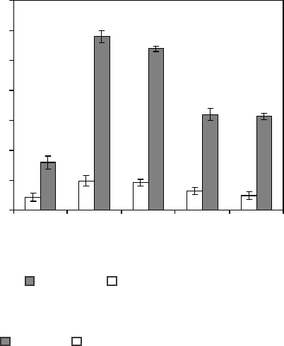
Из рис. 2 видно, что содержание тирозина в
регенерате хряща групп животных с введением
ФКК и “Актовегина” вдвое выше по сравнению с
группой животных, не получавших лечения. В
случае применения ФКК и “Актовегина” уровень
тирозина к 21-м суткам после нанесения травмы
ниже в 1,9 раза, чем в группе здоровых животных.
Содержание оксипролина в регенерате хряща
после применения ФКК в 2 раза выше по сравне-
нию с группой животных, не получавших лечения,
в то время как после введения препарата сравнения
значительных изменений по сравнению с контроль-
ными животными не выявлено. Установлено, что
содержание оксипролина в регенерате хряща после
применения ФКК меньше в 1,2 раза, чем в группе
здоровых животных. Очевидно, что ФКК значи-
тельно стимулирует синтез оксипролина в поражен-
ной хрящевой ткани. Как и при исследовании
протеогликановых компонентов, не выявлены дос-
товерные отличия в содержании данных амино-
кислот в хряще здоровой конечности опытных
животных по сравнению с нормой.
Таким образом, исследование биохимических
по-казателей позволило установить, что внутримы-
шечное введение ФКК стимулирует накопление
основных элементов матрикса и соединительно-
тканных элементов хряща на протяжении всего
срока эксперимента. Это объясняет более быст-
рое восстановление структурно-функциональных
свойств травмированного хряща под влиянием
препарата ФКК по сравнению с контролем.
Для более точного установления эффективности
проведенного лечения изучали динамику двига-
тельной активности экспериментальных животных.
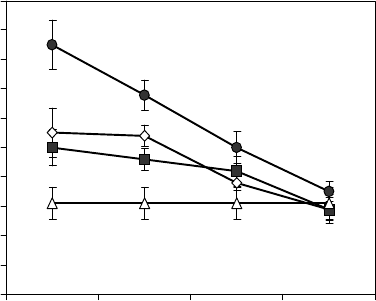
Из рис. 3 видно, что введение ФКК и “Актовегина”
стимулирует нормализацию двигательной актив-
ности значительно быстрее (на 7-е сутки – в 1,7 и
1,6 раза, 14-е сутки – в 1,4 и 1,3 раза соответ-
ственно, 21 и 28-е сутки – 1,2 раза) по сравнению с
группой животных, не получавших лечения.
Достоверные отличия влияния ФКК и “Актове-
гина” по сравнению с группой животных, не полу-
чавших лечения, отмечены уже на 7-е сутки наб-
Рис. 2. Содержание аминокислот в регенерате хряща на
21-е сутки: – тирозин; – оксипролин; Здор. кон. –
здоровая конечность животных опытных групп; * –
р < 0,05 по сравнению с контролем.
Fig. 2. Content of aminoacids in cartilage regenerate to the
21
st
day: – tyrosine; – oxyproline; Non-inj. – non-injuired
limb of animal experimental groups; * – p < 0.05 if compared
with the control.
0
0,5
1
1,5
2
2,5
3
3,5
Контроль Норма Здор. кон. ФКК "Ак то ве гин "
*
*
*
*
*
*
*
Control
Содержание, мг/%
Content, mg/%
Norm Non-inj. CBF “Aktovegin“
To study the effect mechanisms of CBF and
“Actovegin” on cartilage regeneration the content of
main matrix components not only in the cartilage
regenerate itself and in cartilage tissue of rat’s healthy
extremity was studied. The table data show that CBF
and the preparation to compare render no statistically
significant effect on the content of the matrix compo-
nents, likely testifying to their directed active effect
on the impaired cartilage tissue.
Later there was studied the effect of CBF and
“Actovegin” on concentration of oxyproline and tyrosi-
ne in cartilage regenerate, according to the level of
those the content of collagen and non-collagen cartilage
proteins [6, 10, 11], being the criteria of the activity of
fibroblasts and chondrocytes in the traumatized
cartilage may be examined [4, 12].
Fig. 2 demonstrates that the content of tyrosine in
cartilage regenerate of the animals from the groups
injected with CBF and “Actovegin” was twice higher
if compared with those non-treated. In case of applying
the CBF and “Actovegin” tyrosine level to the 21
st
day of trauma initiation was lower in 1.9 times, than in
the group of healthy animals. Oxyproline content in
cartilage regenerate after application of CBF is two
times higher if compared with the group of non-treated
animals, meanwhile after injection of the preparation
to compare no significant changes versus non-treated
animals were found. It has been established that oxy-
proline content in cartilage regenerate after application
of CBF is 1.2 times less than in the group of healthy
animals. It is evident that CBF strongly stimulates the
synthesis of oxyproline in a damaged cartilage tissue.
As well as during studying the proteoglycan compo-

367
PROBLEMS
OF CRYOBIOLOGY
Vol. 19, 2009, ¹3
ÏÐÎÁËÅÌÛ
ÊÐÈÎÁÈÎËÎÃÈÈ
Ò. 19, 2009, ¹3
людения. Достоверных отличий влияния ФКК и
“Актовегина” на двигательную активность крыс
не выявлено. У животных, не получавших лечения,
двигательная активность не восстанавливалась
даже к 28-м суткам.
Выводы
Установлено, что ФКК и “Актовегин” в эквива-
лентных дозах стимулируют накопление основных
компонентов матрикса в регенерате хряща (гексо-
замина, гексуроновых кислот, гиалуроновой
кислоты, гепарина и хондроитинсульфатов), а также
важнейших аминокислот (оксипролина и тирозина),
отражающих содержание коллагеновых и неколла-
геновых элементов хряща. Это позволяет сделать
заключение о положительном влиянии обоих препа-
ратов на репаративную регенерацию хряща. Полу-
ченные экспериментальные данные подтверждены
рентгенологическим исследованием хряща и ре-
зультатами функциональной диагностики конечнос-
ти подопытного животного. При сопоставлении
ФКК и “Актовегина” выявлено, что эффективность
ФКК практически аналогична действию “Актове-
гина” в эквивалентных дозах.
Литература
Гацура В.В. Методы первичного фармакологического
исследования биологической активности.– М.: Медицина,
1974.– 143 с.
Гулевский О.К., Грищенко В.І., Моісєєва Н.М. Властивості
і перспективи викорастання кордової крові в клінічній
практиці // Укр. журн. гематології та трансфузіології.– 2005,
№4.– С. 5–14.
1.
2.
0
2
4
6
8
10
12
14
16
18
20
7142128
*
*
*
*
*
*
*
*
*
Рис. 3. Динамика двигательной активности крыс: –
норма; – контроль; 0 – “Актовегин”; – ФКК; * –
p < 0,05 по сравнению с контролем.
Fig. 3. Dynamics of rats’ motion activity: – norm; –
control; 0 – “Actovegin”; – CBF; * – p < 0.05 if compared
with the control.
Продолжительность наблюдения, суток
Observation term, days
Продолжительность двигательной
активности, мин
Motion activity duration, min
nents no statistically significant differences were found
in the content of these amino acids in the cartilage of
healthy extremity of experimental animals if compared
with the norm.
Thus the investigation of biochemical indices
demonstrates that intramuscular injection of CBF
significantly stimulates the accumulation of the main
elements of the matrix and connective tissue elements
in cartilage within the whole experimental term. This
explain more rapid recovery rates of the traumatized
cartilage if compared with control.
For elucidation of the efficiency of the performed
treatment the dynamics of motor activity of experi-
mental animals was studied. Fig. 3 shows that injection
of CBF and “Actovegin” stimulates the normalization
of motor activity much more rapid (to the 7
th
day in
1.7 times and 1.6 times, to the 14
th
in 1.4 and 1.3 times,
correspondingly, to the 21
st
and 28
th
days in 1.2 times)
if compared with the group of non-treated animals.
Significant differences of the effect of CBF and “Acto-
vegin” if compared with the group of non-treated ani-
mals were found even to the 7
th
observation day.
Statistically significant differences of CBF and “Acto-
vegin” effect on motor activity in rats were not revea-
led. In non-treated animals the motor activity did not
recover even to the 28
th
day.
Conclusions
It has been established that CBF and “Actovegin”
under equivalent doses stimulate the accumulation of
main components of the matrix in cartilage regenerate
(hexosamine, hexuronic acids, hyaluronic acid, heparin
and chondroitin sulfates), as well as the most important
amino acids (oxyproline and tyrosine), reflecting the
content of collagen and non-collagen cartilage ele-
ments. This enables to conclude about positive effect
of both preparations on cartilage reparative regene-
ration. The experimental findings are confirmed with
X-ray examination of the cartilage and the results of
functional diagnostics of the limbs of experimental
animal. When comparing CBF and “Actovegin” there
was found that the efficiency of CBF is quite similar
to the one of “Actovegin” under equivalent doses.
References
Gatsura V.V. Methods of initial pharmacological study of
biological activity.– Moscow: Meditsina, 1974.– 143 p.
Gulevsky O.K., Grischenko V.I., Moiseeva N.M. Peculiarities
and perspectives of cord blood use in clinical practice//
Ukrainskiy Zhurnal Gematologii ta Transfuziologii.– 2005.–
N4.– P. 5–14.
Malyshkina S.V. Structure-metabolic changes of articular
cartilage after local cryoeffect: Authors’ abstract of thesis
of candidate of biol. sciences.– Kharkov, 1985.– 22 p.
Pavlova V.N., Kop’eva T.N., Slutskiy L.I., Pavlov G.G. Carti-
lage.– Moscow: Meditsina, 1988.– 320 p.
1.
2.
3.
4.
368
PROBLEMS
OF CRYOBIOLOGY
Vol. 19, 2009, ¹3
ÏÐÎÁËÅÌÛ
ÊÐÈÎÁÈÎËÎÃÈÈ
Ò. 19, 2009, ¹3
Малышкина С.В. Структурно-метаболические изменения
суставного хряща после локального криовоздействия:
Автореф. дис. ... канд. биол. наук.– Харьков, 1985.– 22 с.
Павлова В. Н., Копьева Т. Н., Слуцкий Л. И., Павлов Г. Г.
Хрящ.– М.: Медицина, 1988.– 320 с.
Риггз Б.Л., Мелтон III Л.Дж. Остеопороз.– М.-СПб.:
Издательство БИНОМ, “Невский диалект”, 2000.– 560 с.
Слуцкий Л.И. Биохимия нормальной и патологически
измененной соединительной ткани. – Л.: Медицина, 1969.–
376 с.
Тарасенко В.И. Криовоздействие при артропластике
тазобедренного сустава: Автореф. дис. ... канд. мед.
наук.– Харьков, 1989.– 18 с.
Boas N.P. Method for the determination of hexosamines in
tissues // J. Biol. Chem.– 1953. – Vol. 204, N2.– P. 553–562.
Dische Z. A new specific color reaction of hexuronic acid //
J. Biol. Chem.– 1947.– Vol. 167, N1.– P. 189–198.
Jordan J.M., Helmick C.G., Renner J.B. et al. Prevalence of
knee symptoms and radiographic and symptomatic knee
osteoarthritis in African Americans and Caucasians: The
Johnston County Osteoarthritis Project // J. Rheumatol.–
2007.– Vol. 34, N1.– P. 172–180.
Kongtawelert P., Francis D.L., Brooks P.M., Ghosh P.
Application of an enzyme-linked immunosorbent-inhibition
assay to quantitate the release of KS peptides into fluids of
the rat sub-cutaneous air pouch model and the effects of
chondroprotective drugs on the release process // Rheumatol.
Int.– 1989.– Vol. 9, N2.– P. 77–83.
Stegemann H., Stalder P. Determination of hydroxiproline //
Clin. Chim. Acta.– 1967.– Vol. 18, N2.– P. 267–273.
Поступила 16.08.2009
Рецензент Н.А. Волкова
3.
4.
5.
6.
7.
8.
9.
10.
11.
12.
Riggz B.L., Melton L.J. Osteoporosis.– Saint Petersburg:
BINOM, Nevsky dialect, 2000.– 560 p.
Slutsky L.I. Biochemistry of normal and pathologically changed
connective tissue.– Leningrad: Meditsina, 1969.– 376 p.
Tarasenko V.I. Cryoeffect at hip joint arthroplasty: Authors’
abstract of thesis of candidate of med. sciences.– Kharkov,
1989.– 18 p.
Boas N.P. Method for the determination of hexosamines in
tissues // J. Biol. Chem.– 1953. – Vol. 204, N2.– P. 553–562.
Dische Z. A new specific color reaction of hexuronic acid //
J. Biol. Chem.– 1947.– Vol. 167, N1.– P. 189–198.
Jordan J.M., Helmick C.G., Renner J.B. et al. Prevalence of
knee symptoms and radiographic and symptomatic knee
osteoarthritis in African Americans and Caucasians: The
Johnston County Osteoarthritis Project // J. Rheumatol.–
2007.– Vol. 34, N1.– P. 172–180.
Kongtawelert P., Francis D.L., Brooks P.M., Ghosh P.
Application of an enzyme-linked immunosorbent-inhibition
assay to quantitate the release of KS peptides into fluids of
the rat sub-cutaneous air pouch model and the effects of
chondroprotective drugs on the release process // Rheumatol.
Int.– 1989.– Vol. 9, N2.– P. 77–83.
Stegemann H., Stalder P. Determination of hydroxiproline //
Clin. Chim. Acta.– 1967.– Vol. 18, N2.– P. 267–273.
Accepted in16.08.2009
5.
6.
7.
8.
9.
10.
11.
12.

369
PROBLEMS
OF CRYOBIOLOGY
Vol. 19, 2009, ¹3
ÏÐÎÁËÅÌÛ
ÊÐÈÎÁÈÎËÎÃÈÈ
Ò. 19, 2009, ¹3
УДК 616.5-001.19:577.115:615.36
А.В. ШИНДЕР, С.Є. ГАЛЬчЕНКО*, Л.В. ОСТАНКОВА, О.П. СИНчИКОВА
Динаміка загоєння холодових ран шкіри, рівень пероксидації
ліпідів та лейкоцитарний профіль крові щурів на фоні введення
екстрактів тваринного походження
UDC 616.5-001.19:577.115:615.36
A.V. SHINDER, S.YE. GALCHENKO*, L.V. OSTANKOVA, O.P. SYNCHYKOVA
Dynamics of Healing of Skin Cold Wounds, Lipid Peroxidation
Level and Leucocyte Profile of Rat’s Blood Against
the Background of Animal Extract Introduction
Досліджено вплив екстракту кріоконсервованих фрагментів селезінки свиней (ЕСС) і екстракту підмору бджіл (ЕПБ) на
динаміку загоєння холодових ран шкіри, рівень ПОЛ та показники крові щурів лінії Вістар і Сфінкс. Встановлено, що холодові
рани у щурів Сфінкс загоюються повільніше, ніж у щурів лінії Вістар. Уведення ЕСС або ЕПБ прискорює загоєння ран,
нормалізує показники крові та знижує рівень пероксидації ліпідів. Екстракт селезінки має більшу біологічну активність, ніж
екстракт бджіл.
Ключові слова: холодова рана, загоєння, екстракт селезінки, екстракт підмору бджіл.
Изучено влияние экстракта криоконсервированных фрагментов селезенки свиней (ЭСС) и экстракта подмора пчел (ЭПП)
на динамику заживления холодовых ран кожи, уровень ПОЛ и показатели крови крыс линии Вистар и Сфинкс. Установлено,
что холодовые раны у крыс Сфинкс заживают медленнее, чем у крыс линии Вистар. Введение ЭСС или ЭПП ускоряет
заживление ран, нормализует показатели крови и снижает уровень пероксидации липидов. Экстракт селезенки обладает
большей биологической активностью, чем экстракт пчел.
Ключевые слова: холодовая рана, заживление, экстракт селезенки, экстракт подмора пчел.
There was examined the effect of the extract of cryopreserved fragments of porcine spleen (PSE) and the one of dead bee bodies
(DBBE) on the dynamics of healing of skin cold wounds, LPO level and the blood indices of Wistar and Sphynx rats. It has been
established that healing of cold wounds in Sphynx rats proceeds slower if compared with those for Wistar ones. The introduction of
PSE and DBBE accelerates the healing of wounds, normalizes blood counts and reduces the level of lipid peroxidation. Porcine spleen
extract has higher biological activity versus the bee’s extract.
Key-words: skin cold wounds, healing, spleen extract, dead bee body extract.
Кількість захворювань злоякісними пухлинами
шкіри неухильно зростає. Вони, як правило, під-
даються комбінованому лікуванню (передопе-
раційний курс хіміо- або променевої терапії з подаль-
шим оперативним втручанням). Для видалення
пухлин часто використовують кріодеструкцію, ос-
кільки цей метод є найменш травматичним, не має
протипоказань для лікування злоякісних новоут-
ворень зовнішніх локалізацій, а в деяких випадках
є методом вибору [8].
Хіміо- і променева терапія пригнічує імуно-
логічну реактивність організму, уповільнює проце-
си репаративної регенерації після оперативних втру-
чань [8, 10]. При цьому активується перекисне окис-
лення ліпідів (ПОЛ), яке також уповільнює процес
загоєння ран [7]. Таким чином, нормалізація проце-
су регенерації шкіри, скорочення термінів лікування
після холодової травми, в тому числі на тлі імуно-
дефіцитного стану організму, є актуальною задачею.
Skin malignant neoplasm morbidity has steadily
increased. As a rule in this case the treatment is com-
bined (pre-operation sessions of chemo- or radiothe-
rapy with following surgery). To remove the tumors
the cryodestruction is often used, because this method
is less traumatic, has no contraindications for treating
malignant neoplasms of outer localizations and in some
cases is the selection method [8].
Chemo- and radiotherapies suppress immunologic
reactivity of an organism, slow the processes of repa-
rative regeneration after surgeries [8, 10]. Herewith
lipid peroxidation (LPO), slowing down the process of
wound healing, activates [7]. Thus, the normalization
of the process of skin regeneration, reduction of the
treatment terms after cold trauma, including those on
the background of immune deficient state of an
organism is an actual task.
It has been shown that the extract of cryopreserved
fragments of porcine spleen (PSE) normalizes an
* Автор, якому необхідно направляти кореспонденцію:
вул. Переяславська, 23, м. Харків, Україна 61015; тел.:+38
(057) 372-74-35, факс: +38 (057) 373-30-84, електронна пошта:
gordienko@gala.net
* To whom correspondence should be addressed: 23,
Pereyaslavskaya str., Kharkov, Ukraine 61015; tel.:+380 57 372
7435, fax: +380 57 373 3084, e-mail: cryo@online.kharkov.ua
Institute for Problems of Cryobiology and Cryomedicine of the Na-
tional Academy of Sciences of Ukraine, Kharkov, Ukraine
Інститут проблем кріобіології і кріомедицини
НАН України, м. Харків
370
PROBLEMS
OF CRYOBIOLOGY
Vol. 19, 2009, ¹3
ÏÐÎÁËÅÌÛ
ÊÐÈÎÁÈÎËÎÃÈÈ
Ò. 19, 2009, ¹3
Показано, що екстракт кріоконсервованих фраг-
ментів селезінки свиней (ЕСС) нормалізує імунний
статус організму, зокрема систему Т-лімфоцитів
[2], які приймають активну участь в регуляції
регенерації [4]. Відомо, що екстракт підмору бджіл
(ЕПБ) має виражену антиоксидантну активність
[5]. Ці факти дозволили припустити, що вказані
вище екстракти сприятимуть прискоренню і нор-
малізації загоєння холодових ран у тварин, у тому
числі з ослабленою імунною системою.
Мета роботи – дослідити вплив екстракту кріокон-
сервованих фрагментів селезінки свиней і екстрак-
ту, одержаного з підмору бджіл, на динаміку загоєн-
ня холодових ран шкіри, рівень ПОЛ та показники
крові щурів лінії Вістар і безшерстих щурів Сфінкс.
Матеріали і методи
Експерименти проведено відповідно до “Загаль-
них принципів експериментів на тваринах”, схвале-
них II Національним конгресом з біоетики (2004 р.,
Київ, Україна) і узгоджених з положеннями “Євро-
пейської Конвенції про захист хребетних тварин, які
використовуються для експериментальних і інших
наукових цілей” (Страсбург, 1985).
Екстракт селезінки свиней одержували за мето-
дом [9], а ЕПБ – екстракцією бджолиного підмору
в апараті Соклета. Екстракти вводили щурам у
черевну порожнину по 1 мл один раз на добу. Кон-
центрація пептидів в ЕСС становила 100 мкг/мл, а
в ЕПБ – 0,25 мг сухої речовини/мл.
Холодову травму шкіри моделювали на 18 щу-
рах лінії Вістар і 18 щурах Сфінкс масою 190–210 г.
Для визначення досліджуваних показників в нормі
було використано по 6 тварин. У дослідних щурів
лінії Вістар видаляли шерсть на стегні. Холодову
травму наносили охолодженим в рідкому азоті
мідним аплікатором діаметром 10 мм, експозиція
становила 60 с. Щури з холодовою травмою були
розділені на групи: контрольні (введення фізіологіч-
ного розчину) та дослідні (введення ЕСС або ЕПБ).
Площу ран визначали за методом [6], а інтен-
сивність ПОЛ – спектрофотометричним методом
за рівнем продуктів реакції з тіобарбітуровою кис-
лотою (ТБК-активні продукти, ТБКАП) у сироватці
крові [1]. При повній епітелізації ранового дефекту
вважали, що рана загоїлася. Для визначення стій-
кості до перекисного окислення в ячейку хемілю-
мінометра, яка містить 1 мл фізіологічного розчину
і 100 мкл сироватки крові, додавали 200 мкл 5%-го
розчину перекису водню і реєстрували світлосуму
в умовних одиницях [3]. Аналіз лейкоцитарної
формули проводили на мазках, забарвлених
азур II – еозином за Романовським-Гімза, підрахо-
вуючи по 500 клітин у світловому мікроскопі
(ЛОМО, об. ×90, ок. ×10) [6]. Кров для досліджень
брали з хвостової вени тварин.
immune status of an organism, in particular, of T-lym-
phocyte system [2], participating actively in regene-
ration regulation [4]. It is known that the extract of
dead bee bodies (DBBE) has manifested antioxidant
activity [5]. These facts enabled the suggestion that
the above mentioned extracts contributed to accele-
ration and normalization of healing of cold wounds in
animals, including those with weakened immune system.
The research aim is to investigate the effect of the
extract of cryopreserved fragments of porcine spleen
and the extract derived from the dead bee bodies, on
the dynamics of healing of skin cold wounds, LPO
level and the blood counts in Wistar and Sphynx rats.
Materials and methods
The experiments were performed according to the
“General principles of experiments in animals”,
approved by the 2
nd
National Congress in Bioethics
(2004, Kyiv, Ukraine) and coordinated with the
guidelines of “European Convention for the protection
of vertebrate animals used for experimental and other
scientific purposes” (Strasbourg, 1985).
Porcine spleen extract was derived with the method
[9], and DBBE one with the extraction of dead bee
bodies in Soxhlet apparatus. The extracts were
introduced to the rats into peritoneal cavity by 1 ml
once a day. The concentration of peptides in PSE made
100 mg/ml and 0.025 mg of dry substance/ml in DBBE.
Skin cold trauma was simulated in 18 Wistar and
18 Sphynx rats of 190–210g. To examine the parame-
ters under study in the norm there were used 6 animals.
In the experimental rats the hair on a hip was removed.
Cold trauma was accomplished with liquid nitrogen-
cooled copper applicator of 10 mm diameter, the
exposure time was 60 seconds. The rats with cold
trauma were divided into the following groups: control
(introduction of physiological solution) and experimental
(introduction of either PSE or DBBE).
The square of wounds was found with the method
[6] and LPO intensity was spectrophotometrically de-
termined on the rate of thiobarbituric acid reactive
substances (TBARS) in blood serum [1]. At a complete
epitheliazation of wound defect there was assumed
that the wound healed. To reveal the steadiness to lipid
peroxidation into the chemiluminometer well with 1 ml
of physiological solution and 100 ml of blood serum
there were added 200 ml 5% hydrogen peroxide solu-
tion and the light sum was recorded in relative units
[3]. Leucocyte formula was analyzed on the smears
stained with azur II-eosin according to Romanovsky-
Giemsa, with counting 500 cells in light microscope
(LOMO, ob.×90, oc. ×10) [6]. Blood for the research
was derived from tail vein of animals.
The results were statistically processed with non-
parametric MANOVA method. The quantitative data
are presented in mean values and mean square errors.
