Wang Zh.M. One-Dimensional Nanostructures
Подождите немного. Документ загружается.

8 Electromagnetic Nanowire Resonances for Field-Enhanced Spectroscopy 193
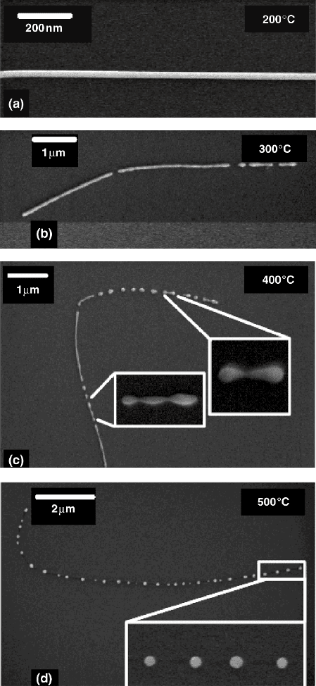
Fig. 8.10 SEM micrographs of (a) an as-prepared wire with diameter 25 nm and (b)–(d) wires
after annealing for 30 min at different temperatures
194 A. Pucci et al.
depicted in Fig. 8.10a. Fig. 8.10b–d display SEM images of Au wires after anneal-
ing for 30 min at different temperatures. The micrographs reveal different states of
the fragmentation process. At 300
◦
C the wires fragment, at 400
◦
C the fragments
become oval, and at 500
◦
C the wires are completely transformed into a chain of
nanospheres. The driving force of the fragmentation is the so-called Rayleigh insta-
bility, i.e., the minimization of the surface energy of the initial cylinder [70]. More
than a century ago, Lord Rayleigh described this process for liquid jets. Nichols and
Mullins extended the model to solid cylinders by [71]. For the nanowires grown in
ion tracks, the distance of adjacent spheres for different wire diameters was found to
be larger than predicted by the Nichols and Mullins model. The deviation between
experiment and theory originates from the fact that the model assumes an initial
cylinder with an isotropic surface energy while solids in general possess crystalline
facets and, thus, have an anisotropic surface energy.
8.3.2 Electron-Beam Lithography
Electron-beam lithography (EBL) makes possible the fabrication of nanostructures
of desired shape, size, and arrangement.
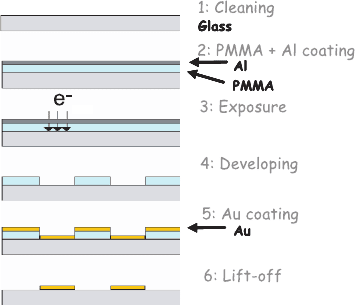
As shown in Fig.8.11, to achieve EBL it is necessary to follow different stages.
1: The first consists in depositing, by spin-coating, on a glass substrate carefully
cleaned the polymer, polymethyl methacrylate (PMMA), whose chains will be
broken during the exposure. The thickness of deposited PMMA is about 150 nm.
The choice of the PMMA thickness depends on the height of the structures which
one wants to design. A ratio 3:1 is generally respected between the thickness of
the layer of polymer and the height of the deposited metal.
2: The purpose of the second stage is to remove the solvent used, methylisobutylke-
ton (MIBK), from the PMMA film while placing the glass substrate covered with
polymer in a drying oven (160
◦
C) during at least 2 h. If the substrate used is glass
or another insulator material, it will accumulate the electrons received during the
Fig. 8.11 Main stages of the
design of a sample by EBL
and lift-off techniques
8 Electromagnetic Nanowire Resonances for Field-Enhanced Spectroscopy 195
exposure in the scanning electron microscope (SEM). That generates an electric
field on the surface of the sample, which disturbs the incident electron beam. To
avoid this problem, an aluminum film of 10 nm could be evaporated on the layer
of PMMA, thus allowing the dissipation of the charges. That requires an addi-
tional stage compared to the use of a conducting substrate like silicon or indium
titanium oxide (ITO).
3: For exposure with the SEM, it is necessary to use a system of control of the elec-
tron beam such as Nanometer Pattern Generation System (NPGS). The designed
nanostructures reproduced on the substrate are drawn using a software package
as DesignCad LT 2000 and the NPGS system transforms the created nanostruc-
tures into a dot matrix. It is then possible to choose the distance between the
points to be exposed as well as the distance between lines of points in a very
precise way. The coordinates of the points to expose are then transmitted to the
system of deflection of the electron beam and to a beam blanker. The exposed
zones have typical size from approximately 100µm.
4: For processing, it is necessary to dissolve the layer of aluminum using a solution
of potassium hydroxide, KOH. The development can then be carried out. This
stage consists of a selective dissolution of the PMMA. Only the exposed zones,
having a lower molecular mass, must be dissolved. One usually uses a mixture
of 1:3 MIBK and isopropanol (IPA).
5: After processing, the metal is evaporated and deposited with the desired thickness
on the surface.
6: The purpose of the last stage is to dissolve the PMMA remaining on the sample
using acetone: the lift-off stage.
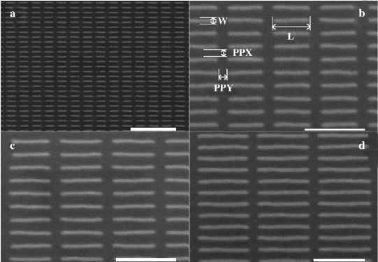
Fig. 8.12 SEM images of arrays of gold nanowires with variable length L (a: L = 420nm, b: L =
620nm, c: L = 720nm, d: L = 1µm),50nmheight,60nmwidth(W). The gap between nanowires
is constant for all arrays and is fixed to 150 nm (PPX and PPY). Scale bars: 2µmfora and 1µm
for b–d
196 A. Pucci et al.
This technique provides homogenous arrays of gold or silver nanowires as it can be
seen in Fig.8.12. The limitation of such technique is given by the SEM resolution. It
then is not possible to get nanostructures with a size lower than the diameter of the
electron beam. Typically, the lowest resolution achieved is few tens of nanometer.
8.4 Spectroscopy of Nanowire Resonances
8.4.1 Infrared Spectroscopy
This subsection reports on the experimental farfield spectroscopy of nanowire
resonances of individual metal nanowires prepared by growth in ion tracks, see
Sect. 8.3.1, supported by a dielectric substrate in the infrared. Different to studies
on ensembles of nanowires [25], one can exclude the influence of wire–wire inter-
action and averaging over locally varying parameters. Therefore, the experimental
results can be compared with existing theories and simulations for individual metal
nanowires (see Sect. 8.2.2).
Spectroscopy was performed on metal nanowires prepared by electrochem-
ical deposition in etched ion-track polycarbonate membranes (see Sect. 8.3.1).
After fabrication the polymer foil was dissolved in dichloromethane and the clean
nanowires were deposited on an infrared transparent substrate, e.g. KBr, ZnS, or
CaF
2
.
Spectroscopic IR microscopy of single metal nanowires was performed with syn-
chrotron radiation with a higher brilliance compared with conventional IR light
sources, e.g. globar. The higher brilliance allows mapping spectral properties of
nanowires. Before IR spectroscopy the individual nanowire on the IR-transparent
substrate was located by optical microscopy with visible light and its length is de-
termined from those images. Estimating the wire length in this way, a rather big
error up to 8% has to be assumed. Operating in the infrared mode of the microscope
a circular area of the IR-beam with a diameter of 8.3µm in the focal plane (sam-
ple position) was selected by inserting a circular aperture in the optical path behind
the sample. After the IR-beam had been centered on the selected nanowire, IR-
transmittance spectra (sample spectra) were recorded. Subsequently, to eliminate ef-
fects from beam profile and from substrate inhomogeneities, reference spectra were
takenatleast10µm away from any nanowire and the ratio of sample and reference
measurement, called relative transmittance spectrum, is calculated. To determine the
lateral position of maximum response of the single nanowire, a grid with a lateral
step size of 1µm is defined and relative transmittance spectra were taken at each
grid point. The spectroscopic measurements were done with a Fourier-transform in-
frared (FT-IR) spectrometer and a LN
2
-cooled mercury-cadmium telluride (MCT)
detector, which collects light normally transmitted through the sample area. A small
fraction of the light scattered away from the normal direction could also be detected
because of the collection lens (Schwarzschild objective, numerical aperture: 0.52).
This can be neglected in further data analysis because most of the intensity scattered
8 Electromagnetic Nanowire Resonances for Field-Enhanced Spectroscopy 197
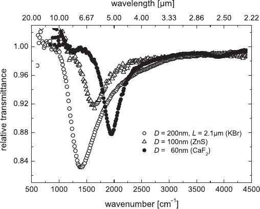
Fig. 8.13 Typical relative transmittance spectra (parallel polarization) of different single gold
nanowires with different lengths and diameters (down to 60 nm) placed on different substrates.
The fundamental mode of the antennas can be seen clearly
by the nanowire is directed outside the cone of collected light. For both sample
and reference measurement IR spectra were recorded by acquisition of at least 10
scans in the spectral range from 600 to 7000cm
−1
.Aresolutionofatleast16cm
−1
was sufficient for fundamental nanowire studies because of the spectrally broad re-
sponse. An even higher resolution of 2cm
−1
was used for the SEIRA measurements
(see Sect. 6.1). For further analysis of the transmitted light a polarizer was inserted
in the optical path before the sample.
The relative transmittance spectra T
rel
of nanowires (Au, Cu) with a length of
a few microns reveal fundamental antenna resonances (see Fig. 8.13). Apart from
the fundamental mode much weaker structures of resonant excitation are detected
at higher wavenumbers. These modes can be attributed to higher order resonances.
Even dipole inactive excitations are observed, which may be due to an electric field
component oblique to the long wire axis [72, 73] or bent nanowires. Since the de-
tailed study of higher order resonances of individual wires was hampered by their
low signal strength when compared with disturbing effects (beam instability, sub-
strate inhomogenities), only the fundamental mode of the antenna resonance (l = 1
in eq. 8.1) is discussed in the following. The main resonance was only observed
for electric field parallel to the long axis of the wire (parallel polarization). For an
electric field perpendicular with respect to the long wire axis (perpendicular polar-
ization) the IR-signal was below the noise level, no plasmon was excited.
In Fig.8.13 the resonance positions (position of minimum transmission) of the
different wires depend on length, diameter, and the used substrate. To separate the
influence of the length on the resonance frequency, wires with comparable diameters

198 A. Pucci et al.
of about 200 nm placed on KBr are considered (Fig.8.2b). Assuming the ideal di-
pole relation as given in eq. 8.1 for a wire completely embedded in vacuum (n = 1),
a shift of the measured data towards higher resonant wavelengths is obvious. Using
the value of the refractive index of KBr in eq. 8.1 the calculated curve seems to fit
the measured data. But the used assumption does not fit to the real configuration.
The wires are not completely embedded in the substrate, rather they are supported
and less than the half of the environment is truly KBr. To get deeper insight, BEM
calculations for diameter D = 200nm and variable lengths (L from 1.5 to 4.5µm)
were performed. But, also these calculations do not fit to the data if vacuum around
the wire is assumed. Only the BEM calculations for wires embedded in an effective
medium with refractive index n
eff
according to eq. 8.2 yield reasonable agreement
to the measured data.
This clearly indicates the influence of the substrate on the resonance position.
The polarizibility of a cover medium gives further effect. To demonstrate this,
individual gold nanowires on different substrates were covered with a thin dielec-
tric layer, e.g. paraffin wax, and IR spectra were taken before and after evapora-
tion. Because of the coverage the resonance wavelength is shifted towards higher
wavelengths, which can be estimated with an effective dielectric constant using
ε
eff
=
1
2
(
ε
c
+
ε
s
) with
ε
c
as the dielectric constant of the cover medium. Together
with eq.8.2 this gives calculated ratios n
air
eff
/n
paraffin
eff
= 0.875 for the KBr substrate
(with
ε
s
= 2.34 and
ε
c
= 2.02 for paraffin) and 0.923 for the ZnS substrate (with
ε
s
= 4.84), respectively. These ratios are consistent with the measured data obtained
from the spectral shift before and after deposition of paraffin [29].
Comparing experimental nanowire data for Au and Cu, it becomes obvious that
the resonance width is proportional to the resonance frequency. This behavior cor-
responds to that of perfectly conducting cylinders in vacuum [26]. It means that
the width of the fundamental resonance in the IR is dominated by the geometry of
the nanowire (length L and diameter D), not by its material properties. In the IR
the dielectric functions of the metals gold and copper show only marginal quantita-
tive differences and the wire diameters are relatively large compared with the skin
depth and to the typical dimensions (a few 10nm) for the onset of size effects in the
conductivity. Deviation could arise if wires are of worse crystalline quality.
From relative transmittance spectra (in Fig.8.13) the ratio of the extinction cross-
section of a single nanowire related to its geometric cross-section was estimated.
Assuming 1 −T
rel
≈ 2(
σ
ext
/A
0
)/(n
s
+ 1), where the influence of the substrate on
the signal strength is taken into account in analogy to the transmittance change by
the substrate of a thin film [74] in comparison with a freestanding film [75], the ratio
σ
ext
σ
geo
≈ A
0
(1−T
rel
) ·(n
s
+ 1) ·
1
2LD
(8.6)
was obtained. Inserting the spot size A
0
, the transmittance change in the spectrum
(1 −T
rel
), the refractive index of substrate n
s
, L from microscopy in the visible
spectral range, and D (known from wire growth process) leads to a
σ
ext
/
σ
geo
shown
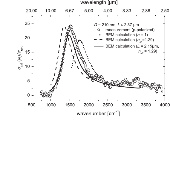
in Fig. 8.14 for a wire with L ≈2.37µm and D = 210nm placed on KBr. Any result
8 Electromagnetic Nanowire Resonances for Field-Enhanced Spectroscopy 199
Fig. 8.14 Extinction cross-section related to the geometric cross-section of a gold nanowire with
length L = 2.37µm and diameter D = 210nm: experimental data (open circles) and BEM calcula-
tions assuming different refractive indices
above 1 means an extinction of intensity above geometric shadowing, which is a
clear indication of the nanowires ability to confine light and therefore an indication
for an enhanced local field in the vicinity of the nanowire. Since the measurements
are performed in the farfield, only a spatially averaged field enhancement factor
σ
ext
/
σ
geo
can be calculated. For the representative spectrum in Fig.8.14 a farfield
enhancement factor of about 5 can be estimated for the resonance position.
The calculated resonance curves of single gold nanowires in Fig. 8.14 are ob-
tained using the boundary element method BEM (see Sect. 8.2.2). Here, the gold
nanowire is modeled as a rod with hemispherical tip ends completely embedded in
a medium. As discussed in Sect. 8.2, the calculated data for a nanowire in vacuum
deviate from the experimental ones because of the polarizibility of the substrate.
Considering an effective refractive index n
eff
= 1.29 (according to eq. 8.2) in order
to describe the influence of the substrate approximately, an even better agreement
in strength and shape of the resonance curve with the experimental data is obtained.
Only the position of the resonance maximum differs slightly. But, a BEM calcu-
lation with a length changed within its error fits the measured spectrum data. The
same does a calculation (not shown) with an unchanged length but a slightly differ-
ent n
eff
of 1.20. At this point the wire-length uncertainty for this sample type limited
a more precise conclusion.
The important role of the substrate polarizability was also discussed by Fumeaux
[76] who studied the resonance length of gold nanoantennas for two fixed IR wave-
lengths and one in the visible range with a special microbolometer setup.
200 A. Pucci et al.
8.4.2 Antenna Resonances in the Visible Range
In the visible to ultraviolet range, below the optical plasma edge of the respective
metal, several types of plasmon resonances of metal nanowires can be observed.
These are the resonances for electric field along the short axis of nanowires, antenna
resonances (LSP) of accordingly short nanowires, and higher orders of antenna res-
onances from wires with micron length.
The LSP resonance position is strongly dependent on the detailed particle shape.
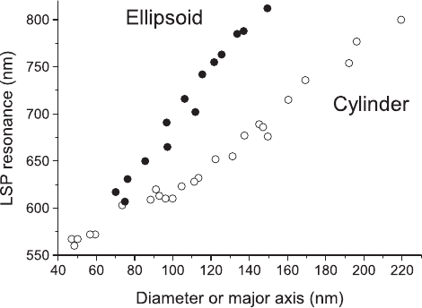
For fixed length that resonance is red-shifted with diameter, and, as shown in
Fig. 8.15, this shift could be of several tens of nanometers and even more when the
size of the nanoparticle increases [2,35,77]. For an ellipsoidal particle the redshift is
faster than for a cylindrical particle as shown in Fig. 8.15. For an increase of 10 nm
in size, the shift of the LSP for an ellipse (e-beam lithographically written structure
with certain height) is of 30 nm whereas it is only of 10 nm for a cylinder-like
structure [2].
The plasmon resonances were measured by extinction (all the losses due to scat-
tering and absorption) spectroscopy in transmission. The e-beam lithographically
prepared samples (Sect. 8.3.2) were illuminated at normal incidence with collimated
white light polarized along the nanowires length. To take into account the opti-
cal transfer function of the experimental set-up (different optical transmission for
each wavelength due to the mirrors, the array or the CCD), it was necessary, in a
first stage, to record the intensity of the light transmission without the nanowires
(I
0
). In a second stage, the intensity of the light transmission is measured consid-
ering the presence of the nanowires (I
t
). The nanowire spectrum corresponding to
Fig. 8.15 Variation of the LSP resonance wavelength with diameter of cylinder-like structures
(open circle) or with length of ellipse-like structures (full circle), respectively. The minor axis of the
ellipsoids is 50 nm and the height of the two kinds of nanoparticles is 50 nm. The gap between two
nanoparticles is kept constant at 200 nm. The particles were made by electron-beam lithography
on glass
8 Electromagnetic Nanowire Resonances for Field-Enhanced Spectroscopy 201
the plasmon resonance was obtained by simply dividing I
0
by I
t
, i.e. it is shown
as (T
rel
)
−1
. Since the experiments were done in transmission configuration, it im-
plies that the nanowires had been deposited on a transparent substrate such as glass
or ITO.
Extinction spectra were recorded on a Jobin–Yvon micro-Raman spectropho-
tometer (Labram) [78]. Each extinction spectrum was recorded, by removing the
notch filter, over an area of 80 ×80µm
2
in diameter selected by the confocal hole.
A 10-fold magnification objective (NA = 0.28) was used. It was important to use
an objective with a small numerical aperture (NA) since only the transmitted light
should be collected and not the light diffusely scattered by the nanowire. The use of
nanoparticle arrays was necessary to get enough signal strength since the extinction
signal of isolated nanoparticles was too low with such experimental set up. (It has
to be noticed that the antenna resonances in the visible range are much weaker than
in the infrared.) With scanning nearfield optical microscopy individual nanoan-
tennas can be studied, however, still with less energy resolution than in farfield
spectroscopy.
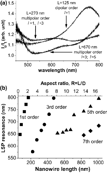
Higher orders of resonance of longer nanowires can be observed as new bands
in extinction spectra at wavelengths considerably lower than the dipolar LSP res-
onance, as shown in Fig. 8.16. Thus, on the spectra, when one resonance band is
Fig. 8.16 (a) Extinction spec-
tra I
o
/I
t
of electron-beam
lithographically made gold
nanowires arrays of differ-
ent lengths L on glass. The
value of l represents the
LSP order. (b) Development
of the LSP resonance with
nanowires length L and re-
spective aspect ratio (in the
array plane) of the nanowire
(ratio R between L and the
width of the nanowires): 1st
order (square), 3rd order (cir-
cle), 5th order (triangle), 7th
order (diamond). The width
and height of the nanowire,
and the gap between two
nanowires were kept con-
stant at 60, 50, and 150 nm,
respectively
202 A. Pucci et al.
red-shifted because of an increasing length, another one appears in the low wave-
length range and is also red-shifted when the length of the nanowire further in-
creases [79]. Schider et al. have assigned these multiorder resonances to odd orders
l ((1)) because the resonance should keep the same symmetry than the nanowire [4].
With nanowires up to 1µm long, we can observe up to the 7th order, as shown in
Fig. 8.16. In the case of multiorder resonances, it should be noticed that the red shift
is not identical for each order, but the variation the LSP resonance position is lower
for higher order [4,31,72,73,79].
For nanowires longer than 1µm, F
´
elidj et al. were able to observe some multi-
order resonances and the associated local maxima by Raman imaging [80]. They
have then measured a LSP beat wavelength of 379 nm. Moreover, Ditlbacher et al.
have proposed to consider the nanowires as LSP resonators [81]. In the case of
nanowires longer than 10µm, they have been able to determine a propagation length
of about 10µm and a nanowire end face reflectivity of 25%. They assume that the
nanowire can be applied as efficient LSP Fabry–Perrot resonator.
Another application takes advantage of the fact that the position of the LSP reso-
nance is both, different for each of the three nonidentical dimension of the nanowire
along their respective directions and strongly dependent on the polarization of the
exciting field. It is the design of a spectral selective polarizing nanowire device for
the visible range proposed by Schider et al. The authors of this work optimized the
extinction ratio between TE and TM polarization through the use of a nanowire grat-
ing and the selection of the appropriate grating parameters and nanowire width [82].
In the IR similar metal-grating polarizers are in practical use since many years.
8.5 Application to Surface Enhanced Raman Scattering
The surface enhanced Raman scattering (SERS) is a powerful tool that has made
possible the observation of small numbers of molecules. Moreover, it has allowed
the observation of individual molecules because of a Raman signal enhancement
estimated to 10
14
[54,83].
This important intensification of the Raman signal is achieved by using some
metallic nanostructures such as nanoparticles, rough thin films, colloidal solutions,
etc. The origin of the Raman scattering enhancement can be interpreted in terms
of two different processes: the first one of electromagnetic nature [23, 84] and the
second process with chemical origin [85,86].
The electromagnetic process is based on the nearfield interaction between a
metallic “particle” (Au, Ag, Cu...) and a molecule. During the illumination of
the SERS surface at the excitation wavelength
λ
0
, the particle interacts with light
and creates a significant local enhancement of the electromagnetic field that can
be achieved under specific conditions: plasmon resonance, lightning rod effect, or
confinement effect. Therefore, a molecule in the vicinity of the resonant particle
is excited because of a huge field. Since the Raman process is proportional to the
excitation field, the molecule scatters a large Raman signal at a shifted wavelength
