Voet D., Voet Ju.G. Biochemistry
Подождите немного. Документ загружается.

Section 8-1. Secondary Structure 231
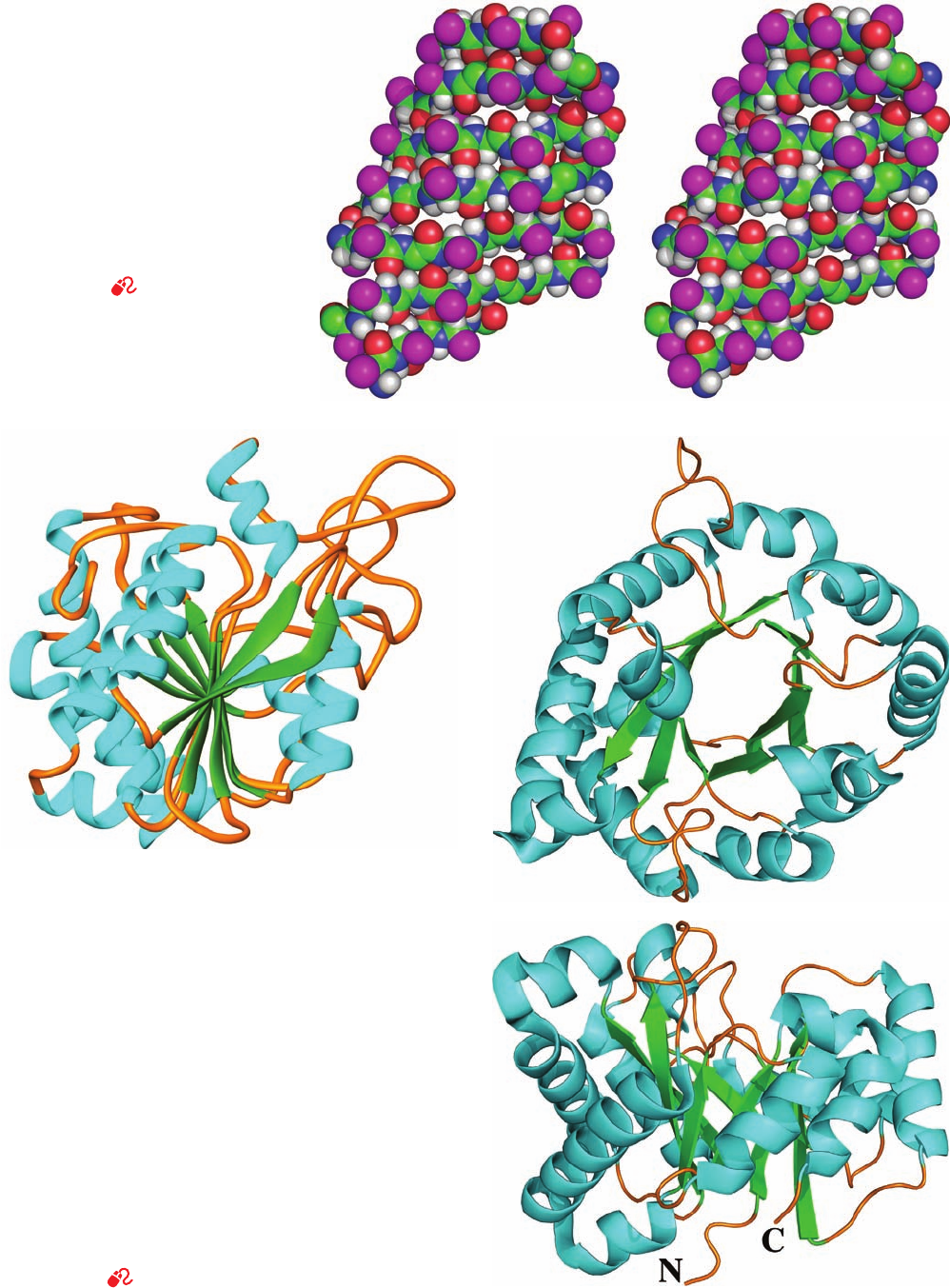
Figure 8-18 Stereo, space-filling
representation of the 7-stranded
antiparallel  pleated sheet in jack bean
concanavalin A as determined by X-ray
crystal structure analysis. The  strands
are approximately horizontal with their
backbone atoms colored according to
type (C green, N blue, O red, and
H white) and their side chains
represented by magenta spheres.
Instructions for viewing stereo drawings
are given in the appendix to this chapter.
[Based on an X-ray structure by Gerald
Edelman,The Rockefeller University.
PDBid 2CNA.]
See Kinemage
Exercise 3-3.
Figure 8-19 Polypeptide chain folding in proteins illustrating
the right-handed twist of  sheets. In these ribbon drawings, the
␣ helices shown as cyan helices, the strands of  sheets are
represented by green arrows pointing toward the C-terminus,
and the remaining portions of the backbone are drawn as orange
worms. Side chains are not shown. (a) Bovine carboxypeptidase
A, a 307-residue protein, contains an 8-stranded mixed  sheet
that forms a saddle-shaped curved surface with a right-handed
twist. (b) Chicken muscle triose phosphate isomerase, a
247-residue enzyme, contains an 8-stranded parallel  sheet that
forms a cylindrical structure known as a  barrel, here viewed
from the top. Note that the crossover connections between
successive strands of the  barrel, which each consist
predominantly of an ␣ helix, are outside the  barrel and have a
right-handed helical sense. (c) Side view of triose phosphate
isomerase. Its N-terminus (N) and C-terminus (C) are indicated.
[Part a based on an X-ray structure by William Lipscomb,
Harvard University. PDBid 3CPA. Parts b and c based on
an X-ray structure by David Phillips, Oxford University, U.K.
PDBid 1TIM.]
See Interactive Exercise 2
(a)
(b)
(c)
JWCL281_c08_221-277.qxd 10/19/10 7:13 AM Page 231
term random coil, which refers to the totally disordered and
rapidly fluctuating set of conformations assumed by dena-
tured proteins and other polymers in solution.
Globular proteins consist largely of approximately
straight runs of secondary structure joined by stretches of
polypeptide that abruptly change direction. Such reverse
turns or  bends (so named because they often connect
successive strands of antiparallel  sheets) almost always
occur at protein surfaces; indeed, they partially define
these surfaces. Most reverse turns involve four successive
amino acid residues more or less arranged in one of two
ways, Type I and Type II, that differ by a 180° flip of the
peptide unit linking residues 2 and 3 (Fig. 8-22). Both types
of  bends contain a hydrogen bond, although deviations
from these ideal conformations often disrupt this hydrogen
bond. Type I  bends may be considered to be distorted
sections of 3
10
helix. In Type II  bends, the oxygen atom of
residue 2 crowds the C

atom of residue 3, which is there-
fore usually Gly. Residue 2 of either type of  bend is often
Pro since it can facilely assume the required conformation.
Many proteins have regions that are truly disordered.
Extended, charged surface groups such as Lys side chains
or the N- or C-termini of polypeptide chains are good ex-
amples: They often wave around in solution because there
are few forces to hold them in place (Section 8-4). Often
entire peptide chain segments are disordered. Such seg-
ments may have functional roles, such as the binding of a
specific molecule, so they may be disordered in one state of
the protein (molecule absent) and ordered in another
(molecule bound). This is one mechanism whereby a pro-
tein can interact flexibly with another molecule in the per-
formance of its biological function.
2 FIBROUS PROTEINS
Fibrous proteins are highly elongated molecules whose sec-
ondary structures are their dominant structural motifs.
Many fibrous proteins, such as those of skin, tendon, and
bone, function as structural materials that have a protective,
connective, or supportive role in living organisms. Others,
such as muscle and ciliary proteins, have motive functions.
In this section, we shall discuss structure–function relation-
ships in two common and well-characterized fibrous pro-
teins: keratin and collagen (muscle and ciliary proteins are
considered in Section 35-3). The structural simplicity of
these proteins relative to those of globular proteins (Sec-
tion 8-3) makes them particularly amenable to understand-
ing how their structures suit them to their biological roles.
Fibrous molecules rarely crystallize and hence are usu-
ally not subject to structural determination by single-crystal
X-ray structure analysis (Section 8-3A). Rather than crys-
tallizing, they associate as fibers in which their long molec-
ular axes are more or less parallel to the fiber axis but in
which they lack specific orientation in other directions.The
X-ray diffraction pattern of such a fiber, Fig. 8-23, for
232 Chapter 8. Three-Dimensional Structures of Proteins
(a) (b)
(c)
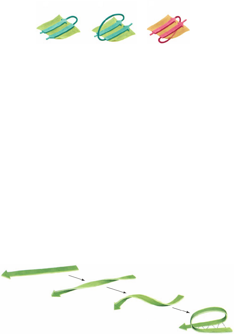
Figure 8-20 Connections between adjacent polypeptide
strands in  pleated sheets. (a) The hairpin connection between
antiparallel strands is topologically in the plane of the sheet. (b)
A right-handed crossover connection between successive strands
of a parallel  sheet. Nearly all such crossover connections in
Figure 8-21 Origin of a right-handed crossover connection. A
possible folding scheme illustrates how right-handed polypeptide
chain twisting favors the formation of right-handed crossover
connections between successive strands of a parallel  sheet.
proteins have this chirality (see, e.g., Fig. 8-19b). (c) A left-handed
crossover connection between parallel  sheet strands.
Connections with this chirality are rare. [After Richardson, J.S.,
Adv. Protein Chem. 34, 290, 295 (1981).]
JWCL281_c08_221-277.qxd 2/23/10 1:58 PM Page 232
example, contains little information, far less than would be
obtained if the fibrous protein could be made to crystallize.
Consequently, the structures of fibrous proteins are not
known in great detail. Nevertheless, the original X-ray
studies of proteins were carried out in the early 1930s by
William Astbury on such easily available protein fibers as
wool and tendon. Since the first X-ray crystal structure of a
protein was not determined until the late 1950s, these fiber
studies constituted the first tentative steps in the elucida-
tion of the structural principles governing proteins and
formed much of the experimental basis for Pauling’s for-
mulation of the ␣ helix and  pleated sheet.
A. ␣ Keratin—A Helix of Helices
Keratin is a mechanically durable and chemically unreac-
tive protein that occurs in all higher vertebrates. It is the
principal component of their horny outer epidermal layer,
comprising up to 85% of the cellular protein, and its related
appendages such as hair, horns, nails, and feathers. Keratins
have been classified as either ␣ keratins, which occur in
mammals, or  keratins, which occur in birds and reptiles.
Mammals have over 50 keratin genes, which are expressed
in a tissue-specific manner and whose products are classi-
fied as belonging to families of relatively acidic (Type I) and
Section 8-2. Fibrous Proteins 233
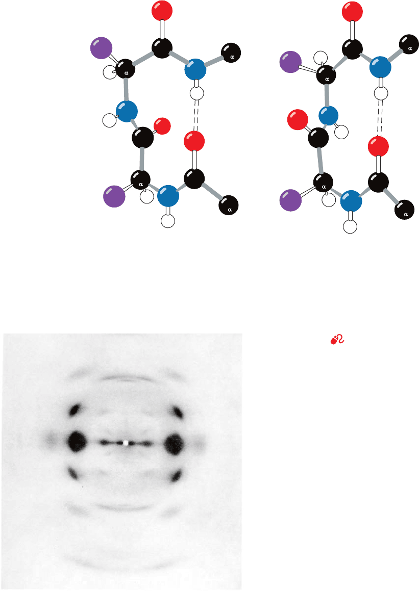
Figure 8-22 Reverse turns in polypeptide chains. (a) A Type I
 bend, which has the following torsion angles:
3
⫽⫺90°
Ⲑ
3
⫽ 0°
2
⫽⫺60°
Ⲑ
2
⫽⫺30°
C
(a) Type I β bend (b) Type II β bend
ψ
3
φ
3
φ
2
ψ
2
ψ
3
φ
3
φ
2
ψ
2
3
C
3
C
4
C
4
C
1
C
1
C
2
C
2
Figure 8-23 X-ray diffraction photograph of a fiber of Bombyx
mori silk. The photograph was obtained by shining a collimated
beam of monochromatic X-rays through the silk fiber and
recording the diffracted X-rays on a sheet of photographic film
placed behind the fiber.The photograph has only a few spots and
thus contains little structural information. [From March, R.E.,
Corey, R.B., and Pauling, L., Biochim. Biophys.Acta 16, 5 (1955).]
(b) A Type II  bend, which has the following torsion angles:
Variations from these ideal conformation angles by as much as
30° are common. Hydrogen bonds are represented by dashed
lines. [Illustration, Irving Geis. Image from the Irving Geis
Collection, Howard Hughes Medical Institute. Reprinted
with permission.]
See Kinemage Exercise 3-4
3
⫽⫺90°
Ⲑ
3
⫽ 0°
2
⫽⫺60°
Ⲑ
2
⫽ 120°
JWCL281_c08_221-277.qxd 8/26/10 7:48 PM Page 233
relatively basic (Type II) polypeptides. Keratin filaments,
which form the intermediate filaments of skin cells (Section
1-2Ae), must contain at least one member of each type.
Electron microscopic studies indicate that hair, which is
composed mainly of ␣ keratin, consists of a hierarchy of
structures (Figs. 8-24 and 8-25).A typical hair is ⬃20 m in
diameter and is constructed from dead cells, each of which
contains packed macrofibrils (⬃2000 Å in diameter) that
are oriented parallel to the hair fiber (Fig. 8-24). The
macrofibrils are constructed from microfibrils (⬃80 Å
wide) that are cemented together by an amorphous protein
matrix of high sulfur content.
Moving to the molecular level, the X-ray diffraction pat-
tern of ␣ keratin resembles that expected for an ␣ helix
(hence the name ␣ keratin). Yet ␣ keratin exhibits a 5.1-Å
spacing rather than the 5.4-Å distance corresponding to
the pitch of the ␣ helix. This observation, together with a
variety of physical and chemical evidence, suggests that
␣ keratin polypeptides form closely associated pairs of ␣ he-
lices in which each pair is composed of a Type I and a Type
II keratin chain twisted in parallel into a left-handed coil
(Fig. 8-25a).The normal 5.4-Å repeat distance of each ␣ he-
lix in the pair is thereby tilted with respect to the axis of
this assembly, yielding the observed 5.1-Å spacing.This as-
sembly is said to have a coiled coil structure because each
␣ helix axis itself follows a helical path.
The conformation of ␣ keratin’s coiled coil is a consequence
of its primary structure: The central ⬃310-residue segment of
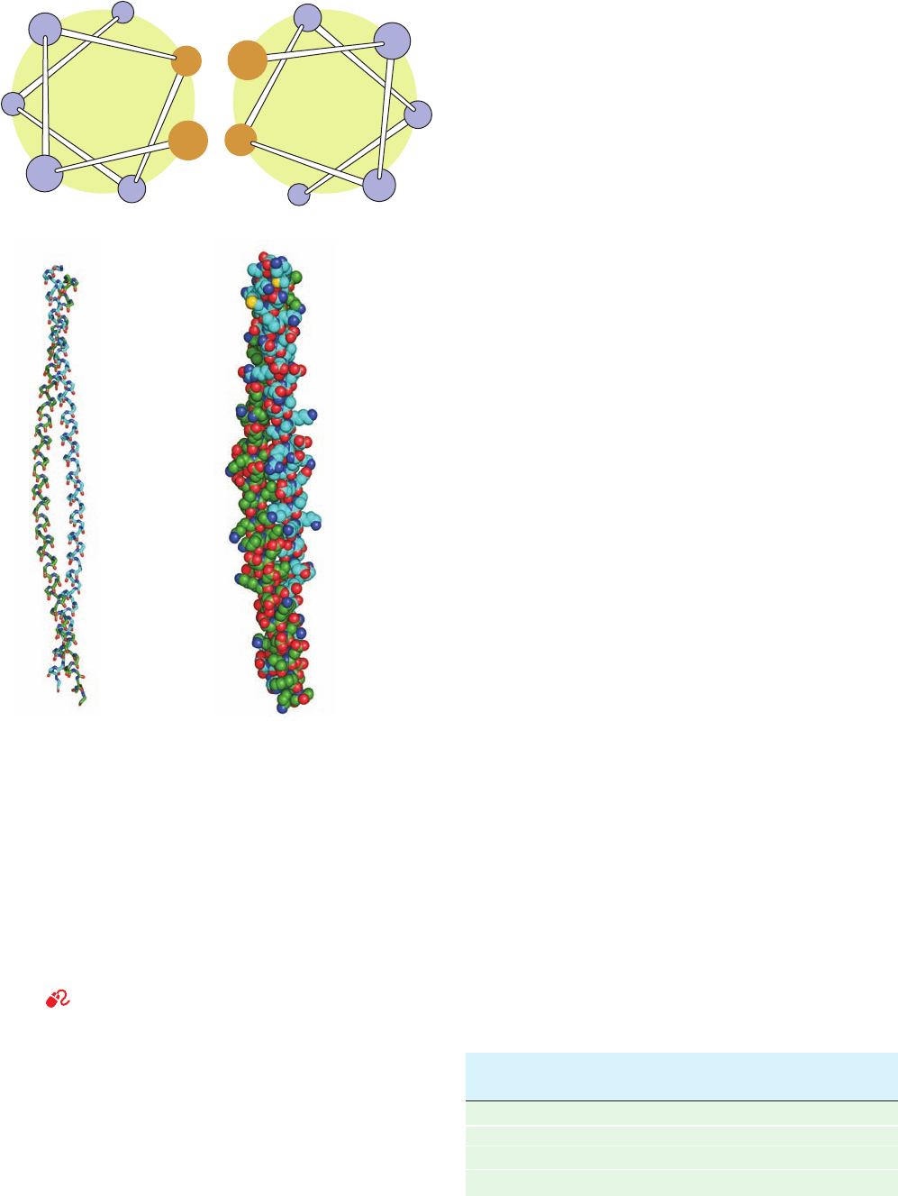
each polypeptide chain has a heptad (7-residue) pseudore-
peat, a-b-c-d-e-f-g, with nonpolar residues predominating at
positions a and d.Since an ␣ helix has 3.6 residues per turn, ␣
keratin’s a and d residues line up on one side of the ␣ helix to
form a hydrophobic strip that promotes its lengthwise associ-
ation with a similar strip on another such ␣ helix (Fig. 8-26;
hydrophobic residues, as we shall see in Section 8-4C, have a
strong tendency to associate). Indeed, the slight discrepancy
between the 3.6 residues per turn of a normal ␣ helix and the
⬃3.5-residue repeat of ␣ keratin’s hydrophobic strip is re-
sponsible for the coiled coil’s coil. The resulting 18° inclina-
tion of the ␣ helices relative to one another permits the heli-
cal ridges formed by the side chains on one helix to fit into
the grooves between these ridges on the other helix, thereby
greatly increasing their favorable interactions. Coiled coils, as
we shall see, are common components of globular proteins as
well as of other fibrous proteins.
The higher order substructure of ␣ keratin is poorly
understood. The N- and C-terminal portions of each poly-
peptide probably have a flexible conformation and facili-
tate the assembly of the coiled coils into ⬃30-Å-wide
protofilaments. These are thought to consist of two stag-
gered antiparallel rows of head-to-tail aligned coiled coils
(Fig. 8-25b). Two such protofilaments are thought to com-
prise an
⬃50-Å-wide protofibril, four of which, in turn, coil
around each other to form a microfibril (Fig. 8-25c).
␣ Keratin is rich in Cys residues, which form disulfide
bonds that cross-link adjacent polypeptide chains. This ac-
counts for ␣ keratin’s insolubility and resistance to stretch-
ing, two of its most important biological properties. The ␣
keratins are classified as “hard” or “soft” according to
whether they have a high or low sulfur content. Hard ker-
atins, such as those of hair, horn, and nail, are less pliable
234 Chapter 8. Three-Dimensional Structures of Proteins
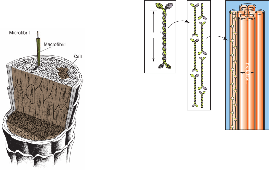
Figure 8-24 The macroscopic organization of hair. [Illustration,
Irving Geis. Image from the Irving Geis Collection, Howard
Hughes Medical Institute. Reprinted with permission.]
Figure 8-25 The structure of ␣ keratin. (a) The central ⬃310
residues of one polypeptide chain each of Types I and II ␣
keratins associate in a dimeric coiled coil.The conformations of
the polypeptides’ globular N- and C-terminal domains are
unknown. (b) Protofilaments are formed from two staggered and
antiparallel rows of associated head-to-tail coiled coils. (c) The
protofilaments dimerize to form a protofibril, four of which form
a microfibril.The structures of these latter assemblies are poorly
characterized but are thought to form helical arrays.
(a) Dimer
N-terminal
heads
Coiled
coil rod
~450 A
C-terminal
tails
(b) Protofilament (c) Microfibril
Protofibril
JWCL281_c08_221-277.qxd 8/10/10 11:48 AM Page 234
than soft keratins, such as those of skin and callus, because
the disulfide bonds resist any forces tending to deform them.
The disulfide bonds can be reductively cleaved with mercap-
tans (Section 7-1B). Hair so treated can be curled and set in
a “permanent wave” by application of an oxidizing agent
which reestablishes the disulfide bonds in the new “curled”
conformation. Although the insolubility of ␣ keratin pre-
vents most animals from digesting it, the clothes moth larva,
which has a high concentration of mercaptans in its digestive
tract, can do so to the chagrin of owners of woolen clothing.
The springiness of hair and wool fibers is a consequence
of the coiled coil’s tendency to untwist when stretched and
to recover its original conformation when the external
force is relaxed. After some of its disulfide bonds have
been cleaved, however, an ␣ keratin fiber can be stretched
to over twice its original length by the application of moist
heat. In this process, as X-ray analysis indicates, the ␣ heli-
cal structure extends with concomitant rearrangement of
its hydrogen bonds to form a  pleated sheet.  Keratin,
such as that of feathers, exhibits a similar X-ray pattern in
its native state (hence the name  sheet).
a. Keratin Defects Result in a Loss of Skin Integrity
The inherited skin diseases epidermolysis bullosa simplex
(EBS) and epidermolytic hyperkeratosis (EHK) are charac-
terized by skin blistering arising from the rupture of epider-
mal basal cells (Fig. 1-14d) and suprabasal cells, respectively,
as caused by mechanical stresses that normally would be
harmless. Symptomatic variations in these conditions range
from severely incapacitating, particularly in early childhood,
to barely noticeable. In families afflicted with EBS, sequence
abnormalities may be present in either keratin 14 or keratin
5, the dominant Types I and II keratins in basal skin cells.
EHK is similarly caused by defects in keratins 1 or 10, the
dominant Types I and II keratins in suprabasal cells (which
arise through the differentiation of basal cells, a process in
which the synthesis of keratins 14 and 5 is switched off and
that of keratins 1 and 10 is turned on). These defects evi-
dently interfere with normal filament formation, thereby
demonstrating the function of the keratin cytoskeleton in
maintaining the mechanical integrity of the skin.
B. Collagen—A Triple Helical Cable
Collagen (Greek: kolla, glue) occurs in all multicellular ani-
mals and is the most abundant protein of vertebrates, com-
prising ⬃30% of their protein mass. It is an extracellular pro-
tein that is organized into insoluble fibers of great tensile
strength. This suits collagen to its role as the major stress-
bearing component of connective tissues such as bone, teeth,
cartilage, tendon, ligament, and the fibrous matrices of skin
and blood vessels. Collagen occurs in virtually every tissue.
Vertebrates have 46 genetically distinct polypeptide
chains comprising 28 distinct collagen types that occur in
different tissues of the same individual. The most promi-
nent of these are listed in Table 8-2. A single molecule of
Section 8-2. Fibrous Proteins 235
(a)
c
g
f
b
e
a
d a′
e′
g′
c′
f ′
b′
d′
(b)
Figure 8-26 The two-stranded coiled coil. (a) View down the
coil axis showing the interactions between the nonpolar edges of
the ␣ helices.The ␣ helices have the pseudorepeating heptameric
sequence a-b-c-d-e-f-g in which residues a and d are predominantly
nonpolar. [After McLachlan,A.D. and Stewart, M., J. Mol. Biol.
98, 295 (1975).] (b) Side view of the polypeptide backbones
drawn in stick form (left) and of the entire polypeptides drawn in
space-filling form (right).The atoms are colored according to
type with C green in one chain and cyan in the other, N blue, O
red, and S yellow. The 81-residue chains are parallel with their
N-terminal ends above. Note that in the space-filling model the
side chains contact each other.This coiled coil is a portion of the
muscle protein tropomyosin (Section 35-3Ac). [Based on an
X-ray structure by Carolyn Cohen, Brandeis University. PDBid
1IC2.]
See Kinemage Exercises 4-1 and 4-2
Source: Eyre, D.R., Science 207, 1316 (1980).
Table 8-2 The Most Abundant Types of Collagen
Chain
Type Composition Distribution
I[␣1(I)]
2
␣2(I) Skin, bone, tendon,
blood vessels, cornea
II [␣1(II)]
3
Cartilage, intervertebral disk
III [␣1(III)]
3
Blood vessels, fetal skin
JWCL281_c08_221-277.qxd 2/23/10 1:58 PM Page 235
Type I collagen is composed of three polypeptide chains
with an aggregate molecular mass of ⬃285 kD. It has a rod-
like shape with a length of ⬃3000 Å and a width of ⬃14 Å.
Collagen has a distinctive amino acid composition:
Nearly one-third of its residues are Gly; another 15 to 30%
of them are Pro and 4-hydroxyprolyl (Hyp) residues:
3-Hydroxyprolyl and 5-hydroxylysyl (Hyl) residues also oc-
cur in collagen but in smaller amounts. Radioactive labeling
experiments have established that these nonstandard hy-
droxylated amino acids are not incorporated into collagen
during polypeptide synthesis: If
14
C-labeled 4-hydroxypro-
line is administered to a rat, the collagen synthesized is not
radioactive, whereas radioactive collagen is produced if the
rat is fed
14
C-labeled proline.The hydroxylated residues ap-
pear after the collagen polypeptides are synthesized, when
certain Pro residues are converted to Hyp in a reaction cat-
alyzed by the enzyme prolyl hydroxylase.
Hyp confers stability on collagen, possibly through in-
tramolecular hydrogen bonds that involve bridging water
molecules. If, for example, collagen is synthesized under
conditions that inactivate prolyl hydroxylase, it loses its na-
tive conformation (denatures) at 24°C, whereas normal
collagen denatures at 39°C (heat-denatured collagen is
known as gelatin). Prolyl hydroxylase requires ascorbic
acid (vitamin C)
to maintain its enzymatic activity. In the vitamin C defi-
ciency disease scurvy, the collagen synthesized cannot form
O
C
H
H
OH
O
OHHO
Ascorbic acid (vitamin C)
CH
2
OH
C
O
CHN
H
H
2
CCH
2
C
12
3
4
5
HO
4-Hydroxyprolyl residue
(Hyp)
N
H
H
2
CC
C
12
3
4
5
OH
3-Hydroxyprolyl residue
H
2
CH
2
NH CCH
O
+
CH
2
CH
CH
2
NH
3
OH
5-Hydroxylysyl residue (Hyl)
1
2
3
4
5
6
C
O
CH
fibers properly. This results in the skin lesions, blood vessel
fragility, and poor wound healing that are symptomatic of
this ultimately fatal vitamin deficiency disease.
a. Collagen Has a Triple Helical Structure
The amino acid sequence of bovine collagen ␣1(I),which
is similar to that of other collagens, consists of monoto-
nously repeating triplets of sequence Gly-X-Y over a contin-
uous 1011-residue stretch of its 1042-residue polypeptide
chain (Fig. 8-27). Here X is often Pro (⬃28%) and Y is of-
ten Hyp (⬃38%). The restriction of Hyp to the Y position
stems from the specificity of prolyl hydroxylase. Hyl is sim-
ilarly restricted to the Y position.
The high Gly, Pro, and Hyp content of collagen suggests
that its polypeptide backbone conformation resembles
those of the polyglycine II and polyproline II helices (Fig.
8-15). X-ray fiber diffraction and model building studies by
Alexander Rich and Francis Crick and by Ramachandran
led them to independently propose, in 1955, that collagen’s
three polypeptide chains, which individually resemble
polyproline II helices, are parallel and wind around each
other with a gentle, right-handed, ropelike twist to form a
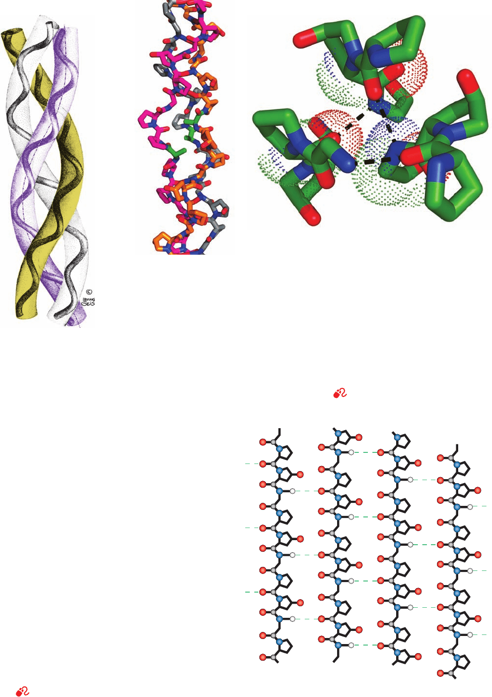
triple helical structure (Fig. 8-28). It was not until 1994,
however, that Helen Berman and Barbara Brodsky con-
firmed this model through their X-ray crystal structure de-
termination of the collagenlike polypeptide (Pro-Hyp-
Gly)
10
in which the fifth Gly is replaced by Ala (Fig. 8-29a).
In this structure, every third residue of each polypeptide
chain passes through the center of the triple helix, which is
so crowded that only a Gly side chain can fit there (Fig.8-29b).
This crowding explains the absolute requirement for a Gly
at every third position of a collagen polypeptide chain (Fig.
8-27). It also requires that the three polypeptide chains be
staggered so that the Gly, X, and Y residues from the three
chains occur at similar levels (Fig. 8-30).The staggered pep-
tide groups are oriented such that the N¬H of each Gly
makes a strong hydrogen bond with the carbonyl oxygen of
236 Chapter 8. Three-Dimensional Structures of Proteins
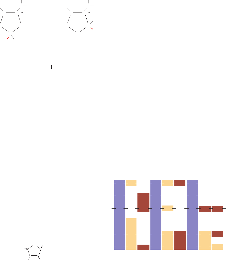
Figure 8-27 The amino acid sequence at the C-terminal end of
the triple helical region of the bovine ␣1(I) collagen chain. Note
the repeating triplets Gly-X-Y, where X is often Pro and Y is
often Hyp. Here Gly is shaded in purple, Pro in tan, and Hyp and
Hyp* (3-hydroxyPro) in brown. [From Bornstein, P. and Traub,
W. , in Neurath, H. and Hill, R.L. (Eds.), The Proteins (3rd ed.),
Vol. 4, p. 483,Academic Press (1979).]
958
Gly
Pro
Ser
Leu
Pro
Pro
Pro
Arg
Hyp
Hyp
Arg
Ala
Hyp
967
Gly
976
Gly
985
Gly
994
Gly
1003
Gly
Gly
Gly
Gly
Gly
Gly
Gly
Gly
Gly
Gly
Gly
Gly
Gly
Pro
Lys
Pro
Arg
Pro
Pro
Hyp
Asp
Ile
Thr
Hyp
Hyp
Ser
Leu
Hyp
*
Asp
Pro
Pro
Ala
Asn
Hyp
Ala
Hyp
Pro
JWCL281_c08_221-277.qxd 2/23/10 1:58 PM Page 236
Section 8-2. Fibrous Proteins 237
Figure 8-28 The triple helix of
collagen. This diagram indicates
how the left-handed polypeptide
helices are twisted together to form
a right-handed superhelical structure.
Ropes and cables are similarly
constructed from hierarchies of
fiber bundles that are alternately
twisted in opposite directions.An
individual collagen polypeptide
helix has 3.3 residues per turn and a
pitch of 10.0 Å (in contrast to
polyproline II’s 3.0 residues per
turn and pitch of 9.4 Å; Fig. 8-15).
The collagen triple helix has 10
Gly-X-Y units per turn and a pitch
of 86.1 Å. [Illustration, Irving
Geis. Image from the Irving Geis
Collection, Howard Hughes
Medical Institute. Reprinted
with permission.]
Figure 8-29 Structure of a collagen model peptide. In this X-ray structure of (Pro-Hyp-Gly)
10
,
the fifth Gly of each peptide has been replaced by Ala. (a) A stick model of the central portion of
the triple helix oriented with its N-termini at the bottom.The C atoms of the three chains are
colored orange, magenta, and gray.The N and O atoms on all chains are blue and red. Note how
the replacement of Gly with the bulkier Ala (C atoms green) distorts the triple helix. (b) This
view from the N-terminus down the helix axis shows the interchain hydrogen bonding associations.
Three consecutive residues from each chain are shown in stick form (C atoms green). Hydrogen
bonds are represented by dashed lines from Gly N atoms to Pro O atoms in adjacent chains. Dots
represent the van der Waals surfaces of the backbone atoms of the central residue in each chain.
Note the close packing of the atoms along the triple helix axis.The substitution of a centrally
located Gly C
␣
atom (CH
2
group) by any other residue would distort the triple helix. [Based on
an X-ray structure by Helen Berman, Rutgers University, and Barbara Brodsky, UMDNJ—
Robert Wood Johnson Medical School. PDBid 1CAG.]
See Kinemage Exercises 4-3 and 4-4
(a)
(b)
N
C
C
C
C
N
N
N
Gly
Gly
Gly
Gly
Gly
Gly
Gly
Gly
Gly
Gly
Gly
Gly
Gly
Chain 1 Chain 2 Chain 3 Chain 1
Figure 8-30 A schematic diagram showing the interchain
hydrogen bonding (dashed lines) in the Gly-containing regions of
the triple helix.This is a cylindrical projection with Chain 1
repeated on the right for clarity. Note that the three chains are
each vertically staggered by one residue so that a Gly, a Pro, and a
Hyp from the three different chains occur on the same level. [After
Bella, J., Eaton, M., Brodsky, B., and Berman, H.M., Science 266, 78
(1994).]
See Kinemage Exercises 4-3 and 4-4
JWCL281_c08_221-277.qxd 8/10/10 11:48 AM Page 237
Although the function of carbohydrates in collagen is un-
known, the observation that they are located in the “hole”
regions of the collagen fibrils suggests that they are in-
volved in directing fibril assembly.
c. Collagen Fibrils Are Covalently Cross-Linked
Collagen’s insolubility in solvents that disrupt hydrogen
bonding and ionic interactions is explained by the observa-
tion that it is both intramolecularly and intermolecularly
covalently cross-linked.The cross-links cannot be disulfide
bonds, as in keratin, because collagen is almost devoid of
Cys residues. Rather, they are derived from Lys and His
side chains in reactions such as those in Fig. 8-33. Lysyl ox-
idase, a Cu-containing enzyme that converts Lys residues
to those of the aldehyde allysine, is the only enzyme impli-
cated in this cross-linking process. Up to four side chains
can be covalently bonded to each other.The cross-links do
not form at random but, instead, tend to occur near the N-
and C-termini of the collagen molecules.
The importance of cross-linking to the normal function-
ing of collagen is demonstrated by the disease lathyrism,
an X (Pro) residue on a neighboring chain. The bulky and
relatively inflexible Pro and Hyp residues confer rigidity
on the entire assembly.
Collagen’s well-packed, rigid, triple helical structure is re-
sponsible for its characteristic tensile strength. As with the
twisted fibers of a rope, the extended and twisted polypep-
tide chains of collagen convert a longitudinal tensional
force to a more easily supported lateral compressional
force on the almost incompressible triple helix.This occurs
because the oppositely twisted directions of collagen’s
polypeptide chains and triple helix (Fig. 8-28) prevent the
twists from being pulled out under tension (note that suc-
cessive levels of fiber bundles in ropes and cables are like-
wise oppositely twisted). The successive helical hierarchies
in other fibrous proteins exhibit similar alternations of
twist directions, for example, keratin (Section 8-2A) and
muscle (Section 35-3Aa).
b. Collagen Is Organized into Fibrils
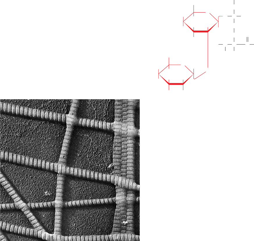
Types I, II, III, V, and XI collagens form distinctive
banded fibrils (Fig. 8-31) that are mostly, if not entirely,
composed of several different types of collagens.These fib-
rils have a periodicity of ⬃670 Å and a diameter of 100 to
2000 Å depending on the types of collagen they contain
and their tissue of origin (the other collagen types form dif-
ferent sorts of aggregates such as networks; we will not dis-
cuss them further). X-ray fiber diffraction studies reveal
that the molecules in fibrils of Type I collagen are packed in
a hexagonal array. Computerized model building studies
further indicate that these collagen molecules are precisely
staggered parallel to the fibril axis (Fig. 8-32). The darker
portions of the banded structures correspond to the 400-Å
“holes” on the surface of the fibril between head-to-tail
aligned collagen molecules. Structural and energetic con-
siderations suggest that the conformations of individual
collagen molecules, much like those of individual ␣ helices
and  sheets, are but marginally stable (Section 8-4). The
driving force for the assembly of collagen molecules into a
fibril is apparently provided by the added hydrophobic in-
teractions within the fibrils in a manner analogous to the
packing of secondary structural elements to form a globu-
lar protein (Section 8-3B).
Collagen contains covalently attached carbohydrates in
amounts that range from ⬃0.4 to 12% by weight, depend-
ing on the collagen’s tissue of origin. The carbohydrates,
which consist mostly of glucose, galactose, and their disac-
charides, are covalently attached to collagen at its Hyl
residues by specific enzymes:
238 Chapter 8. Three-Dimensional Structures of Proteins
Figure 8-31 Electron micrograph of collagen fibrils from skin.
[Courtesy of Jerome Gross, Massachusetts General Hospital.]
HO
OH
CH
2
OH
H
H
H
H
H
O
H
OH
CH
2
OH
H
HO
H
H OH
H
Glucose
Galactose
O
O
OC
C
H
H
H
N
C
O
CH
2
CH
2
CH
2
NH
3
⫹
Hydroxylysine
residue
JWCL281_c08_221-277.qxd 2/23/10 1:58 PM Page 238
which occurs in humans and other animals as a result of the
regular ingestion of seeds from the sweet pea Lathyrus
odoratus.The symptoms of this condition are serious abnor-
malities of the bones, joints, and large blood vessels, which
are caused by an increased fragility of the collagen fibers.
The causative agent of lathyrism, -aminopropionitrile,
-Aminopropionitrile
inactivates lysyl oxidase by covalently binding to its active
site. This results in markedly reduced cross-linking in the
collagen of lathrytic animals.
N ‚ C¬CH
2
¬CH
2
¬NH
3
⫹
Section 8-2. Fibrous Proteins 239
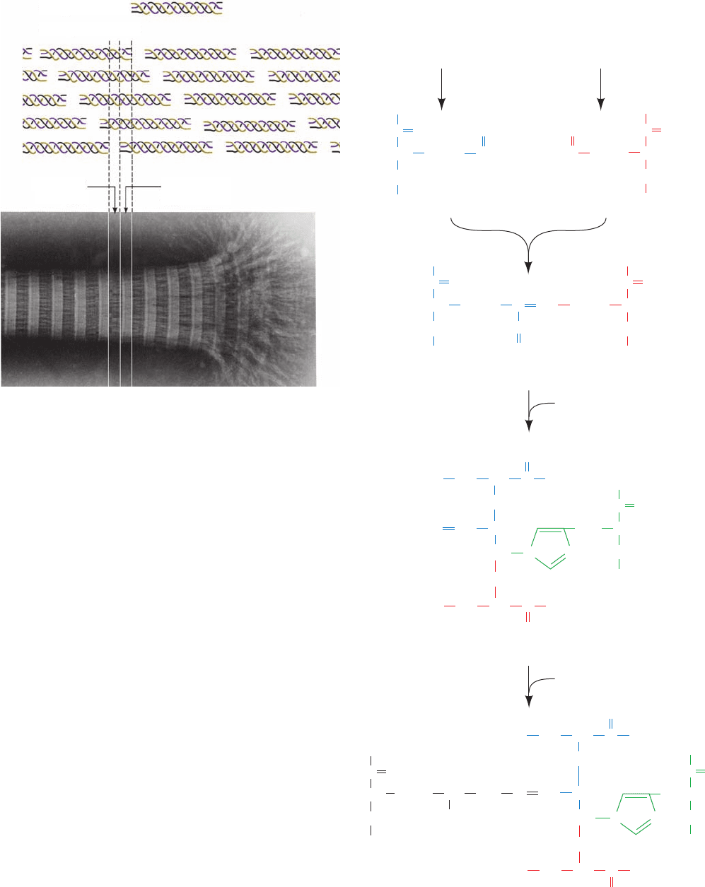
Figure 8-32 Banded appearance of collagen fibrils. The
banded appearance in the electron microscope arises from the
schematically represented staggered arrangement of collagen
molecules (top) that results in a periodically indented surface. D,
the distance between cross striations, is ⬃670 Å, so the length of
a 3000-Å-long collagen molecule is 4.4D. [Courtesy of Karl A.
Piez, Collagen Corporation.]
Figure 8-33 A biosynthetic pathway for cross-linking Lys, Hyl,
and His side chains in collagen. The first step in the reaction is
the lysyl oxidase–catalyzed oxidative deamination of Lys to form
the aldehyde allysine. Two such aldehydes then undergo an aldol
condensation to form allysine aldol. This product can react with
His to form aldol histidine, which in turn, can react with
Hyl to form a Schiff base (an imine bond), thereby cross-linking
four side chains.
Collagen molecule
Packing of molecules
Hole zone
0.6D
Overlap zone
0.4D
(CH
2
)
2
C
CH
O
NH
Lys
lysyl oxidase
Lys
lysyl oxidase
Allysine Allysine
Allysine aldol
His
CH
CH
2
OH
N
O
N
CH
2
CH
C
NHN
N
CH
2
C
CH
O
NH
N
Aldol-His
Histidinodehydrohydroxy-
merodesmosine
C
CH
O
NH
HC
O
(CH
2
)
3
CH
O
C
CH
O
NH
(CH
2
)
3
(CH
2
)
2
C
CH
O
NH
O
C
CH
CH
C
CH
O
NH
(CH
2
)
3
NH CCH
O
(CH
2
)
2
CH
CH
(CH
2
)
3
NH CCH
O
CH
(CH
2
)
3
NH CCH
O
CH
NH CCH
O
(CH
2
)
2
CH CH
5-Hydroxy-Lys (Hyl)
JWCL281_c08_221-277.qxd 2/23/10 1:58 PM Page 239
The degree of cross-linking of the collagen from a par-
ticular tissue increases with the age of the animal. This is
why meat from older animals is tougher than that from
younger animals. In fact, individual molecules of collagen
(called tropocollagen) can only be extracted from the tis-
sues of very young animals. Collagen cross-linking is not
the central cause of aging, however, as is demonstrated by
the observation that lathyrogenic agents do not slow the
aging process.
The collagen fibrils in various tissues are organized in
ways that largely reflect the functions of the tissues (Table
8-3). Thus tendons (the “cables” connecting muscles to
bones), skin (a tear-resistant outer fabric), and cartilage
(which has a load-bearing function) must support stress in
predominantly one, two, and three dimensions, respec-
tively, and their component collagen fibrils are arrayed ac-
cordingly. How collagen fibrils are laid down in these
arrangements is unknown. However, some of the factors
guiding collagen molecule assembly are discussed in Sec-
tions 32-5Aa and 32-5Ba.
d. Collagen Defects Are Responsible for a Variety of
Human Diseases
Numerous heritable disorders of collagen are known.
Mutations of Type I collagen, which constitutes the major
structural protein in most human tissues, usually result in
osteogenesis imperfecta (brittle bone disease). The sever-
ity of this disease varies with the nature and position of the
mutation: Even a single amino acid change can have lethal
consequences. Mutations may affect the structure of the
collagen molecule or how it forms fibrils. For example, the
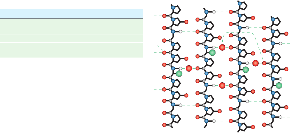
substitution of Ala for the central Gly in each polypeptide
chain of the structure shown in Fig. 8-29a, which reduces
the denaturation temperature of this model compound
from 62°C to 29°C, locally distorts the collagen triple helix.
The need to accommodate the three additional methyl
groups in the tightly packed interior of the triple helix pries
apart the polypeptide chains in the region of the substitu-
tions so as to rupture the hydrogen bonds that would oth-
erwise link the main chain N¬H group of each Ala (nor-
mally Gly) to the carbonyl oxygen of the adjacent Pro in a
neighboring chain (Fig. 8-34). Rather, these hydrogen
bonding groups are bridged by water molecules that insin-
uate themselves into the distorted part of the structure.
Similar distortions almost certainly occur in the Gly S X
mutated collagens responsible for such diseases as osteoge-
nesis imperfecta. Such mutations tend to be dominant be-
cause they affect either the folding of the triple helix or fib-
ril formation even when normal chains are also members
of the triple helix. All known amino acid changes within
Type I collagen’s triple helical region result in abnormali-
ties, indicating that the structural integrity of this region is
essential for proper collagen function.
Many collagen disorders are characterized by defi-
ciencies in the amount of a particular collagen type syn-
thesized or by abnormal activities of collagen-processing
enzymes such as lysyl hydroxylase or lysyl oxidase. One
group of at least 10 different collagen deficiency diseases,
the Ehlers–Danlos syndromes, are all characterized by
hyperextensibility of the joints (really the ligaments hold-
ing them together) and skin. This is because these tissues
also contain large amounts of elastin, a protein with rub-
berlike elastic properties. Consequently, the loss of the
rigidity normally conferred by collagen coupled with the
presence of elastin results in the hyperextensibility of the
affected tissues. Several degenerative diseases exhibit col-
lagen abnormalities in certain tissues, including cartilage in
osteoarthritis and the fibrous atherosclerotic plaques in
human arteries.
240 Chapter 8. Three-Dimensional Structures of Proteins
Table 8-3 The Arrangement of Collagen Fibrils in
Various Tissues
Tissue Arrangement
Tendon Parallel bundles
Skin Sheets of fibrils layered at many angles
Cartilage No distinct arrangement
Cornea Planar sheets stacked crossways so
as to minimize light scattering
Figure 8-34 Distorted structure in abnormal collagen. This
schematic diagram shows hydrogen bonding interactions in the
Ala-containing portions of the X-ray structure of (Pro-Hyp-Gly)
10
in which the fifth Gly is replaced by Ala.This cylindrical
projection is in the style of Fig. 8-30. Note how the Ala side
chains (large green balls) distort the triple helix so as to disrupt
the normally occurring Gly NH Pro O hydrogen bonds and
replace them with water-bridged hydrogen bonds. [After Bella, J.,
Eaton, M., Brodsky, B., and Berman, H.M., Science 266, 78
(1994).]
p
N
N
N
N
CC
C
C
Gly
Gly
Ala
Chain 1 Chain 2 Chain 3 Chain 1
H
2
O
Gly
Gly
Ala
Gly
Gly
Ala
H
2
O
Gly
Gly
Ala
H
2
O
H
2
O
Gly
JWCL281_c08_221-277.qxd 2/23/10 1:58 PM Page 240
