Voet D., Voet Ju.G. Biochemistry
Подождите немного. Документ загружается.

221
CHAPTER 8
Three-Dimensional
Structures of Proteins
1 Secondary Structure
A. The Peptide Group
B. Helical Structures
C. Beta Structures
D. Nonrepetitive Structures
2 Fibrous Proteins
A. ␣ Keratin—A Helix of Helices
B. Collagen—A Triple Helical Cable
3 Globular Proteins
A. Interpretation of Protein X-Ray and NMR Structures
B. Tertiary Structure
C. Structural Bioinformatics
4 Protein Stability
A. Electrostatic Forces
B. Hydrogen Bonding Forces
C. Hydrophobic Forces
D. Disulfide Bonds
E. Protein Denaturation
F. Explaining the Stability of Thermostable Proteins
5 Quaternary Structure
A. Subunit Interactions
B. Symmetry in Proteins
C. Determination of Subunit Composition
Appendix: Viewing Stereo Pictures
The properties of a protein are largely determined by its
three-dimensional structure. One might naively suppose
that since proteins are all composed of the same 20 types
of amino acid residues, they would be more or less alike
in their properties. Indeed, denatured (unfolded) pro-
teins have rather similar characteristics, a kind of homo-
geneous “average” of their randomly dangling side
chains. However, the three-dimensional structure of a
native (physiologically folded) protein is specified by its
primary structure, so that it has a unique set of charac-
teristics.
In this chapter,we shall discuss the structural features of
proteins, the forces that hold them together, and their hier-
archical organization to form complex structures. This will
form the basis for understanding the structure–function re-
lationships necessary to comprehend the biochemical roles
of proteins. Detailed consideration of the dynamic behav-
ior of proteins and how they fold to their native structures
is deferred until Chapter 9.
1 SECONDARY STRUCTURE
A polymer’s secondary structure (2° structure) is defined
as the local conformation of its backbone. For proteins, this
has come to mean the specification of regular polypeptide
backbone folding patterns: helices, pleated sheets, and
turns. However,before we begin our discussion of these ba-
sic structural motifs, let us consider the geometrical proper-
ties of the peptide group because its understanding is
prerequisite to that of any structure containing it.
A. The Peptide Group
In the 1930s and 1940s, Linus Pauling and Robert Corey
determined the X-ray structures of several amino acids
and dipeptides in an effort to elucidate the structural
constraints on the conformations of a polypeptide chain.
These studies indicated that the peptide group has a rigid,
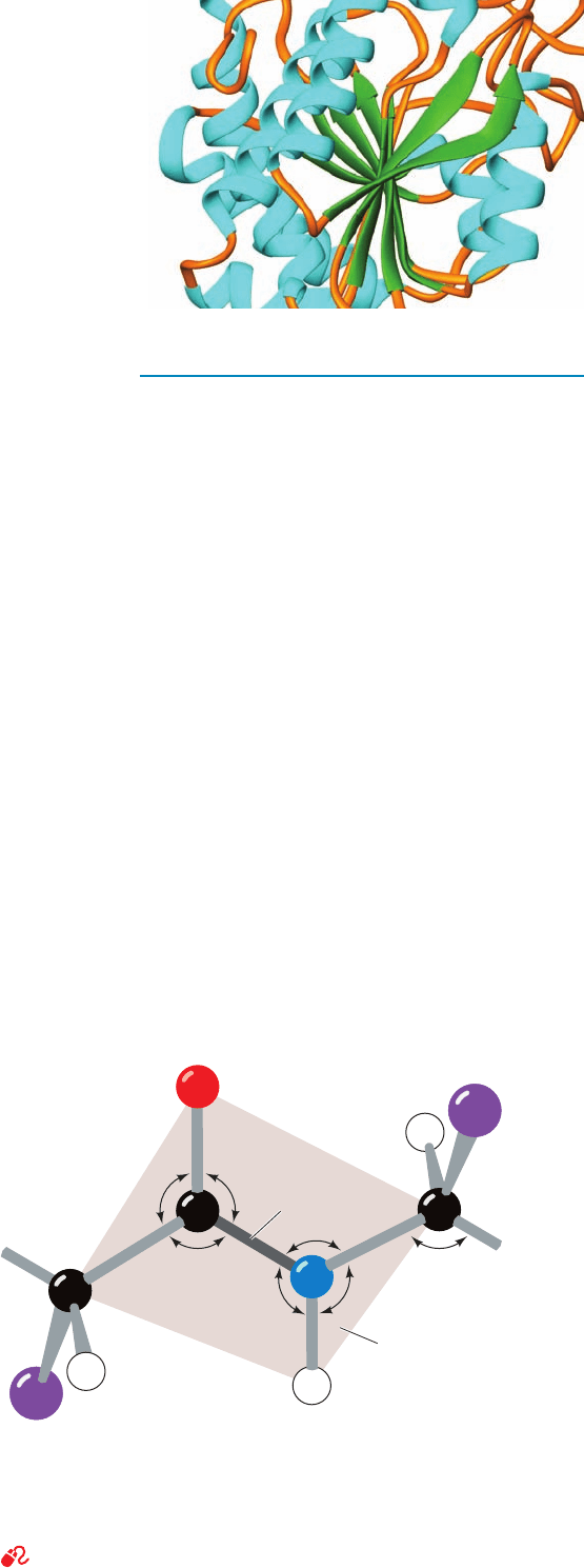
planar structure (Fig. 8-1), which, Pauling pointed out, is a
1.24
1.33
1.46
111°
1.51
123.5°
116°
120.5°
C
α
C
α
Peptide bond
Amide
plane
1.0
118.5°
119.5°
122°
N
H
H
O
C
R
R
H
Figure 8-1 The trans-peptide group. The standard dimensions
(in angstroms, Å, and degrees, °) of this planar group were
derived by averaging the corresponding quantities in the X-ray
crystal structures of amino acids and peptides. [After Marsh,
R.E. and Donohue, J., Adv. Protein Chem. 22, 249 (1967).]
See Kinemage Exercise 3-1
JWCL281_c08_221-277.qxd 2/23/10 1:58 PM Page 221
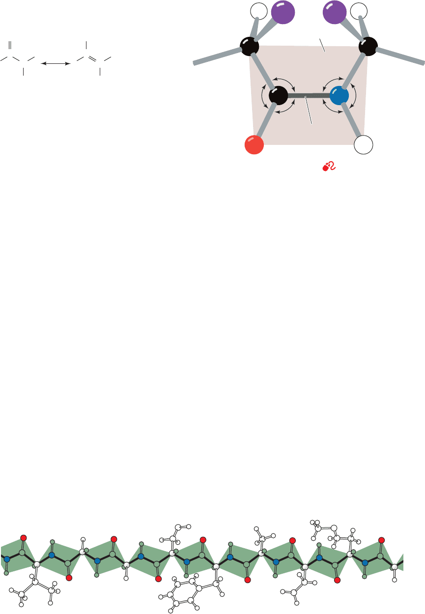
consequence of resonance interactions that give the peptide
bond an ⬃40% double-bond character:
This explanation is supported by the observations that a
peptide’s C¬N bond is 0.13 Å shorter than its N¬C
␣
single bond and that its bond is 0.02 Å longer than
that of aldehydes and ketones. The peptide bond’s reso-
nance energy has its maximum value, ⬃85 kJ ⴢ mol
⫺1
,
when the peptide group is planar because its -bonding
overlap is maximized in this conformation. This overlap,
and thus the resonance energy, falls to zero as the peptide
bond is twisted to 90° out of planarity, thereby account-
ing for the planar peptide group’s rigidity. (The positive
charge on the above resonance structure should be taken
as a formal charge; quantum mechanical calculations in-
dicate that the peptide N atom, in fact, has a partial neg-
ative charge arising from the polarization of the C¬N
bond.)
Peptide groups, with few exceptions, assume the trans
conformation: that in which successive C
a
atoms are on op-
posite sides of the peptide bond joining them (Fig. 8-1).
This is partly a result of steric interference, which causes
the cis conformation (Fig. 8-2) to be ⬃8 kJ ⴢ mol
⫺1
less
stable than the trans conformation (this energy difference
is somewhat less in peptide bonds followed by a Pro
residue and, in fact, ⬃10% of the Pro residues in proteins
follow a cis peptide bond, whereas cis peptides are other-
wise extremely rare).
a. Polypeptide Backbone Conformations May Be
Described by Their Torsion Angles
The above considerations are important because they
indicate that the backbone of a protein is a linked se-
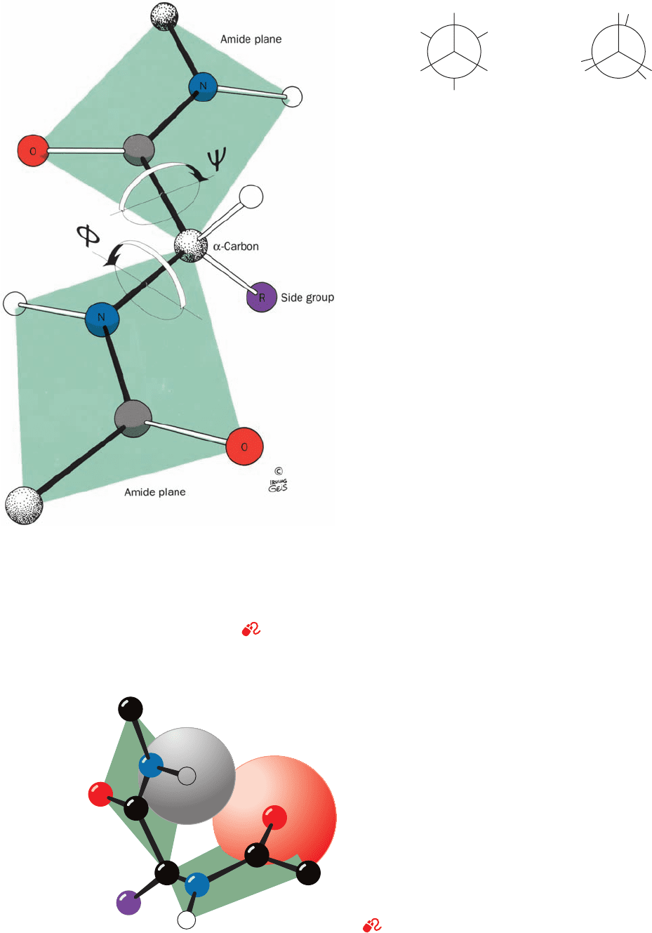
quence of rigid planar peptide groups (Fig. 8-3). We can
therefore specify a polypeptide’s backbone conforma-
tion by the torsion angles (rotation angles or dihedral an-
gles) about the C
␣
¬N bond () and the C
␣
¬C bond ()
C “ O
C
O
O
⫺
N
H
C
N
H
⫹
of each of its amino acid residues. These angles, and ,
are both defined as 180° when the polypeptide chain is in
its planar, fully extended (all-trans) conformation and in-
crease for a clockwise rotation when viewed from C
␣
(Fig. 8-4).
There are several steric constraints on the torsion an-
gles, and , of a polypeptide backbone that limit its con-
formational range. The electronic structure of a single ()
bond, such as a C¬C bond, is cylindrically symmetrical
about its bond axis, so that we might expect such a bond to
exhibit free rotation. If this were the case, then in ethane,
for example, all torsion angles about the C¬C bond would
be equally likely. Yet certain conformations in ethane are
favored due to quantum mechanical effects arising from
the interactions of its molecular orbitals. The staggered
conformation (Fig. 8-5a; torsion angle ⫽ 180°) is ethane’s
most stable arrangement, whereas the eclipsed conforma-
tion (Fig. 8-5b; torsion angle ⫽ 0°) is least stable. The en-
ergy difference between the staggered and eclipsed con-
formations in ethane is ⬃12 kJ ⴢ mol
⫺1
, a quantity that
represents an energy barrier to free rotation about the
C¬C single bond. Substituents other than hydrogen
exhibit greater steric interference; that is, they increase
the size of this energy barrier due to their greater bulk.
222 Chapter 8. Three-Dimensional Structures of Proteins
Figure 8-2 The cis-peptide group. See Kinemage Exercise 3-1
Figure 8-3 A polypeptide chain in its fully extended conformation showing the planarity of
each of its peptide groups. [Illustration, Irving Geis. Image from the Irving Geis Collection,
Howard Hughes Medical Institute. Reprinted with permission.]
113°
126°
121°
123°
119°
118°
1.32
1.24
1.53
1.47
1.0
C
α
C
α
Amide
plane
Peptide
bond
NC
RR
O H
HH
Main chain
Side chain
JWCL281_c08_221-277.qxd 8/10/10 11:47 AM Page 222
Indeed, with large substituents, some conformations may
be sterically forbidden.
b. Allowed Conformations of Polypeptides Are
Indicated by the Ramachandran Diagram
The sterically allowed values of and can be deter-
mined by calculating the distances between the atoms of a
tripeptide at all values of and for the central peptide
unit. Sterically forbidden conformations, such as that
shown in Fig. 8-6, are those in which any nonbonding inter-
atomic distance is less than its corresponding van der Waals
distance. Such information is summarized in a conforma-
tion map or Ramachandran diagram (Fig. 8-7), which was
invented by G.N. Ramachandran.
Figure 8-7 indicates that 77% of the Ramachandran di-
agram (most combinations of and ) is conformationally
inaccessible to a polypeptide chain. The particular regions
of the Ramachandran diagram that represent allowed con-
formations depend on the van der Waals radii chosen to
calculate it. But with any realistic set of values, such as that
in Table 8-1, only three small regions of the conformational
map are physically accessible to a polypeptide chain. Never-
theless, as we shall see, all of the common types of regular
secondary structures found in proteins fall within allowed
regions of the Ramachandran diagram. Indeed, the
Section 8-1. Secondary Structure 223
Figure 8-4 The torsional degrees of freedom in a peptide unit.
The only reasonably free movements are rotations about the
C
␣
¬N bond () and the C
␣
¬C bond ().The torsion angles are
both 180° in the conformation shown and increase, as is indicated,
in a clockwise manner when viewed from C
␣
. [Illustration, Irving
Geis. Image from the Irving Geis Collection, Howard Hughes
Medical Institute. Reprinted with permission.]
See Kinemage
Exercise 3-1
Figure 8-5 Conformations of ethane. Newman projections
indicating the (a) staggered conformation and (b) eclipsed
conformation of ethane.
Figure 8-6 Steric interference between adjacent residues. The
collision between a carbonyl oxygen and the following amide
hydrogen prevents the conformation ⫽⫺60°, ⫽30°.
[Illustration, Irving Geis. Image from the Irving Geis Collection,
Howard Hughes Medical Institute. Reprinted with permission.]
See Kinemage Exercise 3-1.
H
H
H
H
H
H
H
H
H
H
H
H
(a)
Staggered
(b)
Eclipsed
JWCL281_c08_221-277.qxd 8/10/10 11:47 AM Page 223
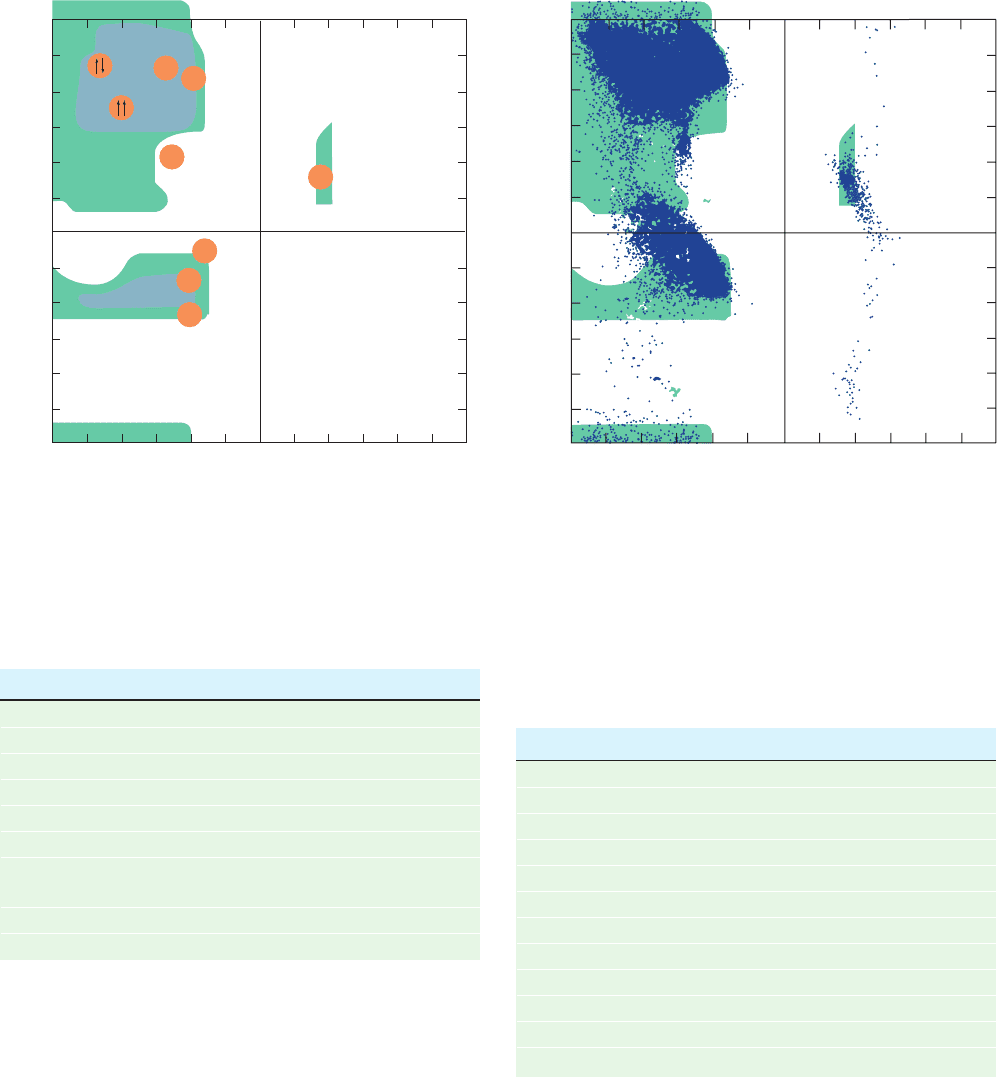
observed conformational angles of most non-Gly residues
in proteins whose X-ray structures have been determined
lie in these allowed regions (Fig. 8-8).
Most points that fall in forbidden regions of Fig. 8-8 lie
between its two fully allowed areas near ⫽0. However,
these “forbidden” conformations, which arise from the col-
lision of successive amide groups, are allowed if twists of
only a few degrees about the peptide bond are permitted.
This is not unreasonable since the peptide bond offers little
resistance to small deformations from planarity.
Gly, the only residue without a C

atom, is much less
sterically hindered than the other amino acid residues.This
is clearly apparent in comparing the Ramachandran dia-
gram for Gly in a polypeptide chain (Fig. 8-9) with that of
other residues (Fig. 8-7). In fact, Gly often occupies posi-
tions where a polypeptide backbone makes a sharp turn
224 Chapter 8. Three-Dimensional Structures of Proteins
Figure 8-8 Distribution of conformation angles in proteins.
The conformation angle distribution of all residues but Gly in
207 high-resolution (ⱕ1.2 Å) X-ray structures comprising 25,327
residues is superimposed on the Ramachandran diagram
(resolution is discussed in Section 8-3Aa). [Courtesy of Scott
Hollingsworth and Andrew Karplus, Oregon State University,
Corvallis, Oregon.]
180
90
–180
0
–90
ψ (deg)
–180 –90 0 90 180
φ (deg)
Source: Ramachandran, G.N. and Sasisekharan,V., Adv. Protein Chem. 23,
326 (1968).
Table 8-1 van der Waals Distances for Interatomic Contacts
Contact Type Normally Allowed (Å) Outer Limit (Å)
2.0 1.9
2.4 2.2
2.4 2.2
2.4 2.2
2.7 2.6
2.7 2.6
2.8 2.7
2.7 2.6
2.9 2.8
3.0 2.9
3.2 3.0
3.2 3.0CH
2
p
CH
2
C
p
CH
2
C
p
C
N
p
C
N
p
N
O
p
C
O
p
N
O
p
O
H
p
C
H
p
N
H
p
O
H
p
H
180
90
0
–90
C
2
3
II
–180
–180 –90 0 90 180
φ (deg)
ψ (deg)
α
π
α
L
Secondary Structure (deg) (deg)
Right-handed ␣ helix (␣) ⫺57 ⫺47
Parallel  pleated sheet (cc) ⫺119 113
Antiparallel  pleated sheet (cT) ⫺139 135
Right-handed 3
10
helix (3) ⫺49 ⫺26
Right-handed helix () ⫺57 ⫺70
2.2
7
ribbon (2) ⫺78 59
Left-handed polyglycine II and ⫺79 150
polyproline II helices (II)
Collagen (C) ⫺51 153
Left-handed ␣ helix (␣
L
)5747
Figure 8-7 The Ramachandran diagram. It shows the sterically
allowed and angles for poly-
L-alanine and was calculated
using the van der Waals distances in Table 8-1. Regions of
“normally allowed” and angles are shaded in blue, whereas
green-shaded regions correspond to conformations having “outer
limit” van der Waals distances.The conformation angles, and ,
of several secondary structures are indicated below:
[After Flory, P.J., Statistical Mechanics of Chain Molecules, p. 253,
Interscience (1969); and IUPAC-IUB Commission on Biochemical
Nomenclature, Biochemistry 9, 3475 (1970).]
JWCL281_c08_221-277.qxd 6/4/10 1:07 PM Page 224
which, with any other residue, would be subject to steric in-
terference.
Figure 8-7 was calculated for three consecutive Ala
residues. Similar plots for larger residues that are un-
branched at C

, such as Phe, are nearly identical. In Ra-
machandran diagrams of residues that are branched at
C

, such as Thr, the allowed regions are somewhat smaller
than for Ala.The cyclic side chain of Pro limits its to the
range ⫺60° ⫾ 25°, making it, not surprisingly, the most
conformationally restricted amino acid residue. The con-
formations of residues in chains longer than tripeptides
are even more restricted than the Ramachandran diagram
indicates because a polypeptide chain with all its and
angles allowed nevertheless cannot assume a conforma-
tion in which it passes through itself. We shall see, how-
ever, that despite the great restrictions that peptide bond
planarity and side chain bulk place on the conformations
of a polypeptide chain, different unique primary struc-
tures have correspondingly unique three-dimensional
structures.
B. Helical Structures
See Guided Exploration 7: Stable helices in proteins: The ␣ helix
Helices are the most striking elements of protein 2° struc-
ture. If a polypeptide chain is twisted by the same amount
about each of its C
␣
atoms, it assumes a helical conforma-
tion.As an alternative to specifying its and angles, a he-
lix may be characterized by the number, n, of peptide units
per helical turn and by its pitch, p, the distance the helix
rises along its axis per turn. Several examples of helices are
diagrammed in Fig.8-10. Note that a helix has chirality;that
Section 8-1. Secondary Structure 225
180
90
–180
0
–90
ψ (deg)
–180 –90 0 90 180
φ (deg)
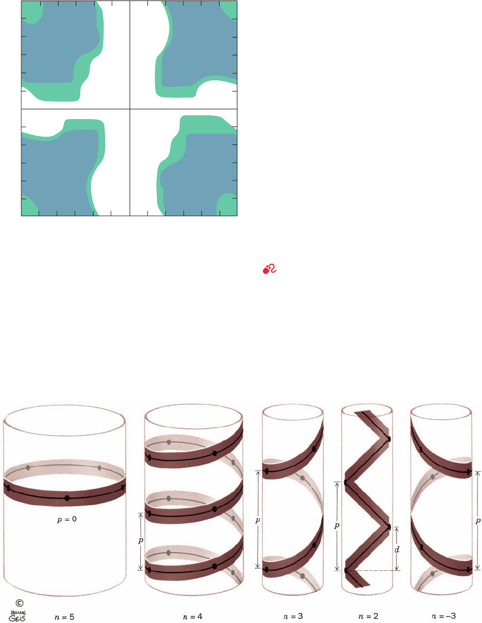
Figure 8-9 The Ramachandran diagram of Gly residues in a
polypeptide chain. “Normally allowed” regions are shaded in
blue, whereas green-shaded regions correspond to “outer limit”
van der Waals distances. Gly residues have far greater
conformational freedom than do other (bulkier) amino acid
residues, as the comparison of this figure with Fig. 8-7 indicates.
[After Ramachandran, G.N. and Sasisekharan, V., Adv. Protein
Chem. 23, 332 (1968).]
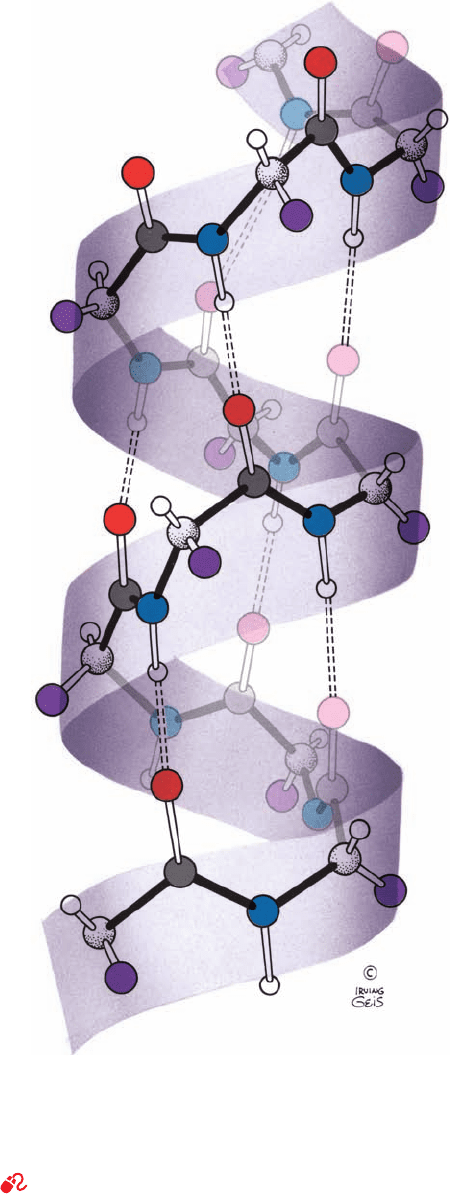
Figure 8-10 Examples of helices. These provide definitions of
the helical pitch, p, the number of repeating units per turn, n, and
the helical rise per repeating unit, d ⫽ p/n. Right- and left-handed
helices are defined, respectively, as having positive and negative
values of n. For n ⫽ 2, the helix degenerates to a nonchiral ribbon.
For p ⫽ 0, the helix degenerates to a closed ring. [Illustration,
Irving Geis. Image from the Irving Geis Collection, Howard
Hughes Medical Institute. Reprinted with permission.]
JWCL281_c08_221-277.qxd 8/10/10 11:47 AM Page 225
is, it may be either right handed or left handed (a right-
handed helix turns in the direction that the fingers of a
right hand curl when its thumb points along the helix axis
in the direction that the helix rises). In proteins, moreover,
n need not be an integer and, in fact, rarely is.
A polypeptide helix must, of course, have conformation
angles that fall within the allowed regions of the Ra-
machandran diagram. As we have seen, this greatly limits
the possibilities. Furthermore, if a particular conformation
is to have more than a transient existence, it must be more
than just allowed, it must be stabilized. The “glue” that
holds polypeptide helices and other 2° structures together
is, in part, hydrogen bonds.
a. The ␣ Helix
Only one helical polypeptide conformation has simulta-
neously allowed conformation angles and a favorable hy-
drogen bonding pattern: the ␣ helix (Fig. 8-11), a particu-
larly rigid arrangement of the polypeptide chain. Its
discovery through model building, by Pauling in 1951,
ranks as one of the landmarks of structural biochemistry.
For a polypeptide made from
L-␣-amino acid residues,
the ␣ helix is right handed with torsion angles ⫽⫺57°
and ⫽⫺47°, n ⫽ 3.6 residues per turn, and a pitch of 5.4 Å.
(An ␣ helix of
D-␣-amino acid residues is the mirror image
of that made from
L-amino acid residues: It is left handed
with conformation angles ⫽⫹57°, ⫽⫹47°, and n ⫽⫺3.6
but with the same value of p.)
Figure 8-11 indicates that the hydrogen bonds of an ␣
helix are arranged such that the peptide N¬H bond of the
nth residue points along the helix toward the peptide
group of the (n ⫺ 4)th residue. This results in a
strong hydrogen bond that has the nearly optimum
distance of 2.8 Å. In addition, the core of the ␣ helix is
tightly packed; that is, its atoms are in van der Waals con-
tact across the helix, thereby maximizing their association
energies (Section 8-4Ab). The R groups, whose positions,
as we saw, are not fully dealt with by the Ramachandran di-
agram, all project backward (downward in Fig. 8-11) and
outward from the helix so as to avoid steric interference
with the polypeptide backbone and with each other. Such
an arrangement can also be seen in Fig. 8-12. Indeed, a ma-
jor reason why the left-handed ␣ helix has never been ob-
served (its helical parameters are but mildly forbidden; Fig.
8-7) is that its side chains contact its polypeptide backbone
too closely. Note, however, that 1 to 2% of the individual
non-Gly residues in proteins assume this conformation
(Fig. 8-8).
The ␣ helix is a common secondary structural element
of both fibrous and globular proteins. In globular proteins,
␣ helices have an average span of ⬃12 residues, which cor-
responds to over three helical turns and a length of 18 Å.
However, ␣ helices with over 140 residues are known.
b. Other Polypeptide Helices
Figure 8-13 indicates how hydrogen bonded polypep-
tide helices may be constructed. The first two, the 2.2
7
rib-
bon and the 3
10
helix, are described by the notation, n
m
,
N
p
O
C “ O
where n, as before, is the number of residues per helical
turn and m is the number of atoms, including H, in the ring
that is closed by the hydrogen bond. With this notation, an
␣ helix is a 3.6
13
helix.
226 Chapter 8. Three-Dimensional Structures of Proteins
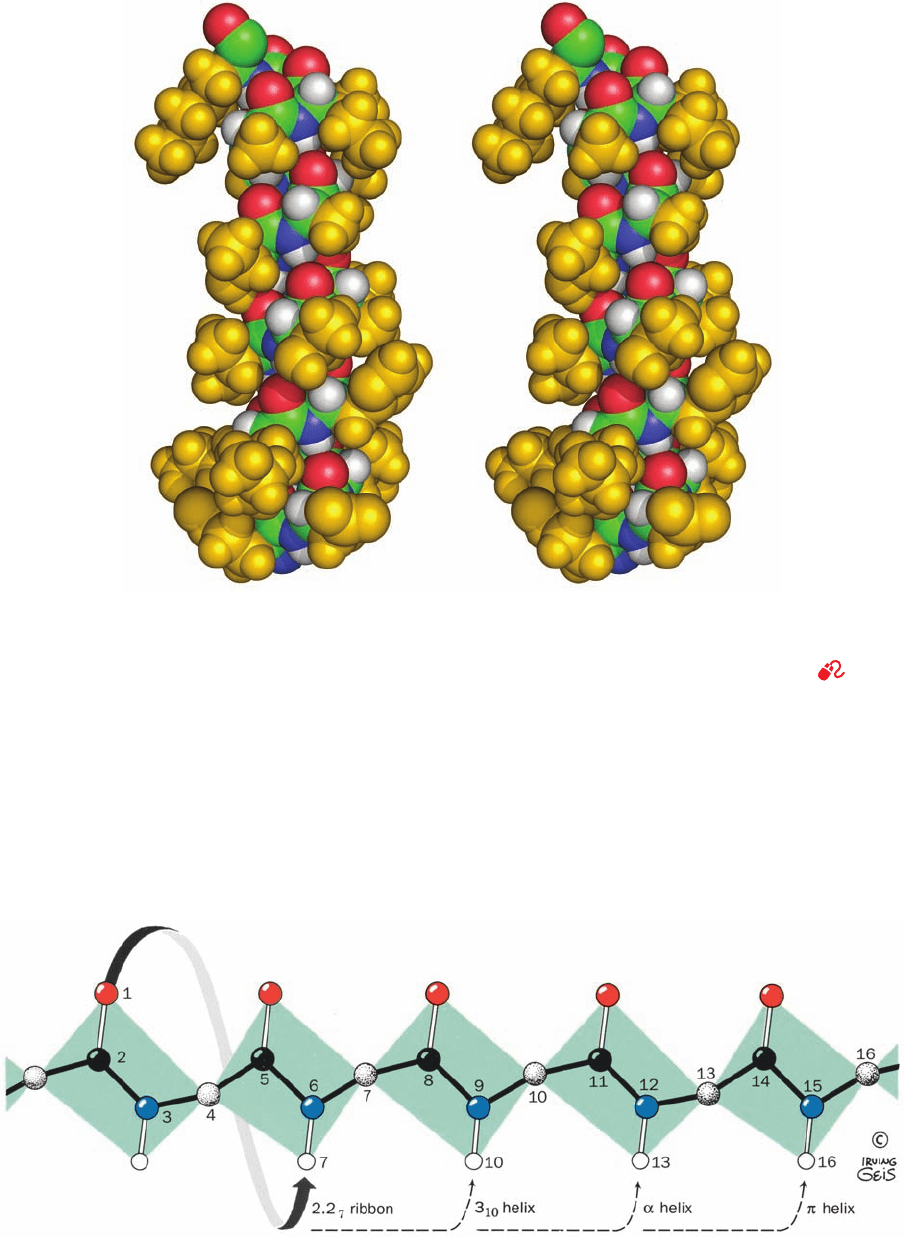
Figure 8-11 The right-handed ␣ helix. Hydrogen bonds between
the N¬H groups and the groups that are four residues
back along the polypeptide chain are indicated by dashed lines.
[Illustration, Irving Geis. Image from the Irving Geis Collection,
Howard Hughes Medical Institute. Reprinted with permission.]
See Kinemage Exercise 3-2 and the Animated Figures
C “ O
JWCL281_c08_221-277.qxd 8/10/10 11:47 AM Page 226
Section 8-1. Secondary Structure 227
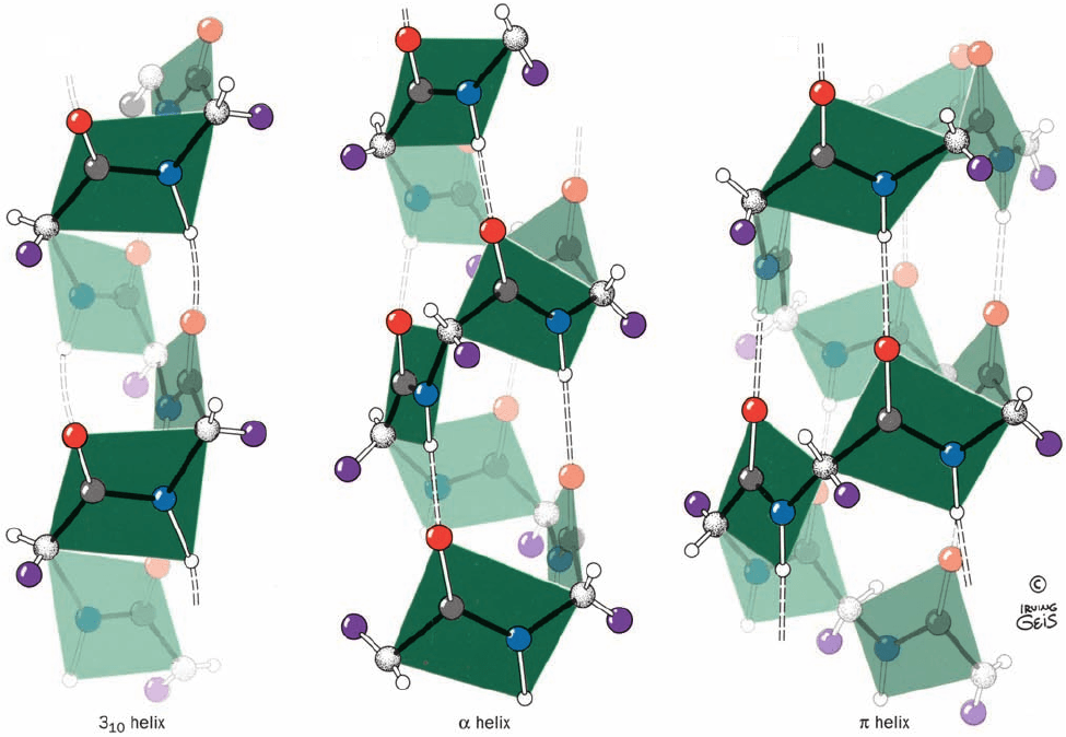
Figure 8-12 Stereo, space-filling representation of an ␣ helical
segment of sperm whale myoglobin (its E helix) as determined
by X-ray crystal structure analysis. Backbone atoms are colored
according to type (C green, N blue, O red, and H white) and the
side chain atoms are gold. Instructions for viewing stereo
Figure 8-13 The hydrogen bonding pattern of several
polypeptide helices. In the cases shown, the polypeptide chain is
helically wound such that the N¬H group on residue n forms a
hydrogen bond with the C“O groups on residues n ⫺ 2, n ⫺ 3, n
diagrams are given in the appendix to this chapter. [Based on an
X-ray structure by Ilme Schlichting, Max Planck Institut für
Molekulare Physiologie, Dortmund, Germany. PDBid 1A6M (for
the definition of “PDBid,” see Section 8-3Ca).]
See Kinemage
Exercise 3-2
⫺ 4, or n ⫺ 5. [Illustration, Irving Geis. Image from the Irving
Geis Collection, Howard Hughes Medical Institute. Reprinted
with permission.]
JWCL281_c08_221-277.qxd 8/10/10 11:47 AM Page 227
The right-handed 3
10
helix (Fig. 8-14a), which has a pitch
of 6.0 Å, is thinner and rises more steeply than does the ␣
helix (Fig. 8-14b). Its torsion angles place it in a mildly for-
bidden zone of the Ramachandran diagram that is rather
near the position of the ␣ helix (Fig. 8-7), and its R groups
experience some steric interference. This explains why the
3
10
helix is only occasionally observed in proteins, and then
mostly in short segments that are frequently distorted from
the ideal 3
10
conformation (the longest known 3
10
helix in a
protein has 15 residues). The 3
10
helix most often occurs as
a single-turn transition between one end of an ␣ helix and
the adjoining portion of a polypeptide chain.
The helix (4.4
16
helix), which also has a mildly forbid-
den conformation (Fig. 8-7), has only rarely been observed
and then only as segments of longer helices. This is proba-
bly because its comparatively wide and flat conformation
(Fig. 8-14c) results in an axial hole that is too small to admit
water molecules but too wide to allow van der Waals asso-
ciations across the helix axis; this greatly reduces its stabil-
ity relative to more closely packed conformations. The 2.2
7
ribbon, which, as Fig. 8-7 indicates, has strongly forbidden
conformation angles, has never been observed.
Certain synthetic homopolypeptides assume conforma-
tions that are models for helices in particular proteins.
Polyproline is unable to assume any common secondary
structure due to the conformational constraints imposed
by its cyclic pyrrolidine side chains. Furthermore, the lack
of a hydrogen substituent on its backbone nitrogen pre-
cludes any polyproline conformation from being knit to-
gether by hydrogen bonding. Nevertheless, under the
proper conditions, polyproline precipitates from solution
as a left-handed helix of all-trans peptides that has 3.0
residues per helical turn and a pitch of 9.4 Å (Fig. 8-15).
This rather extended conformation, which is known as the
polyproline II helix, permits the Pro side chains to avoid
each other. Curiously, polyglycine, the least conformation-
ally constrained polypeptide, precipitates from solution as
a helix whose parameters are essentially identical to those
of polyproline, the most conformationally constrained
polypeptide (although the polyglycine helix may be either
228 Chapter 8. Three-Dimensional Structures of Proteins
(a)
(b)
(c)
Figure 8-14 Comparison of the two polypeptide helices that
occasionally occur in proteins with the commonly occurring ␣
helix. (a) The 3
10
helix, which has 3.0 peptide units per turn and a
pitch of 6.0 Å, making it thinner and more elongated than the ␣
helix. (b) The ␣ helix, which has 3.6 peptide units per turn and a
pitch of 5.4 Å (also see Fig. 8-11). (c) The helix, which has 4.4
peptide units per turn and a pitch of 5.2 Å, making it wider and
shorter than the ␣ helix.The peptide planes are indicated.
[Illustration, Irving Geis. Image from the Irving Geis Collection,
Howard Hughes Medical Institute. Reprinted with permission.]
JWCL281_c08_221-277.qxd 8/10/10 11:47 AM Page 228
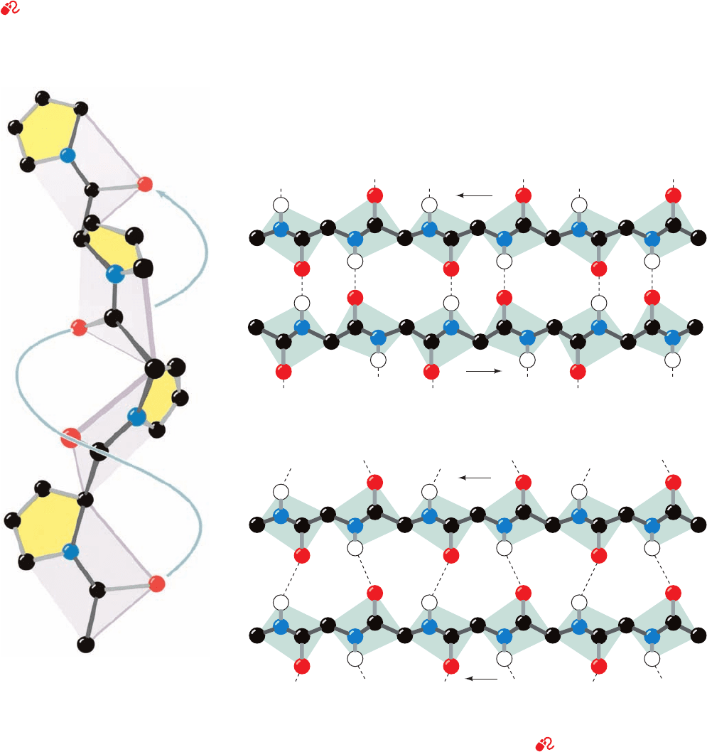
right or left handed because Gly is nonchiral). The structures
of the polyglycine and polyproline helices are of biological
significance because they form the basic structural motif of
collagen, a structural protein that contains a remarkably
high proportion of both Gly and Pro (Section 8-2B).In addi-
tion, the polyproline II helical conformation is commonly
assumed by polypeptide segments of up to 12 residues,
even though it lacks intrahelical hydrogen bonds.
C. Beta Structures
See Guided Exploration 8: Hydrogren bonding in  sheets and
Guided Exploration 9: Secondary structures in proteins
In 1951, the
year that they proposed the ␣ helix, Pauling and Corey also
postulated the existence of a different polypeptide second-
ary structure, the  pleated sheet.As with the ␣ helix, the 
pleated sheet’s conformation has repeating and angles
that fall in the allowed region of the Ramachandran dia-
gram (Fig. 8-7) and utilizes the full hydrogen bonding ca-
pacity of the polypeptide backbone. In  pleated sheets,
however, hydrogen bonding occurs between neighboring
polypeptide chains rather than within one as in ␣ helices.
 Pleated sheets come in two varieties:
1. The antiparallel  pleated sheet, in which neighbor-
ing hydrogen bonded polypeptide chains run in opposite
directions (Fig. 8-16a).
2. The parallel  pleated sheet, in which the hydrogen
bonded chains extend in the same direction (Fig. 8-16b).
The conformations in which these  structures are opti-
mally hydrogen bonded vary somewhat from that of a fully
extended polypeptide (⫽⫽⫾180°), as indicated in
Section 8-1. Secondary Structure 229
Parallel(b)
NC
NC
Antiparallel(a)
NC
NC
Figure 8-15 The polyproline II
helix. Polyglycine forms a nearly
identical helix (polyglycine II).
[Illustration, Irving Geis. Image from
the Irving Geis Collection, Howard
Hughes Medical Institute. Reprinted
with permission.]
Figure 8-16  Pleated sheets. Hydrogen bonds are indicated by dashed lines and
side chains are omitted for clarity. (a) The antiparallel  pleated sheet. (b) The parallel 
pleated sheet. [Illustration, Irving Geis. Image from the Irving Geis Collection, Howard
Hughes Medical Institute. Reprinted with permission.]
See Kinemage Exercise 3-3 and
the Animated Figures
JWCL281_c08_221-277.qxd 8/10/10 11:47 AM Page 229
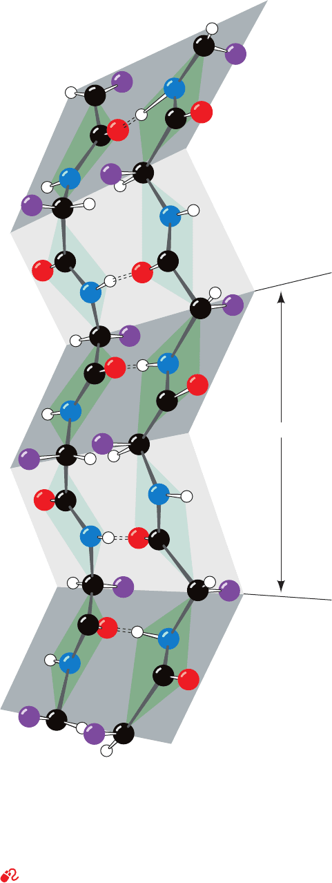
Fig. 8-7. They therefore have a rippled or pleated edge-on
appearance (Fig. 8-17), which accounts for the appellation
“pleated sheet.” In this conformation, successive side chains
of a polypeptide chain extend to opposite sides of the
pleated sheet with a two-residue repeat distance of 7.0 Å.
 Sheets are common structural motifs in proteins. In
globular proteins, they consist of from 2 to as many as 22
polypeptide strands, the average being 6 strands, which
have an aggregate width of ⬃25 Å.The polypeptide chains
in a  sheet are known to be up to 15 residues long, with
the average being 6 residues that have a length of ⬃21 Å.
A 7-stranded antiparallel  sheet, for example, occurs in
the jack bean protein concanavalin A (Fig. 8-18).
Parallel  sheets of less than five strands are rare.
This observation suggests that parallel  sheets are less
stable than antiparallel  sheets, possibly because the hy-
drogen bonds of parallel sheets are distorted in compa-
rison to those of the antiparallel sheets (Fig. 8-16). Mixed
parallel–antiparallel  sheets are common but, neverthe-
less, only ⬃20% of the strands in  sheets have parallel
bonding on one side and antiparallel bonding on the other
(vs an expected 50% for the random mixing of strand
directions).
The  pleated sheets in globular proteins invariably ex-
hibit a pronounced right-handed twist when viewed along
their polypeptide strands (e.g., Fig. 8-19). Such twisted 
sheets are important architectural features of globular pro-
teins since  sheets often form their central cores (Fig. 8-19).
Conformational energy calculations indicate that a 
sheet’s right-handed twist is a consequence of nonbonded
interactions between the chiral
L-amino acid residues in
the sheet’s extended polypeptide chains.These interactions
tend to give the polypeptide chains a slight right-handed
helical twist (Fig. 8-19) which distorts and hence weakens
the  sheet’s interchain hydrogen bonds. A particular
 sheet’s geometry is thus the result of a compromise be-
tween optimizing the conformational energies of its
polypeptide chains and preserving its hydrogen bonds.
The topology (connectivity) of the polypeptide strands
in a  sheet can be quite complex; the connecting links of
these assemblies often consist of long runs of polypeptide
chain which frequently contain helices (e.g., Fig. 8-19). The
link connecting two consecutive antiparallel strands is
topologically equivalent to a simple hairpin turn (Fig.8-20a).
However, tandem parallel strands must be linked by a
crossover connection that is out of the plane of the  sheet.
Such crossover connections almost always have a right-
handed helical sense (Fig. 8-20b), which is thought to better
fit the  sheets’ inherent right-handed twist (Fig. 8-21).
D. Nonrepetitive Structures
Globular proteins consist of, on average, ⬃31% ␣ helix and
⬃28%  sheet. The protein’s remaining polypeptide seg-
ments are said to have a coil or loop conformation. That is
not to say that these nonrepetitive secondary structures are
any less ordered than are helices or  sheets; they are simply
irregular and hence more difficult to describe. You should
therefore not confuse the term coil conformation with the
230 Chapter 8. Three-Dimensional Structures of Proteins
Figure 8-17 A two-stranded  antiparallel pleated sheet
drawn to emphasize its pleated appearance. Dashed lines
indicate hydrogen bonds. Note that the R groups (purple balls)
on each polypeptide chain alternately extend to opposite sides of
the sheet and that they are in register on adjacent chains.
[Illustration, Irving Geis. Image from the Irving Geis Collection,
Howard Hughes Medical Institute. Reprinted with permission.]
See Kinemage Exercise 3-3
7.0 A
o
JWCL281_c08_221-277.qxd 8/26/10 7:47 PM Page 230
