Vlak J.M., de Gooijer C.D., Tramper J., Miltenburger H.G. (Eds.) Insect Cell Cultures: Fundamental and Applied Aspects
Подождите немного. Документ загружается.


112
from the modified transfer vector is then mixed with
circular viral DNA extracted from viral nucleocapsids,
and transfected into insect host cells. Inside the cell,
host enzymes can mediate recombination between the
viral sequences in the transfer vector and the identi-
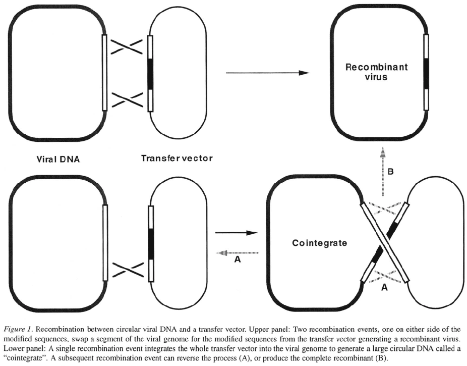
cal sequences in the viral genome. Two recombination
events (crossovers), one on either side of the modifi-
cation, will exchange the modified sequences from the
transfer vector for the wild-type viral sequences, gen-
erating a recombinant virus (Figure 1, upper panel).
However, such double-recombination events occur at
a very low frequency and typically only 0.1–1% of the
viruses released from the transfected cells are recom-
binant. The recombination frequency can be boosted
two- to three-fold by irradiation of the cells with UV
light prior to transfection (Peakman et al
.,
1989) but
this technique is not widely used. The major task is
identifying the rare recombinant viruses. In the clas-
sical method, wild-type viral DNA is used so that the
non-recombinant virus produces plaques that contain
the viral occlusion bodies known as polyhedra, where-
as recombinant viruses in which a foreign gene has
been substituted for the polyhedrin gene give plaques
that lack polyhedra. A trained eye can pick out the
polyhedra-negative plaques from the background of
non-recombinant plaques containing polyhedra. After
picking the putative recombinant plaques, three or four
rounds of plaque purification are required to eliminate
the contaminating wild-type viruses. To obtain a pure
recombinant virus by this method takes three to four
weeks.
A variant of this approach is to use viral DNA from a
virus that expresses such that plaques
of the parental virus stain blue with X-gal, whereas
plaques of a recombinant virus are white (Summers
& Smith, 1987). Although more appealing to novice
users, diffusion of the blue color can obscure the white
plaques, and there is a background of non-recombinant
white plaques due to the relatively high spontaneous
mutation rate of the lac
Z
gene.
Less direct methods have also been employed to
identify the recombinant viruses, including: screen-
ing by nucleic acid hybridization (Summers & Smith,
1987; Fung et al
.,
1988; Pen et al
.,
1989), screening
for protein expression using an antibody (Manns &
Grosse, 1991; Grosse & Manns, 1995), screening for
enzymatic or other activity of the expressed gene, or
fluorescence-activated cell sorting (Peng et al., 1993).
The very low frequency of recombination between
transfer vectors and circular viral DNA makes screen-
ing for recombinant viruses tedious and necessitates
several rounds of purification before a pure recombi-
nant virus is obtained. Although this method is still
commonly used to construct recombinants from some
baculoviruses, for AcMNPV it has largely been super-
seded by the less time-consuming methods described
below.
Use of transfer vectors containing a marker gene
cassette
Rare recombinant plaques can be identified easily if
they express a marker gene not present in the parental
virus. To this end, a cassette consisting of a promot-
er active in insect cells, the E. coli lac
Z
gene, and
a polyadenylation signal has been incorporated into
transfer vectors. The marker gene cassette is inserted
adjacent to the promoter, cloning sites and polyadeny-
lation signal designed for expression of the target gene,
to form a unit that is flanked by viral DNA sequences.
One series of such vectors has been constructed by
Richardson’s group (Vialard et al., 1990; Richardson
et al., 1992a; Richardson et al., 1992b, Lalumière
& Richardson, 1995) (Invitrogen distributes some of
these vectors and their derivatives as pBlueBac vec-
tors), and another series by Vlak’s group (Vlak et al.,
1990; Zuidema et al., 1990). Recombination of such
vectors with viral DNA transfers both expression cas-
settes to the viral genome resulting in a recombinant
virus that expresses and gives a blue
plaque. Although this does not improve the efficiency
of recombination, it is much easier to find rare blue
plaques amongst a background of white plaques than
vice versa.
The presence of polyhedra can also be used to iden-
tify recombinant plaques if DNA from a virus lacking a
functional polyhedrin gene is transfected with a trans-
fer vector containing a polyhedrin expression cassette
(Weyer et al., 1990).
The main drawback to using a marker cassette in the
transfer vector is that cointegrates, in which the whole
transfer vector is inserted into the virus DNA (Fig-
ure 1, lower panel), have a similar plaque phenotype
to recombinant viruses. Many of the putative recombi-
nant plaques will contain cointegrates because a single
crossover integrates the transfer vector into the virus
genome (Figure 1, lower panel) whereas two indepen-
dent crossovers are required to make the desired recom-
binant (Figure 1, upper panel). Cointegrate viruses are
not desirable because they are unstable; recombination
between the repeated copies of viral sequences flank-
ing the transfer vector can excise the plasmid and target
113
gene (Figure 1, lower panel) leading to loss of expres-
sion. In the classical method, cointegrate viruses have
the same plaque phenotype as parental viruses and will
be rejected. The isolation of cointegrate viruses when
using a transfer vector with a marker cassette can be
avoided by transfecting with linear viral DNA (see
below). In this case the product of a single crossover is
a linear cointegrate which will not be viable.
Other disadvantages of using transfer vectors con-
taining a marker cassette are that the second expression
cassette makes the transfer vector large and limits the
unique cloning sites, and recombinant viruses express
an unwanted protein.
Selection against the parental virus
Godeau et al. (1992) constructed viruses that express
the thymidine kinase gene from herpes simplex virus
type 1 (HSV1-tk) so that Ganciclovir can be used to
select against the parental virus. Host cell thymidine
kinases do not metabolize the nucleotide analog Gan-
ciclovir whereas HSV1-tk converts Ganciclovir into a
toxic inhibitor of DNA replication. In AcMNPVIE–
1-tk and AcMNPVIE–1-tk-pl0-SEAP (Godeau et al.,
1992), the polyhedrin gene has been replaced by the
HSV1-tk gene driven by an immediate early AcMN-
PV promoter (IE-1(0)). Expression of HSV1-tk makes
replication of these viruses sensitive to Ganciclovir.
When a standard polyhedrin-based transfer vector is
cotransfected with viral DNA from one of these virus-
es, a double recombination event can replace the
HSV1-tk gene with the foreign gene from the trans-
fer vector. Propagation of the progeny viruses in the
presence of Ganciclovir selects against the parental
viruses and enriches for recombinants. After one cycle
of selection, 85% or more of the viruses are recombi-
nant (Godeau et al., 1992).
114
This method gives a very strong enrichment for
recombinant viruses but sacrifices the ability to confirm
a virus as recombinant by its plaque phenotype. The
major concern when using this method is that mutations
in the thymidine kinase gene of the parental virus will
give rise to a background of non-recombinant viruses
that survive the Ganciclovir selection.
Recombination between a transfer vector and linear
viral DNA
Viral DNA that has been linearized at a unique site is
100- to 1000-fold less infectious than circular DNA,
but retains the capacity to recombine with transfer vec-
tors that contain homology to the viral DNA on either
side of the break (Kitts et al., 1990). By cotransfecting
transfer vectors with linearized viral DNA, the back-
ground of non-recombinant plaques is greatly reduced
but the number of recombinant plaques is affected to
a much lesser extent. Consequently, about 25% of the
resulting plaques are recombinant (Kitts et al., 1990).
The original single-cut virus, AcRP6-SC, has a unique
Bsu
36I
site in place of the polyhedrin gene (Kitts et
al., 1990), hence it is not possible to visually identify
recombinant plaques by the absence of polyhedra. Sub-
sequently, other viruses have been engineered to con-
tain a unique restriction site that allows linearization
of the viral DNA but also permits a visual screen for
recombinant plaques. Viruses that contain the E. coli
lac
Z
gene have a unique Bsu
36
I site that lies within the
coding sequences. By using linearized
viral DNA from a lacZ virus, plaques of the parental
virus are blue and can easily be rejected, raising the
proportion of white plaques that are recombinant to 30
to 100% (Kitts et al., 1990, Copeland & Wang, 1993;
Kitts, 1995). Two linearizable AcMNPV derivatives
that express an intact polyhedrin gene are also avail-
able; one, AcV EPA, has a unique Bsu36I site follow-
ing the polyhedrin coding sequences (Hartig & Cardon,
1992), the other has a unique Sse8387I site upstream
of the polyhedrin gene (Day et al., 1995). Using linear
viral DNA from these viruses, plaques of the parental
virus contain polyhedra and can be rejected; 50 to
80% of the polyhedra-negative plaques are recombi-
nant (Hartig & Cardon, 1992;Day et al., 1995). Not all
plaques with a non-parental phenotype obtained from
cotransfections using these linear DNAs are recombi-
nant; the non-parental non-recombinant plaques pre-
sumably result from imperfect recircularization of the
viral DNA that deletes bases from the junction so that
a functional or polyhedrin gene is no
longer expressed (Kitts et al., 1990).
Cotransfections using linear viral DNA give a high
proportion of plaques that are recombinant; 10 to 25%
without a visual screen, or 30 to 100% when plaques of
the parental virus can be rejected because of their phe-
notype. This makes identifying and purifying recom-
binant plaques much easier and less time consuming
compared with using circular viral DNA. Linear viral
DNA can profitably be combined with the use of trans-
fer vectors containing a marker cassette because it
eliminates the problem of isolating cointegrate viruses
(see above).
The linear viral DNA method has been found to
work for either the polyhedrin or pl0 loci of AcMN-
PV, provided a cognate transfer vector is used (Kitts
et al., 1990; Kitts, 1995; Martens et al., 1995). AcM-
NPV viral DNA linearized at either the polyhedrin or
p10 locus is commercially available from Invitrogen or
PharMingen.
Recombination between linear viral DNA and a
transfer vector containing a dominant selectable
marker
Lerch & Friesen (1993) investigated the use of the
neomycin-resistance gene or the p35 gene from AcM-
NPV as dominant selectable markers and found that
the p35 gene provided the strongest selection. The p 35
gene suppresses premature cell death due to induc-
tion of apoptosis in Sf21 cells infected with AcMN-
PV (Clem et al., 1991); consequently production of
budded virus from a virus that has a deletion in the
p
35
gene, is greatly reduced. Insertion of a
functional p
35
cassette at another location in the virus
restores the production of budded virus to wild-type
levels (Hershberger et al., 1992). Thus a transfer vec-
tor containing a p35 cassette adjacent to the polyhedrin
promoter driving expression of the target gene can be
used to allow selection of recombinant viruses. Trans-
fection of this vector with virus yields recom-
binants as 30% of the total progeny (Lerch & Friesen,
1993). The proportion of recombinant viruses can be
further improved by replacing the virus with
linearized viral DNA from another virus with a deletion
in the p
35
gene, Cotransfection of the
p
35
transfer vector and this linear viral DNA yields 82
to 96% recombinant viruses (Lerch & Friesen, 1993).
A pure recombinant virus can be obtained after a single
round of plaque purification.

115
This method gives a very high proportion of recom-
binant viruses. Its major limitation is that it requires a
specialized transfer vector. Another drawback to using
this system is that the virus needed to pre-
pare the linearized viral DNA is produced at a 10-fold
lower level than wild-type virus.
Recombination between a transfer vector and linear
viral DNA with a “lethal deletion”
A very high proportion of recombinant viruses,
approaching 100%, can also be obtained by using
a modified linear viral DNA from which essential
sequences have been removed such that recombina-
tion with a transfer vector is necessary to generate a
viable virus (Kitts & Possee, 1993). In AcMNPV the
gene downstream of polyhedrin, ORF1629, encodes a
phosphoprotein that is associated with the viral nucleo-
capsid or virion envelope (Vialard & Richardson, 1993;
Pham et al., 1993) and is essential for production of
infectious virus (Possee et al., 1991). A Bsu36I site
was engineered into the C-terminus of ORF1629 with-
out altering the amino acid sequence of the protein,
and a second Bsu36I site was introduced in ORF603
upstream of the polyhedrin promoter (Kitts & Possee,
1993). The best results were obtained using a virus that
also had the E. coli lacZ gene in place of the polyhedrin
gene and thus contained a third site for Bsu36I (Kitts &
Possee, 1993). This virus, BacPAK6, is viable; how-
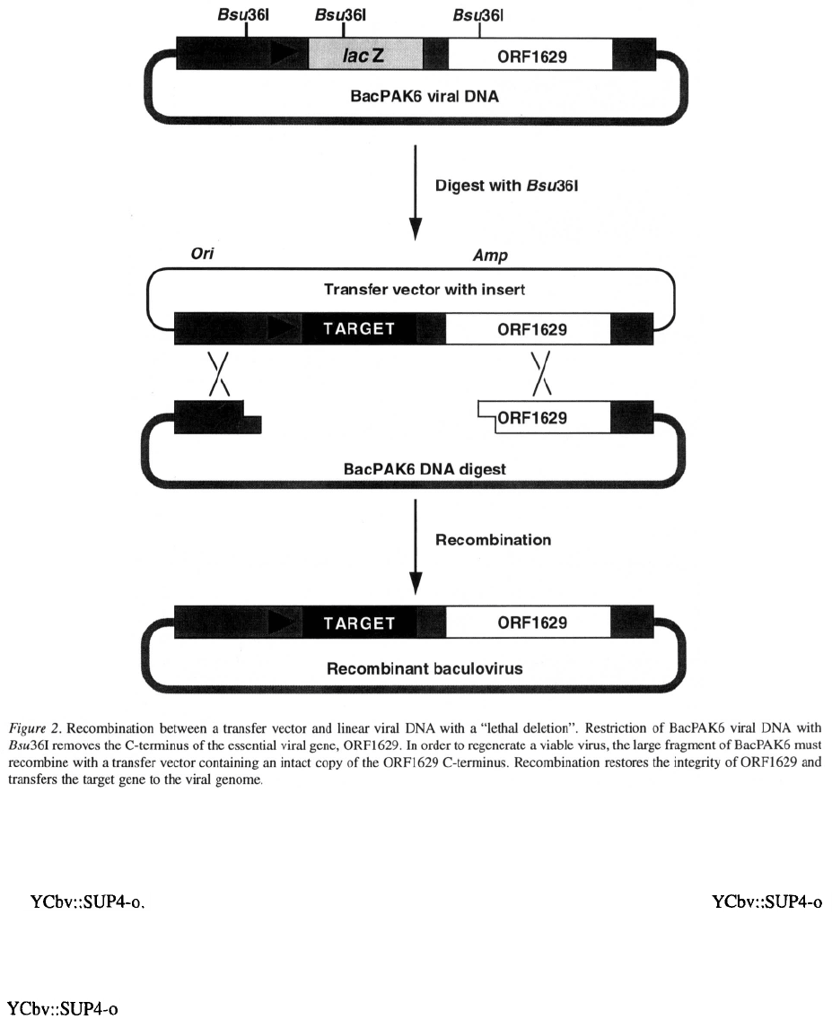
ever, restriction of the viral DNA with Bsu36I removes
a fragment containing part of the essential ORF1629
gene (Figure 2). Recombination between the large frag-
ment of viral DNA and a transfer vector that contains
an intact copy of the C-terminus of ORF1629 can res-
cue the viral DNA by regenerating an intact ORF1629
gene (Figure 2). In this process the foreign gene is also
transferred to the viral genome (Figure 2). 90 to 100%
of the viruses produced by cotransfecting a transfer
vector and BacPAK6 viral DNA digested with Bsu36I
express the gene from the transfer vector (Kitts & Pos-
see, 1993). This proportion is sufficiently high that it
is possible to use the progeny virus stock for protein
expression without the need to plaque purify a recom-
binant virus. Even if a clonal recombinant virus is
desired it is easy to isolate a pure recombinant plaque.
The major advantage of this method is the very
low background of non-recombinant viruses. A sec-
ond significant advantage is that it is compatible with
a wide variety of transfer vectors that are based on
the polyhedrin locus of AcMNPV and hence con-
tain the sequences necessary to rescue restricted Bac-
PAK6 viral DNA. Although the yield of virus from
this method is low, it is more than adequate for con-
verting a transfer vector containing a unique insert to
a recombinant virus.
BacPAK6 viral DNA digested with Bsu36I to
remove the ORF1629 C-terminus is commercially
available from CLONTECH or as BaculoGold™ from
PharMingen.
Generation of recombinant viruses in alternative
hosts
To circumvent the low efficiency of recombination
inside insect cells, viruses have been modified to con-
tain elements that allow them to replicate as episomes
in either yeast or E. coli. This allows the viral genome
to be manipulated in these more tractable hosts. Once
the desired recombinant viral genome has been con-
structed, DNA can be prepared and transfected into
insect cells where the virus will replicate to generate a
stock of infectious virus.
Recombination in yeast
Recombination in Saccharomyces cerevisiae is highly
efficient, especially when one of the participating DNA
molecules has a double-strand break, and markers are
also available that facilitate selection and screening for
recombinant molecules. Patel et al. (1992) developed
a method to generate recombinant viruses in yeast that
takes advantage of these properties.
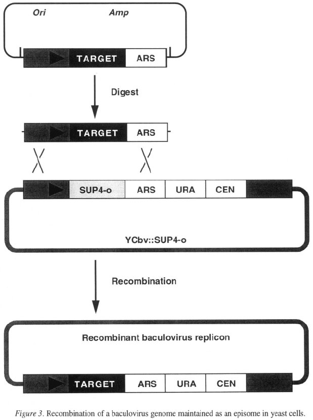
was constructed by inserting into the polyhedrin locus
of AcMNPV the elements required for replication and
selection in yeast (Figure 3). When yeast cells are
transformed with DNA, the ARS and
CEN elements enable the viral genome to replicate as
an episome, and the URA marker can be used to select
for cells in which replication of the viral DNA has been
established. The SUP4-o ochre suppressing allele of a
tRNA gene provides a marker that can either be select-
ed or counterselected. The host strain has an ochre
mutation in the ADE2 gene which makes it depen-
dent on adenine for growth and causes the colonies to
be pink. A second ochre mutation in the CAN1 gene
makes the cells resistant to canavanine. Expression of
the SUP4-o gene from suppresses the
ochre mutations of the host so that growth of cells con-
taining this vector is adenine independent but sensitive
to canavanine, and the colonies are white. A comple-
mentary transfer vector was constructed that contains
116
a cloning site flanked on one side by the AcMNPV
polyhedrin promoter and upstream viral sequences,
and on the other by the same ARS sequences present
in The target gene is cloned into
this vector and DNA from the resulting plasmid is
digested with restriction enzymes that release a lin-
ear DNA fragment carrying the target gene and flank-
ing sequences (Figure 3). If yeast cells containing
are transformed with the transfer vec-
tor digest, homologous recombination can replace the
SUP4-o gene in the viral genome with the target gene
(Figure 3). In theory, yeast cells in which this has
occurred can be selected as pink colonies that are resis-
tant to canavanine; however, a significant background
of canavanine resistant mutants necessitates a modi-
fied procedure. Yeast cells containing
are cotransformed with the transfer vector digest and
a plasmid containing a TRP gene that complements
a mutation making the host dependent on tryptophan.
Yeast cells that have taken up DNA are selected on
medium lacking tryptophan, and the resulting colonies
are screened for canavanine resistance resulting from
the loss of the SUP4-o allele. Because SUP4-o makes
the cells grow slower, choosing the largest colonies
117
or pink colonies enriches for the desired clones so
that approximately 80% of the selected colonies will
be resistant to canavanine (Patel et al., 1992). These
colonies harbor a recombinant YCbv vector containing
the target gene. After restreaking to obtain a clonal iso-
late containing a recombinant YCbv, DNA is prepared
from the yeast cells , fractionated on a sucrose gra-
dient, and fractions containing viral DNA are located
using PCR. Insect cells are then transfected with these
fractions, initiating viral replication that generates a
stock of recombinant virus.
The advantages of this method are that the recombi-
nation frequency is high, identification of colonies con-
taining recombinant virus replicons is relatively easy,
and a pure clone can be obtained quickly from a sin-
gle yeast colony. Limitations of this method are that it
requires a specialized transfer vector, and significant
labor is required to purify the viral DNA over a sucrose
gradient before it can be transfected into insect cells.
Although yeast is easy to work with, laboratories not
already working with Saccharomyces cerevisiae may
be deterred by the need to work with another host.
Recombination in Escherichia coli.
Luckow and coworkers have developed a method for
generating recombinant baculoviruses in E. coli that
uses the transposon Tn7 to insert the target gene into
the viral genome (Luckow et al., 1993). A baculovirus-
plasmid hybrid (bacmid), capable of being propagated
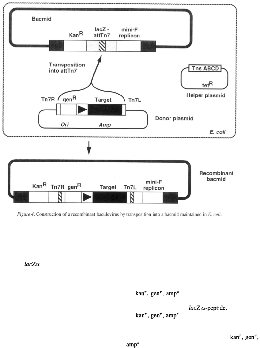
118
in E. coli as a single-copy plasmid was constructed by
inserting a minimal replicon derived from the E. coli
F plasmid and a kanamycin-resistance gene into the
polyhedrin locus of AcMNPV (Figure 4). The bacmid
also contains the peptide region from a pUC-
based plasmid with an in-frame insertion of the tar-
get site for Tn7 (att-Tn7). Consequently, colonies of
an appropriate host transformed with the bacmid stain
blue with X-gal. A specialized donor plasmid carries a
mini-Tn7 containing
a
gentamycin-resistance gene, a
copy the AcMNPV polyhedrin promoter followed by
a multiple cloning site, and a polyadenylation signal
(Figure 4). The target gene is cloned into the donor
plasmid downstream from the polyhedrin promoter.
To construct a recombinant bacmid, the donor plas-
mid containing the target gene is transformed into an
E. coli strain containing both the bacmid and a helper
plasmid that expresses the Tn7 proteins necessary for
transposition of the mini-Tn7 (Figure 4). Transposi-
tion of the mini-Tn7 from the donor plasmid into the
att-Tn7 of the bacmid generates a recombinant bacmid
(Figure 4). Bacteria harboring the desired product can
be identified by screening for colonies which express
kanamycin resistance from the bacmid and continue to
express gentamycin resistance from the mini-Tn7, but
are ampicillin-sensitive because they no longer con-
tain the donor plasmid itself. Approximately half of
the colonies will be white because
the mini-Tn7 will have inserted into the att-Tn7 site in
the bacmid disrupting the The remain-
ing colonies will be those in which
the mini-Tn7 inserted into the att-Tn7 site in the bac-
terial chromosome and these will be blue. Typically,
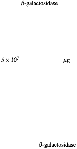
5–25% of all the colonies have the white,
phenotype indicative of a recombinant bacmid
(Luckow et al., 1993). After restreaking to obtain a
clonal isolate containing a recombinant bacmid, plas-
mid DNA is prepared and transfected into insect cells.
119
Once inside insect cells the bacmid replicates as a virus
and yields a stock of recombinant baculovirus.
An advantage of this method is that E. coli is a
familiar host which grows fast and is easy to work
with. Once the target gene has been inserted into the
donor plasmid, one can quickly generate a recombinant
bacmid, obtain a pure clone by restreaking the bacteria,
and transfect insect cells to produce a stock of recom-
binant virus. The method is limited by the requirement
for a specialized transfer vector. In addition, distin-
guishing between colonies containing recombinant and
non-recombinant bacmids can be difficult because the
bacmid is a single-copy plasmid and generates a much
fainter blue color than the familiar multicopy plasmid
cloning vectors.
A version of the bacmid system is distributed by
Life Technologies, Inc. under the trade name Bac-To-
Bac™.
Generation of recombinant viruses
in vitro
Another way of circumventing the limitations of
recombination inside insect cells is to modify the viral
DNA in vitro. Two different methods for constructing
a recombinant virus in vitro have been described.
Recombination mediated by Cre
The bacteriophage P1 encodes a recombinase, Cre,
that mediates recombination between two copies of a
specific site, loxP, in the phage genome to circular-
ize the viral DNA after infection and to allow stable
inheritance when the viral DNA is replicating as a plas-
mid (Sternberg & Hoess, 1983). In vitro, purified Cre
recombinase is sufficient to mediate efficient recom-
bination between any two copies of the 34-bp loxP
recombination site (Abremski et al., 1983; Hoess &
Abremski, 1984). Peakman et al. (1992) have made
use of the Cre-loxP recombination system to generate
recombinant baculoviruses in vitro. This system uses a
derivative of AcMNPV, v Aclox, that has a copy of loxP
in place of the polyhedrin gene (Figure 5). A special-
ized transfer vector, ploxZ, also contains a loxP site and
the AcMNPV polyhedrin promoter followed by a mul-
tiple cloning site into which target genes are cloned.
ploxZ also carries a marker cassette consisting of the
AcMNPV p10 promoter, the E. coli lacZ gene and a
polyadenylation signal (Figure 5). vAclox viral DNA
is mixed with plasmid DNA from ploxZ containing the
target gene and incubated with Cre in vitro. Recombi-
nation between the loxP site on the plasmid and the
loxP site in the viral DNA integrates the whole plas-
mid into the viral genome, generating a recombinant
virus (Figure 5). A second round of recombination can
reverse the reaction and excise the plasmid. Incubation
with Cre therefore generates a mixture of the two input
DNAs and recombinant viral DNAs. The recombina-
tion products are transfected into insect cells, and the
progeny viruses are harvested and plated out to pro-
duce individual plaques. Plaques of recombinant virus
express from the marker cassette and
stain blue with X-gal. Up to 50% of the viruses pro-
duced are recombinant (Peakman et al., 1992). Blue
recombinant plaques are picked, purified by repeated
plaque assays and then amplified to produce a stock of
recombinant virus.
This method very efficiently converts the input viral
DNA into recombinant viruses, yielding as many as
recombinants per of plasmid DNA (Peak-
man et al., 1992). Moreover, plaques of recombinant
virus are easily identified and may constitute up to
50% of the total plaques. An additional advantage
is that no intermediate host is involved. Despite the
efficiency with which this system produces recombi-
nant viruses, a plaque assay is still required to identify
recombinant plaques and at least one round of plaque
purification is required to obtain a clone free of conta-
minating parental virus. Other disadvantages are that
the recombination reaction generates some viruses that
have multiple plasmids inserted, and the recombinant
viruses express in addition to the tar-
get protein. This method also requires a specialized
transfer vector.
Direct cloning into baculovirus DNA
All the methods described above require that the target
gene be cloned into a transfer vector as an intermediate
step in the construction of a recombinant baculovirus. It
is possible to avoid this extra step by directly ligating a
DNA fragment into viral DNA that has been linearized
at a unique restriction site (Ernst et al., 1994). Viral
DNA from Ac-omega, which has a unique site for
the intron-encoded endonuclease I-SceI downstream of
the polyhedrin promoter, is linearized with I-SceI and
dephosphorylated. The target gene is modified so that it
is flanked by I-SceI sites that after restriction generate
ends compatible with the non-palindromic ends of the
linear viral DNA. After ligating the viral DNA and
insert for 60 hours, the ligation products are transfected
into insect cells, and virus harvested a few days later.
120
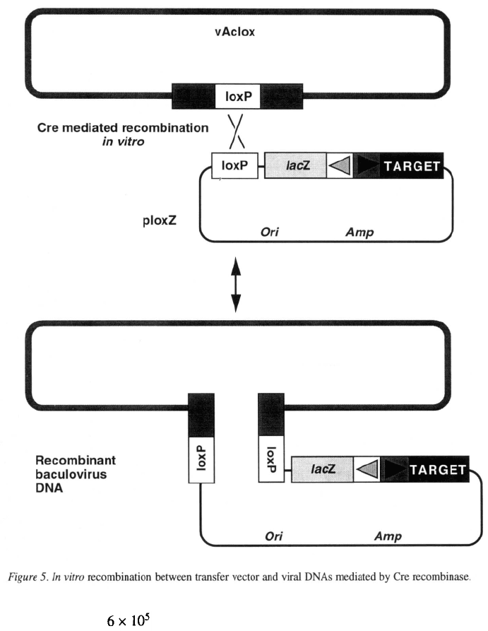
It is possible to obtain more than recombinant
viruses per mg of viral DNA, with the non-recombinant
background estimated to be less than 5% (Ernst et al.,
1994).
Although simple, this direct approach is limited to
the use of a few endonucleases, Bsu36I, SrfI, Sse8387I
and I-SceI, that do not cut the wild-type AcMNPV
genome (Ayres et al., 1994). Furthermore it lacks the
experimental flexibility provided by a transfer vector.
However, direct cloning into the viral DNA may be
suitable for constructing libraries of recombinant bac-
uloviruses.
Appropriate methods for particular applications
Expression of foreign genes
Several of the methods described above can be used
to generate one or more individual recombinant virus-
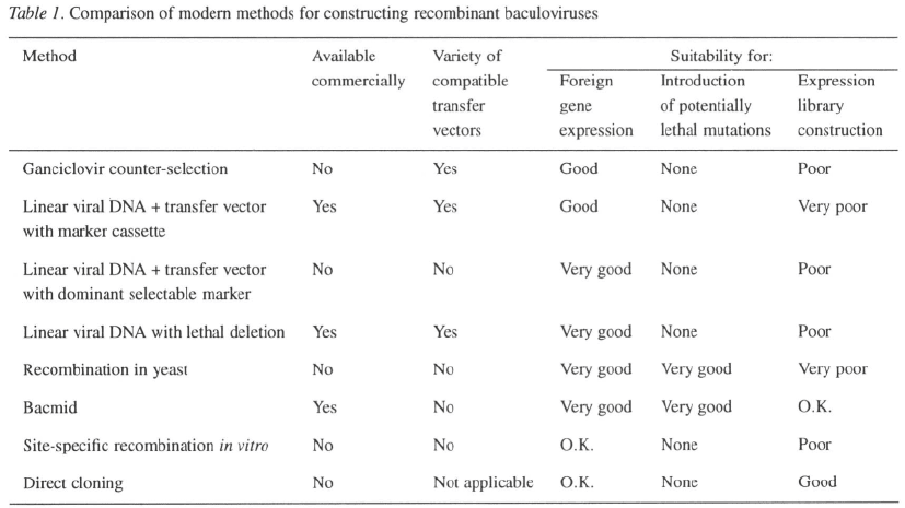
es quickly and efficiently (Table 1). Many users will
find the convenience of a commercially available sys-
tem appealing since this avoids the labor involved in
preparing high-quality linear viral DNA. On the other
hand, the bacmid and yeast systems use a host strain
carrying the baculovirus as an episome and avoid the
need for viral DNA altogether.
Another important factor to consider is the avail-
ability of compatible transfer vectors providing options
121
such as secretion signals, tags for affinity purifica-
tion, multiple promoters, or alternative promoters (for
review see López-Ferber et al., 1995). For convenience
and versatility it is hard to beat the use of linearized
viral DNA with a lethal deletion. The methods that
require a specialized transfer vector are currently lim-
ited to vectors with the standard polyhedrin promoter
without any fusion tags or secretion signals, although
modified versions could easily be constructed.
The methods that use E. coli or yeast as an alter-
native host and yield a pure clone of recombinant
virus without any plaque assays may appeal to the
novice user who frequently has difficulty obtaining
good plaques. However, plaque assays are generally
required later to get the accurate virus titer needed to
optimize expression of the recombinant protein.
Ultimately, the decision over which system to use
will be based on personal preference, experience, and
the availability of local expertise.
Introduction of potentially lethal mutations
In the study of baculovirus biology, it is frequently
desirable to knock out a viral gene in order to under-
stand its function. If the target gene is essential, it will
be impossible to construct such a mutation using meth-
ods that rely on virus replication inside insect cells. The
only methods that allow the introduction of potentially
lethal mutations are those that use an alternative host,
either E. coli or yeast, to propagate the recombinant
viral genome (Table 1).
Expression library construction
For some purposes it may be desirable to convert a
mixed population of target genes, e.g. a pool of mutants
or a cDNA population, into a library of recombinant
baculoviruses. This requires a method that (i) efficient-
ly converts the input population of fragments or trans-
fer vectors into recombinant virus, and (ii) produces
progeny viruses with a sufficiently high proportion of
recombinants that the pool of virus can be used without
any further selection. None of the existing methods are
ideally suited for this purpose (Table 1). Linear viral
DNA with a lethal deletion, and linear viral DNA with
a transfer vector using p35 as a dominant selectable
marker, produce close to 100% recombinants; how-
ever, the low efficiency with which transfer vector
is converted to virus will preclude the use of these
methods for complex pools. Conversely, in vitro site-
specific recombination is very efficient at converting
the input plasmid to virus, but at best yields only 50%
recombinants. The bacmid system should consistent-
ly give about 50% recombinants with good efficiency
hence this method may be acceptable for generating
libraries. Direct cloning into linearized baculovirus
DNA is analogous to the construction of libraries in
phage or plasmid vectors and is probably the existing
method most suitable for constructing libraries.
