Venables J. Introduction to Surface and Thin Film Processes
Подождите немного. Документ загружается.


lasers. Laser ablation, sometimes called pulsed laser deposition (PLD), has typically
been used to deposit ceramic materials, including high temperature superconductors
(Dijkamp et al. 1987, Venkatasan et al. 1988, Morimoto & Shimizu 1995); it produces
very rapid deposition in which whole chunks of material can break off and be depos-
ited during the immense peak powers which typically last for 10–20 ns. A particular
advantage of rapid evaporation is the control of stoichiometry, since the different
species do not have time to segregate to the surface during the evaporation phase.
2.5.3 Molecular beam epitaxy and related methods
The large scale use of such evaporation sources, especially for depositing semiconduc-
tor or metallic multilayers, has become known as molecular beam epitaxy (MBE). This
acroymn has spawned several sub-cases, such as GSMBE (Gas Source MBE), and
MOMBE (Metal–Organic MBE) which is sometimes called CBE (chemical beam
epitaxy). Thus MBE really spans a range of techniques which range from being merely
a fancy name for thermal evaporation, to much more chemically oriented flow process
techniques such as chemical vapor deposition (CVD) and MOCVD which we describe
briefly in section 2.5.5. MBE is covered in several books (e.g. Parker 1985, Tsao 1993,
Glocker & Shah 1995) and in many review articles and ongoing conference series. Only
a few general points can usefully be made here, but this preparation method lies behind
much of the science discussed later in chapters 5–8.
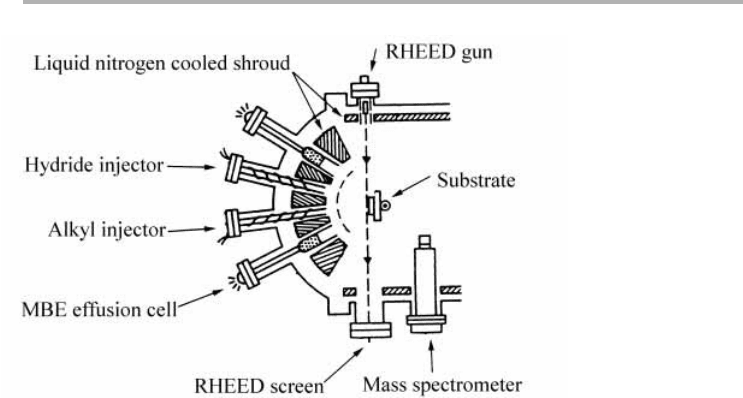
The growth end of such a chamber used for GSMBE or MOMBE is shown sche-
matically in figure 2.8 (Abernathy 1995). The halfspace in front of the substrate is a
bank of effusion cells and various forms of gas injector cells, which transport reacting
chemicals to the substrate. The growth of III–V compounds such as GaAs or AlGaAs
has been pursued using such techniques, starting from metal–alkyl compounds such as
triethylaluminum (TEA) and triethylgallium (TEG) and hydrides such as AsH
3
. The
group five hydrides are injected via cracker cells which convert them catalytically into
As
2
and hydrogen, which then impinge on the substrate to react with the alkyl beam to
produce the growing film (Panish & Sumski 1984, Abernathy 1995).
These cells are surrounded by liquid nitrogen cooled shrouds, which both condense
unwanted evaporants and improve the vacuum in the sample region, which is monitored
by the mass spectrometer. One of the advantages of MBE and related methods is the rel-
ative ease by which in situ diagnostic tools can be incorporated into the vacuum system.
The most widely used are RHEED, shown in figure 2.8 and described in section 3.2.2,
plus various optical techniques, some of which can also work in higher pressure environ-
ments. A compendium of these ‘real-time diagnostics’, particularly in the context of
semiconductor growth, is given by several authors in Glocker & Shah (1995, part D).
2.5.4 Sputtering and ion beam assisted deposition
There are many uses for ions in connection with the production of thin films.
Sputtering using relatively low energy (100 eV–2 keV) ions is often used for cleaning
samples, while higher energy (5–200 keV) ions can be used for doping layers with
2.5 Thin film deposition procedures 57

electrically active impurities. Ions can also be used for deposition, where individual
ions, or charged clusters can be deposited at a range of energies. Clearly, the fact that
the ions are charged allows extra control, and there are various methods by which this
can be done. Directed ion beams from an accelerator form the most obvious possibil-
ity, but plasma and magnetron sources are also widely used (Bunshah 1991, Vossen &
Kern 1991, Shah 1995, Smith 1995).
One of the main claims for IBAD (Ion Beam Assisted Deposition) or IBSD (Ion
Beam Sputter Deposition) is that better quality deposits can be obtained at lower sub-
strate temperatures, thus avoiding large scale interdiffusion which results from high tem-
perature processing. However, the deposited films adhere well to the substrate because
of the localized limited mixing caused by the ion impacts (Itoh 1989, 1995, Marton
1994). This is one example of device engineers trying to reduce the ‘thermal budget’ and
so produce sharper interfaces between dissimilar components. Another possibilty is to
produce clusters in a supersonic expansion source, and to ionize them, controlling their
flight during cluster depostion with applied voltages. This has been termed ICB (Ionized
Cluster Beam) technology by the inventers (Takagi 1986, Takagi & Yamada 1989). Yet
another possibility is to use ion beams to react with the substrate and grow compounds
such as oxides or nitrides at and near surfaces (Herbots et al. 1994).
All of these procedures are inherently complex; for almost all purposes the question
is whether they produce ‘better’ films for particular applications, i.e. whether they give
a large enough improvement over existing methods to justify the considerable invest-
ment involved. Although our aim here is to give an outline description, we will not
pursue models of how of these methods work in any detail; they are all rather specific
to the combination of material and ion beam technique, and whether the processes are
reactive, in the sense of involving (ion) chemistry, or physical, meaning that only colli-
sions and clustering are involved. Nonetheless, ion beam assisted methods are very
58 2 Surfaces in vacuum
Figure 2.8. GSMBE or MOMBE growth chamber containing effusion and cracker cells, and
RHEED diagnostics (after Abernathy 1995, reproduced with permission). See text for
discussion.

widespread and are of great economic importance; sputter deposition in particular has
high throughput for the production of (textured) polycrystalline thin films of a wide
variety of materials (Bunshah 1991, Vossen & Kern 1991, Shah 1995). The language
used to describe such ion based processes (see Greene 1991) necessarily starts from a
similar vantage point to that used to describe thermal evaporation; the latter topic is
treated here in detail in chapter 5.
2.5.5 Chemical vapor deposition techniques
The growth of semiconductor layers for device production is most frequently done by a
variant of CVD (Chemical Vapor Deposition). This is usually implemented as a flow
technique in which the reacting gases pass over a heated substrate, as indicated in figure
2.9(a) for growth of silicon from silane and hydrogen. CVD reactors are either of the hot
wall or cold wall type, and are surrounded by an extended gas handling and pumping
system with rigorous control of the (often highly toxic) gases (Carlsson 1991, Vescan
1995). Because the pressure may be up to an atmosphere in certain cases (APCVD:
Atmospheric Pressure CVD) the growth rate can be high, and UHV technology is not
an absolute necessity; but control of impurities is a dominant problem, as pointed out
in section 2.4.4 (O’Hanlon 1994). Thus UHV-CVD is becoming more widespread, where
the total pressure is , 1 mbar. However, most commercial processing corresponds to
2.5 Thin film deposition procedures 59
Figure 2.9. (a) Schematic diagram of reactions involved in the CVD of silicon from silane
(SiH
4
) and hydrogen (H
2
); (b) typical variation of the growth rate on gas velocity with rate
limiting steps indicated (after Vescan 1995, reproduced with permission).
gas velocitygas velocity
Growth rateGrowth rate
thermodynamics
surface
kinetics
surface
kinetics
gas
transport
gas
transport
(b)
(a)
substrate
reactor wallreactor wall
input
output
H
2
SiH
4
H
2
SiH
4
gas phase reactionsgas phase reactions
diffusion
diffusion
adsorption desorption
surface
processes
surface
processes

LPCVD (Low Pressure CVD) where the pressure is 0.1–1 mbar. MOCVD
(Metal–Organic CVD) or OMVPE (Organo–Metallic Vapor Phase Epitaxy) is also a
widely used technique, as is PECVD (Plasma Enhanced CVD); there are many variants
on this theme (Vossen & Kern 1991).
The question of reaction mechanisms and rate limiting steps in CVD is highly
complex. Under LPCVD conditions diffusion processes in the gas are typically not the
dominant effect, so that at the growth temperatures, kinetic processes on the growing
surface are rate limiting, as indicated in figure 2.9(b) (Vescan 1995). However, we can
see from figure 2.9(a) that all the reactions, in the gas phase and on the surface of the
growing film, are in series and that there is typically very little information on the inter-
mediate states of the reaction. Thus understanding CVD in atomic and molecular terms
is very much an ongoing research project, which we will return to later in section 7.3.
Further reading for chapter 2
Dushman, S. & J. Lafferty (1992) Scientific Foundations of Vacuum Technique (John
Wiley).
Glocker, D.A. & S.I. Shah (Eds) (1995) Handbook of Thin Film Process Technology
(Institute of Physics), especially parts A and B.
Hudson, J.B. (1992) Surface Science: an Introduction (Butterworth-Heinemann) chap-
ters 8 and 9.
Lüth, H. (1993/5) Surfaces and Interfaces of Solid Surfaces (3rd Edn, Springer) chap-
ters 1 and 2.
O’Hanlon, J.F. (1989) A Users Guide to Vacuum Technology (John Wiley).
Matthews, J.W. (Ed.) (1975) Epitaxial Growth, part A (Academic).
Moore, J.H., C.C. Davis & M.A. Coplan (1989) Building Scientific Apparatus (2nd Edn,
Addison-Wesley) chapters 3 and 5.
Roth, A. (1990) Vacuum Technology (3rd Edn, North-Holland).
Smith, D.L. (1995) Thin-Film Deposition: Principles and Practice (McGraw-Hill).
Tsao, J.Y. (1993) Materials Fundamentals of Molecular Beam Epitaxy (Academic).
Problems for chapter 2
These problems are to practice and test ideas about vacuum systems, design problems
and surface preparation techniques.
Problem 2.1. Design of vacuum systems for specific purposes
Use your knowledge of (and appendices on) conductances of standard size tubes, and
the characteristics of vacuum pumps, to suggest (and justify semi-quantitatively)
design choices in the following situations.
60 2 Surfaces in vacuum

(a) Pumping an approximately spherical chamber of diameter 0.5 m. The chamber is
to be let up to air infrequently, and we want to achieve as good a pressure as pos-
sible (, 10
210
mbar).
(b) Pumping a cylindrical chamber of length about 10 m and diameter 0.1 m. The
chamber is to be periodically flooded with rare gases up to about 10
23
mbar pres-
sure, and the important point is to be able to achieve pressures below 10
28
mbar
quickly and economically.
(c) Pumping a state of the art particle accelerator from sections of pipe of length
about 50 m and diameter 0.1 m, with a total length in excess of 50 km (kilometers)
at a pressure of ,10
211
mbar.
Problem 2.2. Design of a Knudsen source for depositing elemental
metal films
A Knudsen source is an evaporation furnace which relies on the establishment of the
vapor pressure above a solid or liquid source material. A small hole in the furnace
above the source material, plus collimating holes in front of the source, allow a beam
of the source material to be directed at the sample. Use your knowledge of vapor
pressures and kinetic theory to design a source which will deposit one monolayer per
minute on a sample held 0.15 m away from the exit of the source, will be uniform on
the sample within 1% for the central 0.01 m diameter, and will not deposit any
material on the sample outside a radius of 0.02 m. Do this in stages, with discussion,
as follows.
(a) Consider the formula R5 nv/4 for the number of atoms hitting unit area per second
of an enclosure, and how this formula applies to a Knudsen source. Derive
the formula by considering the relevant integrations over angles and the
Maxwell–Boltzmann velocity distribution.
(b) Consider the geometry of the design, and the constraints on the uniformity and
area of the deposit. Show that this will limit the size of the hole in the furnace, and
suggest a suitable size for holes in both the furnace and the collimator.
(c) Choose an elemental metal of interest to you, and find out the formula for the
vapor pressure as a function of furnace temperature. Using the relationship
between density n and pressure p for this material, coupled with your design from
part (b) work out the temperature at which the source will have to operate to satisfy
the deposition rate requirement, explaining your assumptions.
(d) If you actually want to design a real source for this material, consider carefully the
materials of which the source can be made, whether you should be using a Knudsen
or some other type of source, and how to power the furnace to achieve sufficient
temperature uniformity, etc.
Note: short descriptions of most possible deposition techniques are given by Smith (1995)
and by Glocker & Shah (1995); some specific designs are in Yates (1997).
Problems for chapter 2 61

Problem 2.3. Some questions on surface preparation and related
techniques
Questions about surface preparation are always very specific to the materials con-
cerned, but here are a few which may be relevant and which spring from the text of this
section.
(a) Why should one either cool the sample slowly through a surface phase transition
(e.g. as in the case of Si(111)), or not anneal the sample above a bulk phase transi-
tion (e.g. in preparing b.c.c. Fe surfaces)?
(b) What is the main reason why Si(111) produces a 231 reconstruction after cleav-
age, when the equilibrium surface structure is the 737?
(c) Device engineers always grow a ‘buffer layer’ on Si(100) before attempting to grow
a device, e.g. by molecular beam epitaxy. Why is this precaution taken, and how
does it improve the quality of the devices grown on such surfaces?
(d) Mass spectrometry shows a range of mass numbers (M/e ratios) for the contents
of the vacuum system, but they don’t seem to be simply related to the molecules,
e.g. O
2
,N
2
,CO,H
2
O, CO
2
which are present. What range of processes are respon-
sible for this discrepancy?
(e) GaAs often evaporates to leave small liquid Ga droplets on the surface. Why does
this happen, and how can it be prevented?
62 2 Surfaces in vacuum

3 Electron-based techniques for
examining surface and thin film
processes
This book presumes that the reader is interested in experimental techniques for exam-
ining surface and thin film processes; however, there are many books devoted to surface
physics and chemistry techniques, some of which are given as further reading at the
end of the chapter. There are even several books which are just about one technique,
such as Pendry (1974) or Clarke (1985), both on low energy electron diffraction
(LEED) in relation to surface crystallography. By the mid-1980s it was already stretch-
ing the limits of the review article format to compare the capabilities of the available
surface and thin film techniques (Werner & Garten 1984).
Since then, the various sub-fields have proliferated, so we cannot be comprehensive,
or give all the latest references here. In section 3.1 we discuss ways of classifying the
large number of techniques which exist, and thereafter the chapter is restricted to tech-
niques based on the use of electron beams. Section 3.2 discusses the most widely used
(elastic scattering) diffraction techniques used for studying surface structure. Section
3.3 discusses forms of electron spectroscopy based on inelastic scattering, which are
used to obtain composition and chemical state information. Individuals can look in
more detail into a particular technique. Students have been asked to present a talk to
the class, followed by questions and discussion; some of the topics considered in this
way are listed, along with selected problems, at the end of the chapter. As examples,
some case studies are given in section 3.3 on Auger electron spectroscopy, in section 3.4
on quantification of AES, and in section 3.5 on the development of secondary and
Auger electron microscopy. Stress is placed on the relationship between microscopy
and analysis: microstructure and microanalysis. The frontier is at nanostructures and
nanometer resolution analysis.
3.1 Classification of surface and microscopy techniques
3.1.1 Surface techniques as scattering experiments
Most physics techniques can be classified as scattering experiments: a particle is inci-
dent on the sample, and another particle is detected after the interaction with the
sample. Surface physics is no exception: we can think of an incident probe and a
response as set out in table 3.1.
63

The probe will be formed from a particular type of particle, and typically will have
a well defined energy E
0
, and often a well defined wave vector k
0
, or equivalently
momentum p
0
5"k
0
. The response can either be the same or a different particle, and,
depending on the detection system, its energy E and/or its wave vector (momentum)
k(p), and maybe other attributes, can be measured. If we understand the nature of the
scattering process, then we can interpret the experiment and deduce the corresponding
characteristics of the sample. It is easy to see from table 3.1 how the number of tech-
niques, and the corresponding acronyms, can be very large, especially once one realizes
that any probe particle can give rise to several responses, and that we may have different
names for essentially the same technique used at different energy, and different wave-
vector (momentum or angular) regimes.
3.1.2 Reasons for surface sensitivity
The next question is ‘which techniques are actually useful for studying surfaces?’. There
are two cases. In the first case, the emergent (i.e. response) particle or the probe parti-
cle has a short mean free path,
l
. This leads to a useful ‘single surface’ technique.
Examples are Auger electron spectroscopy (AES), where the emerging electrons in
the energy range 100–2000 eV have
l
for inelastic scattering in solids typically in the
range 0.5–2.5nm. Using an energy analyzer to measure only those Auger electrons
which have not lost energy, attenuates the signal from subsurface layers strongly. A
cruder form of energy discrimination is used in observing LEED patterns, where
both the incident and the emergent electrons have short mean free paths for energy
loss processes. In SIMS (secondary ion mass spectrometry), the emergent ions have
a very high probability of being neutralized if they do not originate very near the
surface. ICISS (impact collision ion scattering spectroscopy) is surface sensitive
because the incident ion will be neutralized, and thereby not detected, if the probe
particle penetrates the solid. An introduction to ion based techniques can be found
in Feldman & Mayer (1986), in Rivière (1990), and in several chapters contained in
Walls (1990); many other books can be unearthed via the web.
In the second case, the sample has a large surface to volume ratio. This condition
allows us to extract surface information from techniques which are not particularly
surface sensitive. We can perform heat capacity or other thermodynamic measure-
ments, or study structures and dynamics by X-ray or neutron scattering. Here we need
to know the signal from the bulk, and maybe subtract it in a differential measurement.
Much of physical chemistry work on surfaces has been done this way, on powdered, or
exfoliated, samples.
64 3 Electron-based techniques
Table 3.1. Particle scattering techniques
Electrons (E
0
, k
0
, . . . spin s) Electrons (E, k, . . . spin s)
Probe
{
Radiation (
v
0
, k
0
,...polarization)
Response
{
Radiation (
v
, k, . . . polarization)
Atoms (E
0
, k
0
, atomic number Z) Atoms (E, k, atomic number Z9)
Ions (E
0
, k
0
,Z, charge state 6n) Ions (E, k,Z9, charge state 6n9)

3.1.3 Microscopic examination of surfaces
Microscopy can be categorized into fixed beam, scanned beam and scanned probe
techniques. A typical fixed beam technique is transmission electron microscopy
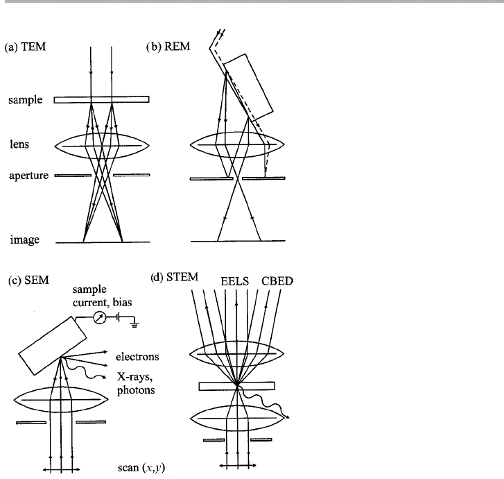
(TEM); the same instrument can often be used for reflection electron microscopy
(REM). These techniques are illustrated schematically in figures 3.1(a) and (b).
Examples will be given later which show that it is not essential to have these instru-
ments operating at UHV in order to produce useful surface related information: UHV
experiments followed by ex situ examination can be very informative, provided the final
samples are not too reactive in air.
The central element of TEM or REM is the objective lens, a cylindrically symmet-
ric strong magnetic field positioned just after the sample. As the equivalent of the first
stage of an amplifier in electronics, this element is critical to the performance of the
microscope, and the aberrations and phase transfer characteristics of this lens deter-
mine both the resolution and contrast that are seen in the image. A particularly useful
feature is the use of the aperture situated in the back focal plane of the objective lens
3.1 Classification of surface and microscopy techniques 65
Figure 3.1. Schematic geometries for TEM, STEM, SEM and REM (after Venables et al.
1987, redrawn with permission).

to select individual diffraction features and to relate these features to the image. In the
case of REM, the image is strongly foreshortened, but this does not mean that the
images are particularly difficult to interpret, as we experience the same sort of image
foreshortening when we look ahead driving a car along the road. Both TEM and REM
have been able to image surface steps, particularly on high atomic number relatively
inert materials such as Au(111) and Pt(111), without requiring that the surfaces were
truly clean.
There are many books on electron microscopy, and TEM in particular has the rep-
utation for being difficult to understand, primarily due to the need for a dynamical
theory of electron diffraction to interpret the images of crystalline samples. For an
overview of the field, see Buseck et al. (1988), which includes a chapter on surfaces
(Yagi 1988); recent surveys of high resolution (HR)-TEM, describing the approach to
atomic resolution at surfaces and interfaces, are given by Smith (1997) and Spence
(1999), both with extensive references. I have attempted a ten-minute sketch of the
various techniques in Venables et al. (1987).
A few groups have converted their instruments to, or constructed instruments for,
UHV operation, and in situ experiments. These instruments, which can also be used for
transmission high energy electron diffraction (THEED) and reflection high energy
electron diffraction (RHEED), have produced highly valuable information on surface
studies, as reviewed, for example, by Yagi (1988, 1989, 1993). More recently low energy
electron microscopy (LEEM) has been developed, which can be combined with LEED,
and is making a major contribution (Bauer, 1994). This instrument can also be used
for photoemission microscopy (PEEM), which has been developed in several different
versions. A specialist form of microscopy with a venerable history is field ion micros-
copy (FIM), which is especially useful for studying individual atomic events such as
diffusion and cluster formation, as discussed by Bassett (1983), Kellogg (1994), Ehrlich
(1991, 1994, 1995, 1997) and Tsong & Chen (1997).
The great virtue of fixed beam techniques is that the information from each picture
element (pixel) is recorded at the same time, in parallel. This leads to relatively rapid
data acquisition, and the ability to study dynamic events, often in real time, e.g. via
video recording. In contrast, data in a scanned beam technique, such as scanning elec-
tron microscopy (SEM) or scanning transmission electron microscopy (STEM), is col-
lected serially, point by point, with the sample placed after the objective lens as
illustrated in figures 3.1(c) and (d).
This configuration means that multiple signals (not just electrons at the probe energy
as in TEM or REM) can be used, which makes the instruments very versatile. It also
makes them ideally adapted for computer control and computer-based data collection,
but can have a corresponding disadvantage: the need to concentrate a very high current
density into a small spot means that not all forms of information can be obtained
rapidly, that there will be substantial signal to noise ratio (SNR) problems, and that the
beam can cause damage to sensitive specimens. Nonetheless SEM and STEM form the
basis of a very useful class of techniques; UHV-SEM has been developed in several
laboratories, including the University of Sussex, and UHV-STEM especially at
Arizona State University. We examine particular developments in section 3.5.
The above techniques have been available for several decades, and have been
66 3 Electron-based techniques
