Tsoulfanidis N. Measurement and detection of radiation
Подождите немного. Документ загружается.

438
MEASUREMENT
AND
DETECITON OF RADIATION
Thickness, mg/crna Thickness, rng/crna
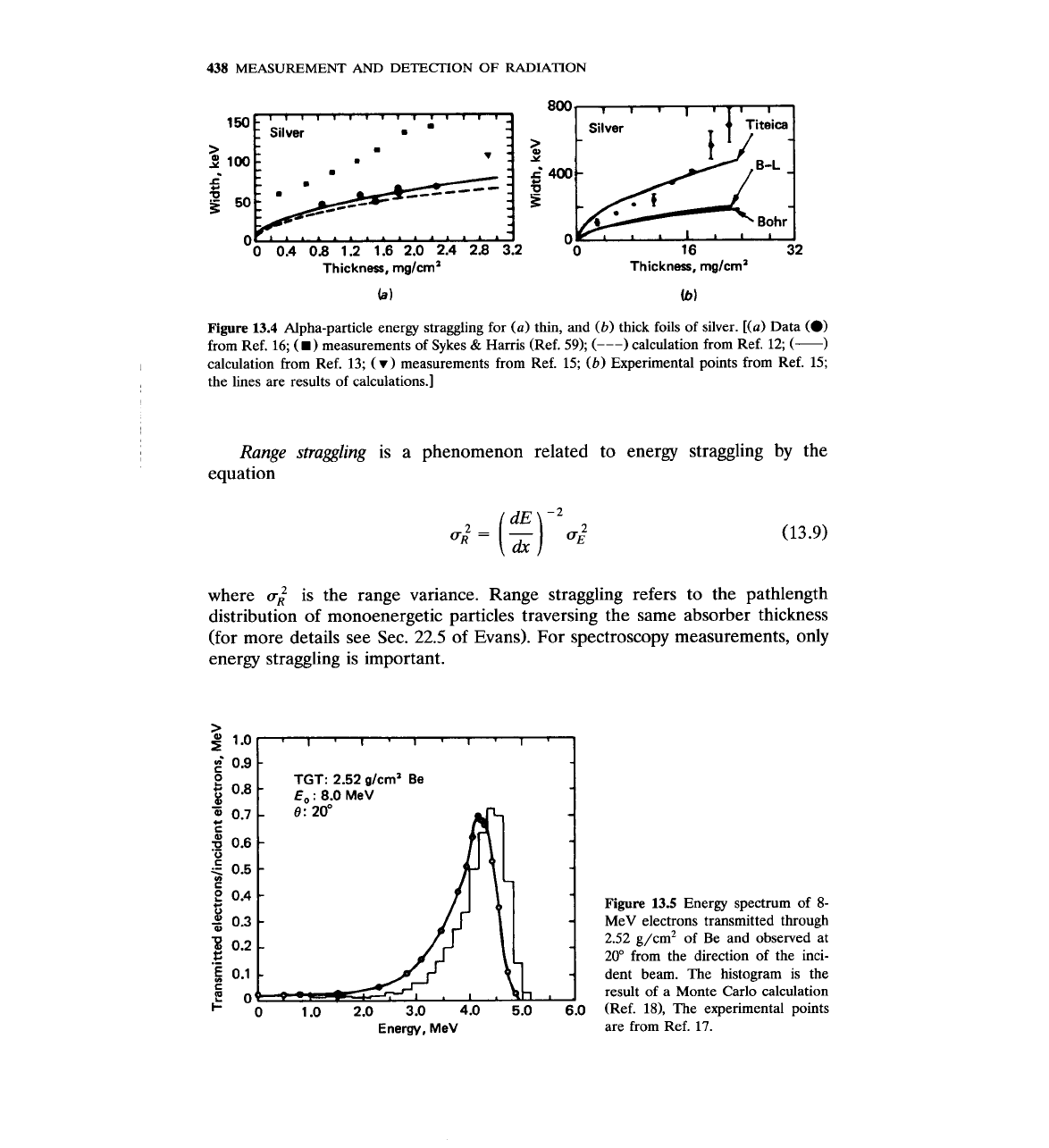
Figure
13.4
Alpha-particle energy straggling for (a) thin, and
(b)
thick foils of silver. [(a) Data
(0)
from Ref.
16;
(.)
measurements of Sykes
&
Harris (Ref. 59);
(---)
calculation from Ref.
12;
(-)
calculation from Ref.
13;
(r)
measurements from Ref. 15;
(b)
Experimental points from Ref. 15;
the lines are results of calculations.]
Range straggling
is a phenomenon related to energy straggling by the
equation
where
u;
is the range variance. Range straggling refers to the pathlength
distribution
of
monoenergetic particles traversing the same absorber thickness
(for more details see Sec.
22.5
of Evans). For spectroscopy measurements, only
energy straggling is important.
>
0
1.0
2.0
3.0
4.0
5.0
6.0
Energy,
MeV
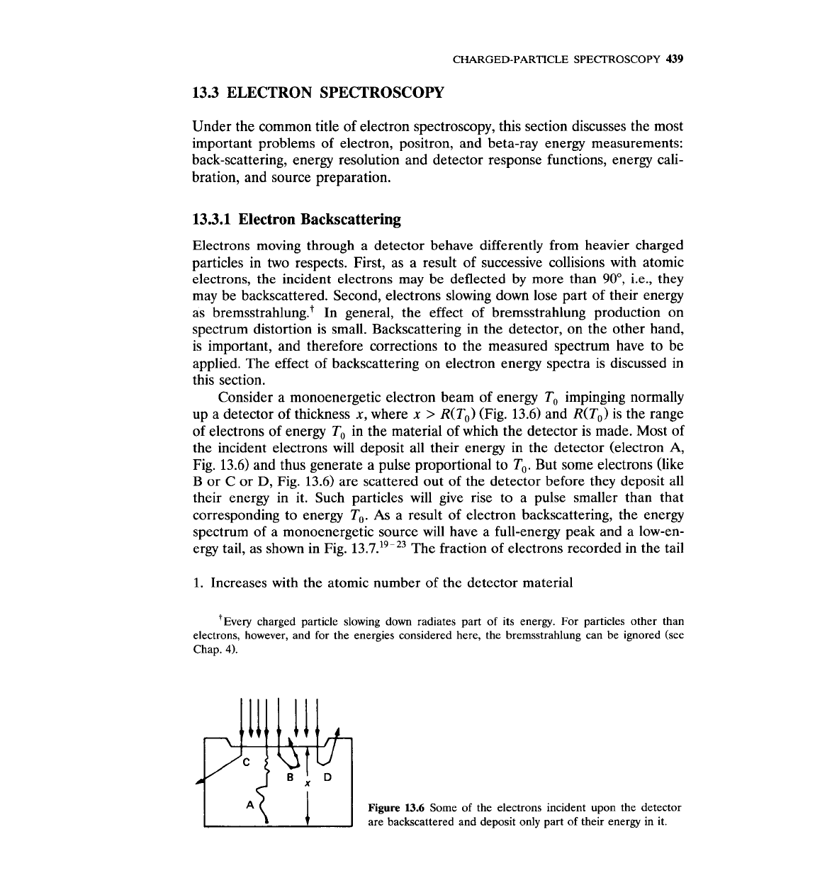
Figure
13.5
Energy spectrum of
8-
MeV electrons transmitted through
2.52 g/cm2 of Be and observed at
20" from the direction of the inci-
dent beam. The histogram is the
result of a Monte Carlo calculation
(Ref.
18),
The experimental points
are from Ref.
17.
CHARGED-PARTICLE
SPECTROSCOPY
439
13.3 ELECTRON SPECTROSCOPY
Under the common title of electron spectroscopy, this section discusses the most
important problems of electron, positron, and beta-ray energy measurements:
back-scattering, energy resolution and detector response functions, energy cali-
bration, and source preparation.
13.3.1 Electron Backscattering
Electrons moving through a detector behave differently from heavier charged
particles in two respects. First, as a result of successive collisions with atomic
electrons, the incident electrons may be deflected by more than
90°,
i.e., they
may be backscattered. Second, electrons slowing down lose part of their energy
as bremsstrahlung.+ In general, the effect of bremsstrahlung production on
spectrum distortion is small. Backscattering in the detector, on the other hand,
is important, and therefore corrections to the measured spectrum have to be
applied. The effect of backscattering on electron energy spectra is discussed in
this section.
Consider a monoenergetic electron beam of energy
To
impinging normally
up a detector of thickness
x,
where
x
>
R(To) (Fig. 13.6) and R(To) is the range
of electrons of energy To in the material of which the detector is made. Most of
the incident electrons will deposit all their energy in the detector (electron
A,
Fig.
13.6)
and thus generate a pulse proportional to To. But some electrons (like
B
or
C
or
D,
Fig. 13.6) are scattered out of the detector before they deposit all
their energy in it. Such particles will give rise to a pulse smaller than that
corresponding to energy
To.
As
a result of electron backscattering, the energy
spectrum of a monoenergetic source will have a full-energy peak and a low-en-
ergy tail, as shown in Fig. 13.7.'~-'~ The fraction of electrons recorded in the tail
1.
Increases with the atomic number of the detector material
v very
charged particle slowing down radiates part of its energy. For particles other than
electrons, however, and for the energies considered here, the bremsstrahlung can be ignored (see
Chap.
4).
Figure
13.6
Some
of
the electrons incident upon the detector
are backscattered and deposit only part of their energy in it.
440
MEASUREMENT AND
DETECTION
OF
RADIATION
A
'I'
:I!
!It
i
I
i?
C
3
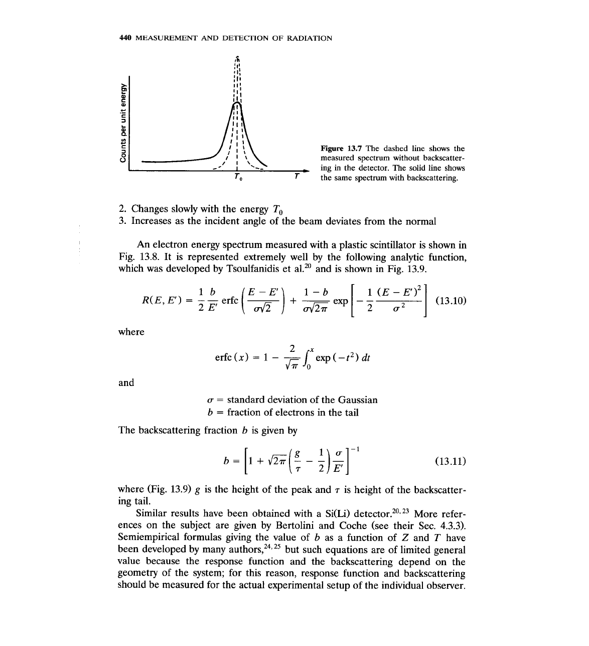
Figure
13.7
The dashed line shows the
measured spectrum without backscatter-
-
I
'--
-
ing
in
the detector. The solid line shows
To
T
-
the
same spectrum with backscattering.
2.
Changes slowly with the energy
To
3. Increases as the incident angle of the beam deviates from the normal
An
electron energy spectrum measured with a plastic scintillator is shown in
Fig. 13.8. It is represented extremely well by the following analytic function,
which was developed by Tsoulfanidis et
a1." and is shown in Fig. 13.9.
lb E
-
E'
1-b
1
(E
-
E')~
R(E,
E')
=
-
-
erfc
-
2
E'
()exp[
u2
]
(13.10)
where
and
u
=
standard deviation of the Gaussian
b
=
fraction of electrons in the tail
The backscattering fraction b is given by
where (Fig. 13.9)
g
is the height of the peak and
7
is height of the backscatter-
ing tail.
Similar results have been obtained with a
Si(Li) detect~r.~~,~~ More refer-
ences on the subject are given by Bertolini and Coche (see their Sec. 4.3.3).
Semiempirical formulas giving the value of b as a function of
Z
and
T
have
been developed by many a~thors,~~,~~ but such equations are of limited general
value because the response function and the backscattering depend on the
geometry of the system; for this reason, response function and backscattering
should be measured for the actual experimental setup of the individual observer.
CHARGED-PARTICLE
SPECTROSCOPY
441
I
1
Channel
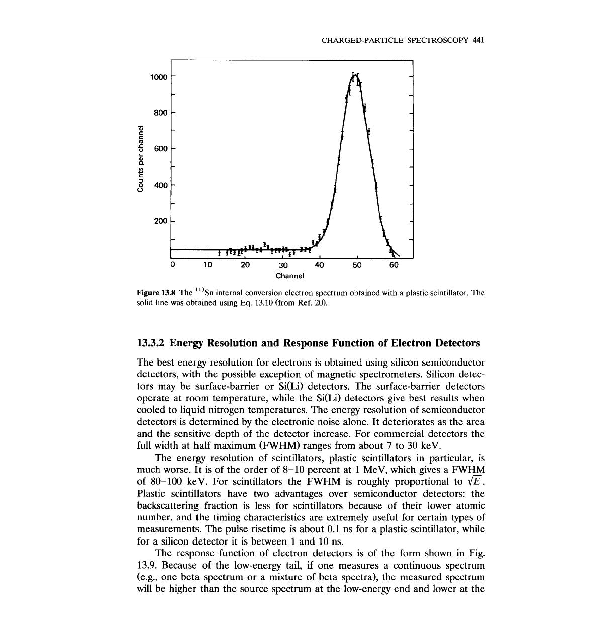
Figure
13.8
The '13sn internal conversion electron spectrum obtained with a plastic scintillator. The
solid line was obtained using
Eq.
13.10
(from Ref.
20).
13.3.2
Energy Resolution and Response Function of Electron Detectors
The best energy resolution for electrons is obtained using silicon semiconductor
detectors, with the possible exception of magnetic spectrometers. Silicon detec-
tors may be surface-barrier or
Si(Li) detectors. The surface-barrier detectors
operate at room temperature, while the Si(Li) detectors give best results when
cooled to liquid nitrogen temperatures. The energy resolution of semiconductor
detectors is determined by the electronic noise alone. It deteriorates as the area
and the sensitive depth of the detector increase. For commercial detectors the
full width at half maximum (FWHM) ranges from about
7
to 30 keV.
The energy resolution of scintillators, plastic scintillators in particular, is
much worse. It is of the order of 8-10 percent at
1
MeV, which gives a FWHM
of 80-100 keV. For scintillators the FWHM is roughly proportional to
I@.
Plastic scintillators have two advantages over semiconductor detectors: the
backscattering fraction is less for scintillators because of their lower atomic
number, and the timing characteristics are extremely useful for certain types of
measurements. The pulse risetime is about 0.1 ns for a plastic scintillator, while
for a silicon detector it is between
1
and 10 ns.
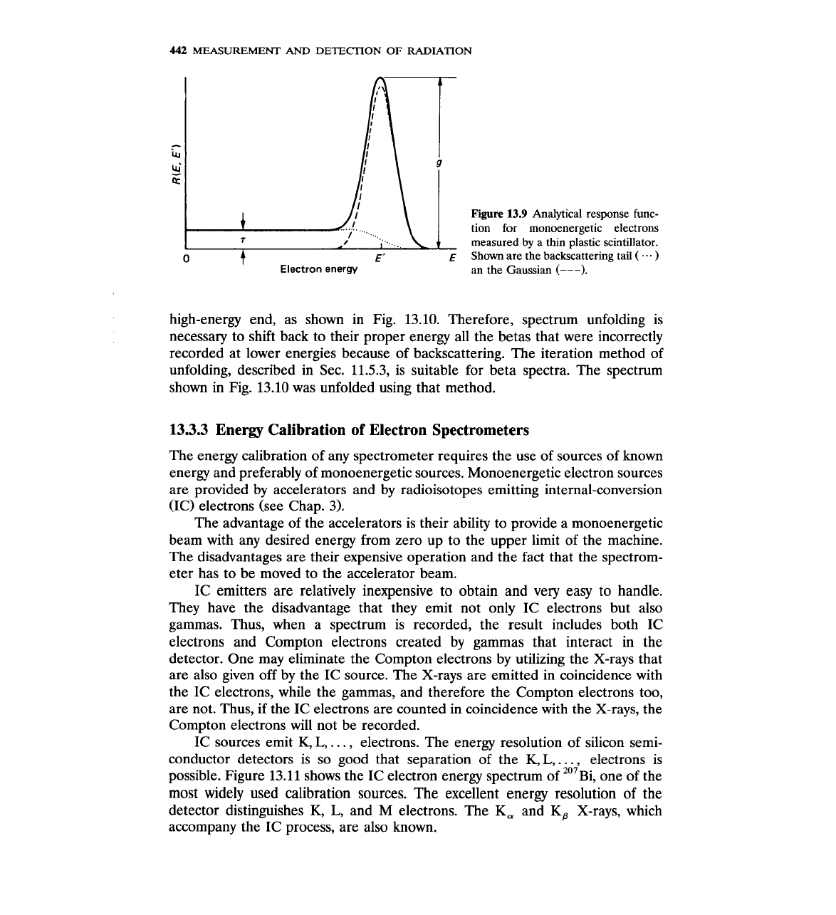
The response function of electron detectors is of the form shown in Fig.
13.9. Because of the low-energy tail, if one measures a continuous spectrum
(e.g., one beta spectrum or a mixture of beta spectra), the measured spectrum
will be higher than the source spectrum at the low-energy end and lower at the
442
MEASUREMENT
AND
DETECTION OF RADIATION
0
t
E'
E
Electron energy
Figure
13.9
Analytical response func-
tion for monoenergetic electrons
measured
by
a thin plastic scintillator.
Shown are the backscattering tail
(
...
)
an the Gaussian
(---).
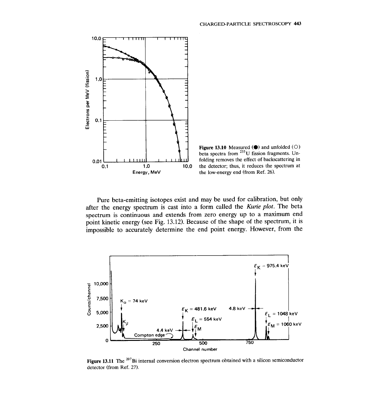
high-energy end, as shown in Fig. 13.10. Therefore, spectrum unfolding is
necessary to shift back to their proper energy all the betas that were incorrectly
recorded at lower energies because of backscattering. The iteration method of
unfolding, described in Sec. 11.5.3, is suitable for beta spectra. The spectrum
shown in Fig. 13.10 was unfolded using that method.
13.3.3
Energy Calibration of Electron Spectrometers
The energy calibration of any spectrometer requires the use of sources of known
energy and preferably of monoenergetic sources. Monoenergetic electron sources
are provided by accelerators and by radioisotopes emitting internal-conversion
(IC) electrons (see Chap.
3).
The advantage of the accelerators is their ability to provide a monoenergetic
beam with any desired energy from zero up to the upper limit of the machine.
The disadvantages are their expensive operation and the fact that the spectrom-
eter has to be moved to the accelerator beam.
IC emitters are relatively inexpensive to obtain and very easy to handle.
They have the disadvantage that they emit not only IC electrons but also
gammas. Thus, when a spectrum is recorded, the result includes both IC
electrons and Compton electrons created by gammas that interact in the
detector. One may eliminate the Compton electrons by utilizing the X-rays that
are also given off by the IC source. The X-rays are emitted in coincidence with
the IC electrons, while the gammas, and therefore the Compton electrons too,
are not. Thus, if the IC electrons are counted in coincidence with the X-rays, the
Compton electrons will not be recorded.
IC sources emit K,
L,
. . .
,
electrons. The energy resolution of silicon semi-
conductor detectors is so good that separation of the K,
L,
. . .
,
electrons is
possible. Figure 13.11 shows the IC electron energy spectrum of 'O'B~, one of the
most widely used calibration sources. The excellent energy resolution of the
detector distinguishes K,
L,
and M electrons. The
K,
and Kp X-rays, which
accompany the IC process, are also known.
CHARGED-PARTICLE
SPECTROSCOPY
443
Figure
13.10
Measured
(0)
and unfolded
(0)
beta spectra from 235~ fission fragments. Un-
0.01
folding removes the effect of backscattering in
0.1
1
.O
10.0
the detector; thus, it reduces the spectrum at
Energy, MeV
the low-energy end (from Ref. 26).
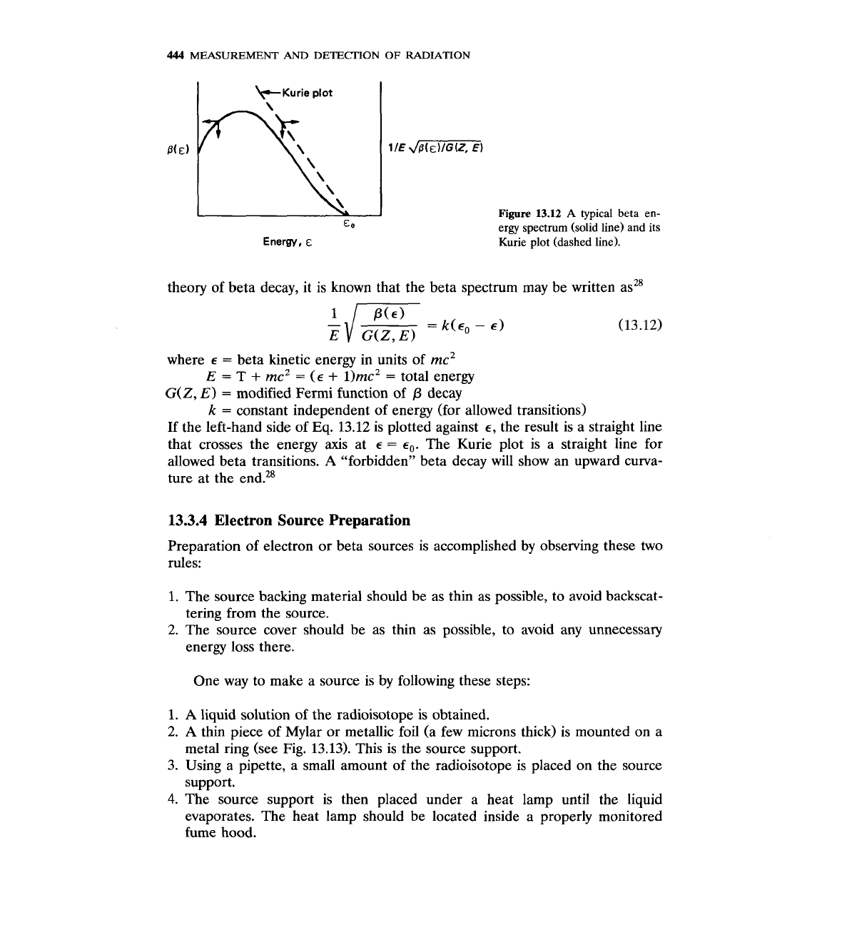
Pure beta-emitting isotopes exist and may be used for calibration, but only
after the energy spectrum is cast into a form called the
Kurie
plot.
The beta
spectrum is continuous and extends from zero energy up to a maximum end
point kinetic energy (see Fig.
13.12).
Because of the shape of the spectrum, it is
impossible to accurately determine the end point energy. However, from the
EL
=
554
keV
=
10$0
keV
4.4
keV
4
Compton edge
'-)
$1;.
0
250 500
7
50
Channel number
Figure
13.11
The '''~i internal conversion electron spectrum obtained with a silicon semiconductor
detector (from Ref. 27).
444
MEASUREMENT
AND
DETECTION
OF
RADIATION
+Kurie
plot
Figure
13.12
A
typical beta en-
ergy spectrum (solid line) and
its
Kurie
plot
(dashed
line).
theory of beta decay, it is known that the beta spectrum may be written asz8
where
E
=
beta kinetic energy in units of mc2
E
=
T
+
meZ
=
(E
+
1)mc2
=
total energy
G(Z,
E)
=
modified Fermi function of
P
decay
k
=
constant independent of energy (for allowed transitions)
If the left-hand side of
Eq.
13.12 is plotted against
E,
the result is a straight line
that crosses the energy axis at
E
=
E,.
The Kurie plot is a straight line for
allowed beta transitions.
A
"forbidden" beta decay will show an upward curva-
ture at the end.%
13.3.4
Electron
Source
Preparation
Preparation of electron or beta sources is accomplished by observing these two
rules:
1.
The source backing material should be as thin as possible, to avoid backscat-
tering from the source.
2. The source cover should be as thin as possible, to avoid any unnecessary
energy loss there.
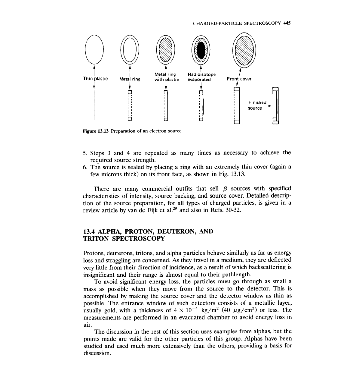
One way to make a source is by following these steps:
1.
A
liquid solution of the radioisotope is obtained.
2.
A
thin piece of Mylar or metallic foil (a few microns thick) is mounted on a
metal ring (see Fig. 13.13). This is the source support.
3. Using a pipette, a small amount of the radioisotope is placed on the source
support.
4.
The source support is then placed under a heat lamp until the liquid
evaporates. The heat lamp should be located inside a properly monitored
fume hood.
CHARGED-PARTICLE
SPECTROSCOPY
445
t
Thin plastic
I
t
Figure
1
I
Metal ring
~etal'
ring
with plastic
3.13
Preparation of an electron source.
~adiokotope
evaporated
4
!
Front cover
5.
Steps 3 and 4 are repeated as many times as necessary to achieve the
required source strength.
6.
The source is sealed by placing a ring with an extremely thin cover (again a
few microns thick) on its front face, as shown in Fig. 13.13.
There are many commercial outfits that sell
P
sources with specified
characteristics of intensity, source backing, and source cover. Detailed descrip-
tion of the source preparation, for all types of charged particles, is given in a
review article by van de
Eijk et al.29 and also in Refs. 30-32.
13.4
ALPHA, PROTON, DEUTERON,
AND
TRITON SPECTROSCOPY
Protons, deuterons, tritons, and alpha particles behave similarly as far as energy
loss and straggling are concerned.
As
they travel in a medium, they are deflected
very little from their direction of incidence, as a result of which backscattering is
insignificant and their range is almost equal to their pathlength.
To avoid significant energy loss, the particles must go through as small a
mass as possible when they move from the source to the detector. This is
accomplished by making the source cover and the detector window as thin as
possible. The entrance window of such detectors consists of a metallic layer,
usually gold, with a thickness of 4
x
lop4
kg/m2 (40 pg/cm2) or less. The
measurements are performed in an evacuated chamber to avoid energy loss in
air.
The discussion in the rest of this section uses examples from alphas, but the
points made are valid for the other particles of this group. Alphas have been
studied and used much more extensively than the others, providing a basis for
discussion.
446
MEASUREMENT
AND
DETECTION
OF
RADIATION
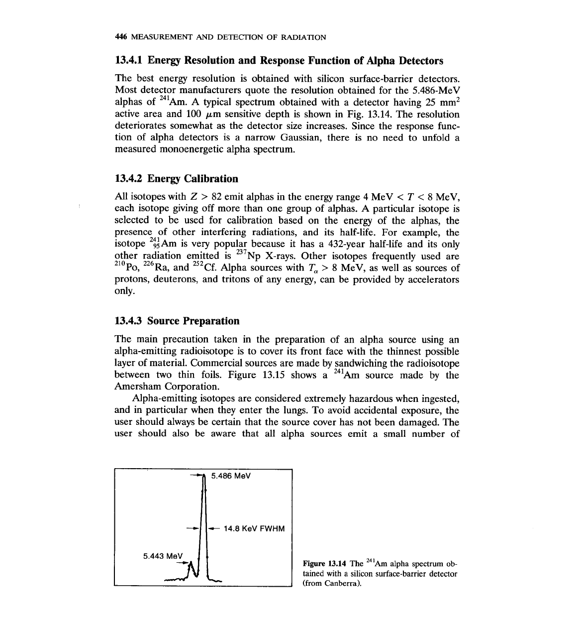
13.4.1 Energy Resolution and Response Function of Alpha Detectors
The best energy resolution is obtained with silicon surface-barrier detectors.
Most detector manufacturers quote the resolution obtained for the 5.486-MeV
alphas of 241Am.
A
typical spectrum obtained with a detector having 25 mm2
active area and 100 pm sensitive depth is shown in Fig. 13.14. The resolution
deteriorates somewhat as the detector size increases. Since the response func-
tion of alpha detectors is a narrow Gaussian, there is no need to unfold a
measured monoenergetic alpha spectrum.
13.4.2 Energy Calibration
All isotopes with
Z
>
82
emit alphas in the energy range 4 MeV
<
T
<
8 MeV,
each isotope giving off more than one group of alphas.
A
particular isotope is
selected to be used for calibration based on the energy of the alphas, the
presence of other interfering radiations, and its half-life. For example, the
isotope '$Am is very popular because it has a 432-year half-life and its only
other radiation emitted is
237~p
X-rays. Other isotopes frequently used are
210~o, 226~a, and 252~f. Alpha sources with
T,
>
8 MeV, as well as sources of
protons, deuterons, and tritons of any energy, can be provided by accelerators
only.
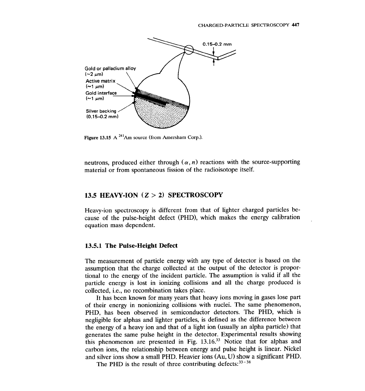
13.4.3 Source Preparation
The main precaution taken in the preparation of an alpha source using an
alpha-emitting radioisotope is to cover its front face with the thinnest possible
layer of material. Commercial sources are made by sandwiching the radioisotope
between two thin foils. Figure 13.15 shows a
241Am source made by the
Amersham Corporation.
Alpha-emitting isotopes are considered extremely hazardous when ingested,
and in particular when they enter the lungs. To avoid accidental exposure, the
user should always be certain that the source cover has not been damaged. The
user should also be aware that all alpha sources emit a small number of
Figure
13.14
The
241~m
alpha spectrum ob-
tained with
a
silicon surface-barrier detector
(from Canberra).
CHARGED-PARTICLE
SPECTROSCOPY
447
i
Gold or palladium alloy
(-2
prn)
\
Active matrix
Figure
13.15
A
"'Am source (from Amersham Corp.).
neutrons, produced either through
(a,
n)
reactions with the source-supporting
material or from spontaneous fission of the radioisotope itself.
13.5
HEAVY-ION
(Z
>
2)
SPECTROSCOPY
Heavy-ion spectroscopy is different from that of lighter charged particles be-
cause of the pulse-height defect (PHD), which makes the energy calibration
equation mass dependent.
13.5.1
The Pulse-Height Defect
The measurement of particle energy with any type of detector is based on the
assumption that the charge collected at the output of the detector is propor-
tional to the energy of the incident particle. The assumption is valid if all the
particle energy is lost in ionizing collisions and all the charge produced is
collected,
i.e., no recombination takes place.
It has been known for many years that heavy ions moving in gases lose part
of their energy in nonionizing collisions with nuclei. The same phenomenon,
PHD, has been observed in semiconductor detectors. The PHD, which is
negligible for alphas and lighter particles, is defined as the difference between
the energy of a heavy ion and that of a light ion (usually an alpha particle) that
generates the same pulse height in the detector. Experimental results showing
this phenomenon are presented in Fig.
13.16.33
Notice that for alphas and
carbon ions, the relationship between energy and pulse height is linear. Nickel
and silver ions show a small PHD. Heavier ions (Au,
U)
show a significant PHD.
The PHD is the result of three contributing
defect^:^^-^^