Tadrous P.J. Diagnostic criteria handbook in histopathology: a surgical pathology vade mecum
Подождите немного. Документ загружается.


JWBK208-04 December 8, 2007 15:59 Char Count= 0
Cut-Up and Reporting Guidelines 39
r
The term ‘glottis’ refers to the true vocal cords and space between them (called the rima glottidis).
r
The laryngeal spaces are based on the natural fibroelastic membrane and lymphatic territories that are
relevant to the local spread of tumour. The fibroelastic membranes (akin to the muscularis mucosae in
other sites) mark the boundary between mucosa and submucosa and penetration of this membrane by
tumours limited to the true cord contraindicates local ablation Rx.
r
The laryngeal regions are for description of tumour subsite and distribution. The lower limit of the
glottic region is not universally agreed but one should state if a glottic tumour extends >1cm below
the true cord (= ‘glottic tumour with subglottic extension’) and distinguish these from tumours that
are entirely subglottic (i.e. no true cord involvement).
Axilla (Lymph node levels)
r
I (low axilla and intramammary): low axillary LN lie below and lateral to the pectoralis minor
r
II (mid axilla) lie superficial and deep to the pectoralis minor
r
III (apical axilla region) lie above and medial to the pectoralis minor. The actual apical nodes lie just
lateral to the 1
st
rib below the axillary vein
r
Other named regions are: internal mammary, infraclavicular (= subclavicular) and supraclavicular
r
All the above count as locoregional LN for breast carcinoma by TNM6 if they are ipsilateral
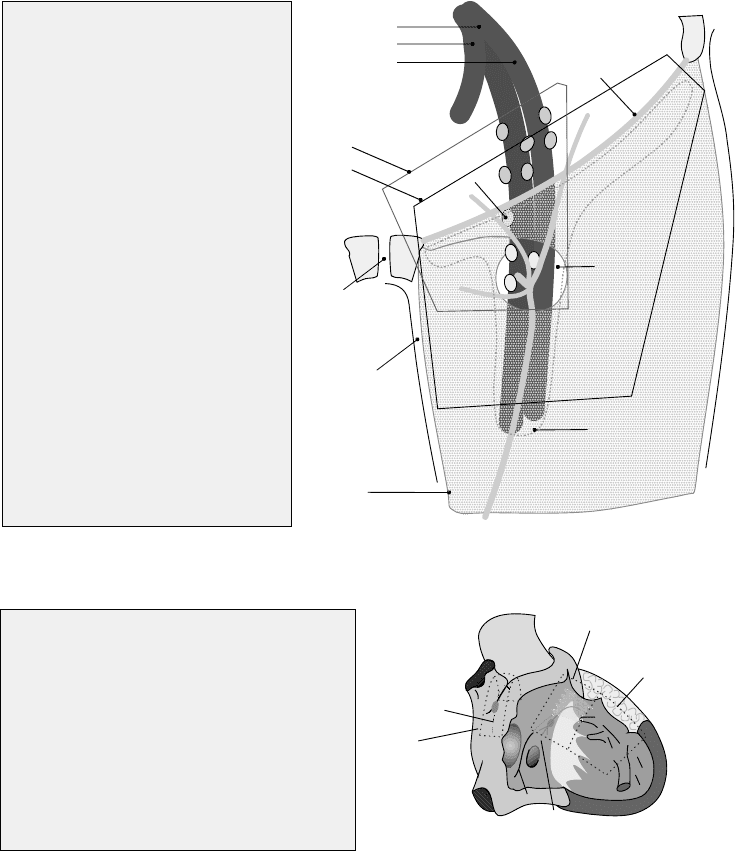
Inguinal Lymphadenectomy
pubic
symphysis
anterior superior
iliac spine
inguinal
ligament
iliac vessels:
common
internal
external
limits of dissection for
inguinal lymphadenectomy:
modified
standard
fossa ovalis (filled with
the loose cribriform fascia)
great saphenous vein
superficial fascia
(= subcutaneous fat
of Camper plus deeper
membranous layer
of Colles)
deep fascia
(fascia lata)
CR node
region of superficial
inguinal nodes
Points to Note:
•
The femoral artery begins as
the continuation of the external iliac
at the midpoint between the anterior
superior iliac spine & pubic symphysis.
•
The fossa ovalis here refers to the
saphenous opening in the fascia lata.
•
The external iliac nodes are shown
in grey.
•
The deep inguinal nodes (in the
fossa ovalis) are shown in yellow and
one is constant, labelled as 'CR Node',
at the medial high point of the femoral
vein (= Cloquet's node of Rosenm
ü
ller).
•
The superficial inguinal nodes lie near
Colles' fascia, are arranged in vertical
& horizontal groups and total upto 25.
The lateral part of the horizontal group
receive lymph from the lower back.
The region marked 'SN' is where the
sentinel nodes of penile
CA
are found.
•
The modified lymphadenectomy field
is that of Catalona and is a saphenous
vein-sparing method with fewer complic-
ations used prophylactically in clinically
node negative patients - but may be
converted to the standard radical
dissection if positive nodes are found
(incl. by the use of frozen section).
SN
FIGURE 4.5 Inguinal lymphadenectomy
Heart: Conducting system
Key (View of heart from the right)
SVC
IVC
aorta
1
7
10
9
8
6
5
4
3
2
position of
crista terminalis
strip to be taken
for SA node
block to be taken
for AV node & His bundle
block to be taken
for left & right
bundle branches
1. Rt. atrial appendage
2. fossa ovalis
3. coronary sinus
4. eustachian valve (of IVC), the continuation of which
(shown as a dotted grey line) overlies the tendon
of Todaro
5. annulus (attachment) of the septal cusp of the tricuspid
6. triangle of Koch (base = 3, sides = 4 & 5)
7. septal cusp of tricuspid
8. membranous septum
9. septal papillary muscle(s)
10. moderator band
FIGURE 4.6 Heart: Conducting system
For the dissection method, block-taking and relevant bibliography, see pp. 389–390.

JWBK208-04 December 8, 2007 15:59 Char Count= 0
Cut-Up and Reporting Guidelines 40
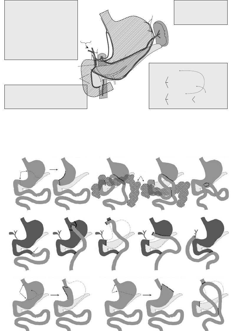
Upper GI Anatomy
Pancreatic Components
1. Head (to the Rt. of the Lt. border of the SMV)
2. Uncinate process of the head
3. Body (between the head & the Lt. border of the aorta )
4. Tail (to the Lt. of the Lt. border of the aorta)
Left gastric
Oeso
phag
eal
bran
c
hes
Short gastric arteries
Right & Left gastroe
p
iploics
Inferior mesenteric vein
(joining the splenic vein)
IVC
Superior mesenteric vein (SMV)
and artery
Hepatic arteries
Portal Vein
Common bile duct
Gastroduodenal artery
(goes behind D1)
Superior and Inferior
pancreaticoduodenal
arteries
Porta Hepatis Contents
1. Portal veins (Lt. & Rt.)
2. Hepatic ducts (Lt. & Rt.)
3. Hepatic arteries (Lt. & Rt.)
4. LNs (draining liver & gallbladder)
Operative Specimens (Pancreas)
Whipple's:
1. head of pancreas
2. distal bile ducts & gallbladder
3. duedenum
4. stomach (distal 2/3)
Pylorus-preserving pancreaticoduodenectomy:
1.- 3. above
Total pancreaticoduodenectomy:
as for subtotal ± spleen
Sub-total pancreaticoduodenectomy:
+ body of pacreas
± stomach
1.- 3. above
Distal pancreatectomy:
body & tail of pancreas ± spleen
Branches of the Coeliac Axis
1. Lt. gastric artery
2. Splenic
3. Hepatic
Lt. gastroepiploic
pancreatic branches
short gastrics
Rt. & Lt. hepatic
gastroduodenal
Rt. gastric
Rt. gastroepiploic
superior
pancreaticoduodenal
Main Vessels Labelled
FIGURE 4.7 Upper GI anatomy
r
The minor duodenal papilla, when present, is 2cm proximal to the ampulla of Vater ± a little anterior
r
The Lt gastric artery is most commonly involved with gastric ulcers; the gastroduodenal with DU
Surgical alterations
Normal
Normal
Proximal gastrectomy with gastric tube reconstruction
Proximal gastrectomy with direct oesophageal anastomosis
Total gastrectomy
CUT HERE
CUT HERE
CUT HERE
CUT HERE
Billroth 1 (Hofmeister anastomosis) Billroth 2
Colon
Gastric bypass
Total gastrectomy
Distal gastrectomy
Choledochojejunostomy
Billroth 2 Anterior gastrojejunostomy
FIGURE 4.8 Surgical alterations
r
The Billroth distal gastrectomies have many modifications e.g. Hofmeister anastomoses only allow a
patent lumen near the greater curve part of the cut stomach while Polya anastomoses allow lumenal
continuity over the whole length (a Polya Billroth 2 also tends to have a retrocolic anastomosis).
r
The middle row shows some procedures for which a Roux-en-Y jejunal anastomosis is used.
r
A pyloroplasty is often performed with proximal gastrectomies.
r
A jejunal pouch (e.g. ‘U’-shaped) may be used to restore continuity after proximal or total gastrectomy.
r
A segment of colon or jejunum may be used to reconstruct after a high oesophagogastrectomy.

JWBK208-04 December 8, 2007 15:59 Char Count= 0
Cut-Up and Reporting Guidelines 41
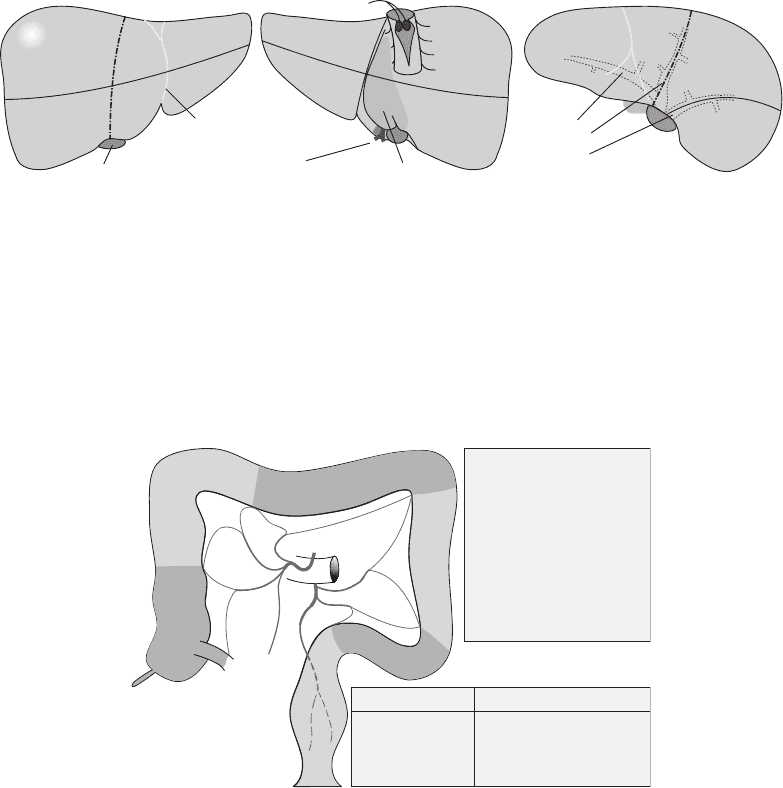
Liver (Segmental anatomy)
2
3
4a
4b
5
8
1
2
3
6
7
falciform ligament
gallbladder
caudate lobe
hepatic artery &
portal vein branches
IVC
hepatic veins
ANTERIOR VIEW
POSTERIOR VIEW SUPERIOR VIEW
hepatic veins (projected)
4a
8
7
2
1
left
middle
right
IVC
FIGURE 4.9 Liver (Segmental anatomy)
r
Although Rt and Lt lobes are defined by the falciform ligament; segments are vascular compartments
useful for planning surgery and are precisely defined for any individual by pre-op radiology.
r
The bold broken line in the figure joins the left side of the groove for the IVC to the mid-part of the
gallbladder fossa and divides the liver into left and right hemilivers.
r
The falciform ligament divides the left hemiliver into segments 4 and 2+3.
r
The caudate lobe is segment 1.
Lower GI Anatomy (blood supply)
SMA
IMA
1
2
3
4
5
6
ILEO
RIGHT
MIDDLE
LEFT
SIGMOID
SUPERIOR
RECTAL
Key to Arteries:
1. ileocolic
2. right colic
3. middle colic
4. left colic
5. sigmoid
6. superior rectal
NB: The IMA arises
≈
4 cm
above the aortic bifurcation
& may be small.
Operation Vascular Ties
Rt. hemicolectomy ileocolic, Rt. colic ± mid colic
Transverse colectomy midcolic ± Lt. colic
Lt. hemicolectomy
Lt. colic, sigmoid, ± superior rectal
Anterior resection/APR superior rectal
FIGURE 4.10 Lower GI anatomy (blood supply)
r
Some lesions (e.g. carcinoids) may be categorised according to the part of the gut they arise in using
embryonic terminology as: foregut, midgut, hindgut, tailgut. The first 3 correspond to those parts of
the adult GIT supplied by the coeliac axis, SMA and IMA.
r
The tailgut (a diverticulum of the hindgut under the tail-root of some animals) may persist to form a
tailgut cyst (multilocular retrocaecal hamartoma with smooth muscle in the wall and a variable lining
from columnar to transitional/squamous) or adenocarcinoma.
Kidney (Hilar structures)
r
The renal vein is (usu.) the most anterior structure (anterior to the renal arteries and pelvis/ureter).
r
The renal artery usu. has three branches at the pelvis – two go anterior to the pelvis/ureter and 1
posterior.
r
The renal sinus is that part of the pelvis lateral to the hilar plane, its fat envelops the collecting system
and abuts the renal cortex without an intervening fibrous capsule.
r
Invasion (incl. microscopic invasion) of the sinus fat by carcinoma constitutes stage pT3a.
r
The sinus contains muscular tributaries of the renal vein, gross invasion of which by carcinoma consti-
tutes stage pT3b (renal vein involvement).
Urinary Bladder
r
The superior posterior aspect is covered by serosa.
r
The urethral aspect is tapered.

JWBK208-04 December 8, 2007 15:59 Char Count= 0
Cut-Up and Reporting Guidelines 42
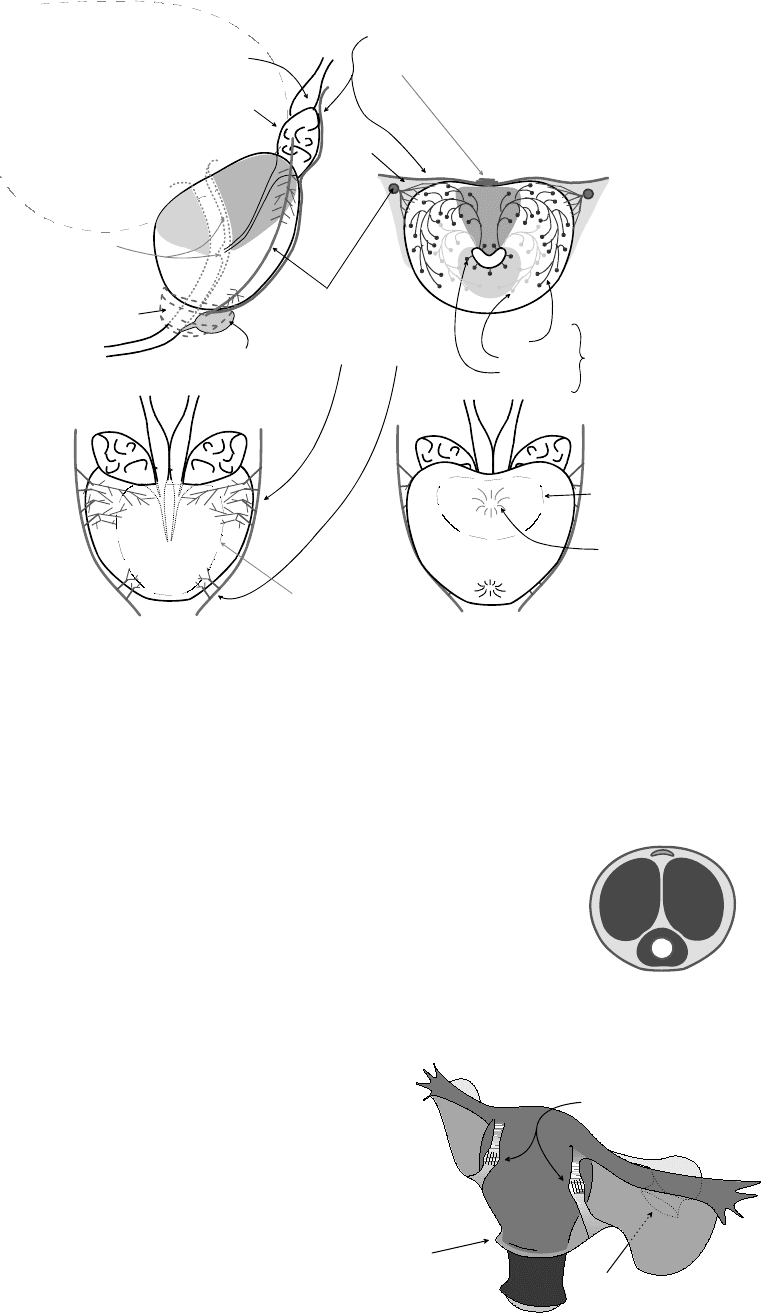
Prostate
Bladder
TZ
CZ
PZ PZ
B
A S E
A P E X
Internal
urethral
meatus
External
urethral
sphincter
Urethral crest and
Verumontanum
(with openings for
ejaculatory ducts
& prostatic utricle)
Ampulla of vas deferens
Seminal vesicle
Fascia of Denonvilliers with its
median (longitudinal smooth) muscular
thickening - ! not rectal muscularis propria
Adipose
Neurovascular
bundle (capsular invasion
is most common in the
regions of nerve penetration
at the superior and inferior
pedicles)
Glands
Main
Submucosal
Periurethral
(= mucosal)
CZ
TZ
P O S T E R I O R
Apex
Base
Posterior View
Attachment of
bladder base
Internal
urethral
meatus
Anterior View
Attachment of
rectum
Oblique
transverse
slice of the
superior half
Cowper's
bulbourethral
gland
FIGURE 4.11 Prostrate
r
For TNM staging the prostate may be divided into Lt. and Rt. halves (=‘lobes’) ignoring the anatomical
middle lobe as a distinct entity; involvement of the external sphincter urethrae (striated muscle) or
bladder neck (smooth muscle) = pT4.
r
pT2b (tumour restricted to >1/2 of 1 lobe) is interpreted as >50% volume by some (in which case it
is vanishingly rare) or tumour linear dimension >1/2 of the lobe’s linear dimension by others.
Penis
Ventral
Dorsal
FIGURE 4.12 Penis
r
Specimens may be distorted by disease and fixation artefact.
r
The most dorsal aspect contains the dorsal vein overlying the paired corpora
cavernosa, the latter enclosed in a thick fibrous coat – the tunica albuginea
corporum cavernosum.
r
The urethral is most ventral and surrounded by the corpus spongiosum
invested in a thinner tunica albuginea corporis spongiosi.
r
The spongiosum overshoots the cavernosa and expands to cover the distal
cavernosa in the form the conical glans penis. Its oblique basal rim, the
corona glandis, overhangs the shaft and the groove be-
tween the shaft and corona is the coronal sulcus. The
obliquity is such as to allow the dorsal corona to be more
proximal than the ventral part.
r
The overlying skin contains a sub-dermal dartos smooth
muscle layer with Buck’s vascular fibroelastic fascia sep-
arating this muscle from the tunica albuginea of the 3
corpora.
1.
(anterior
peritoneal
reflection)
2.
(posterior ovarian pedicle)
3. (anterior round ligaments
with forcep marks)
UTERUS ANTEROLATERAL VIEW
FIGURE 4.13 Uterus
Uterus (Orientation)
r
There are three main landmarks, some of which may not
be present in every specimen):
1. the peritoneal reflection is higher anteriorly
(bladder) than posteriorly (pouch of Douglas)

JWBK208-04 December 8, 2007 15:59 Char Count= 0
Cut-Up and Reporting Guidelines 43
2. the ovarian pedicles (or ovaries if present) are on the posterior surface of the broad ligament
3. the round ligaments are anterior (these usu. have forceps marks imprinted on them as the
surgeon grasps the uterus and pulls it forward by the round ligaments during an abdominal
hysterectomy).
Specimen Handling and Reporting Guidelines
Interpretation of LN Metastases w.r.t. the TNM6 Classification
r
Tumour in a LN is considered LN +ve whether it got there by metastasis or direct extension
r
Tumour deposit without residual LN structure is LN +ve if it has a smooth outline consistent with the
form of a LN (else = vascular +/ discontinuous tumour spread). In the UK this is controversial (2007)
and some (e.g. GI pathologists) advocate continued use of the TNM5 rule based on size not contour
(viz. a deposit >3mm
in a lymph draining region is a LN metastasis, if ≤3mm = discontinuous 1
◦
tumour spread and is included in the pT category). See also Neck Dissection below
r
Tumour nodules in a LN that are >2mm in max. dimension are LN +ve with no further qualification
r
If ≤2mm but >0.2mm =‘micrometastasis’: use the ‘mi’ qualifier, e.g. pN1(mi), only if there are none
bigger
r
If ≤0.2mm then = ‘isolated tumour cells’ (ITC) and staged as pN0(i+)
1
r
The ITC concept is also valid for the pM classification in bone marrow (pM0(i+))
r
That ITC results in a pN0 stage is controversial in the UK (2007) so some advocate adding a comment
like ‘this would previously have been called pN +ve’
r
In some pN stages, size of the LN metastasis is important (not the size of the whole LN)
r
LN mets distant to the regional lymph drainage fields are included in the pM category (not pN)
r
Use of pTNM implies a resection sufficient to assess the most extreme scores
Central Nervous System (CNS)
r
Record volume of sample and nature of procedure (incl. whether CUSA was used)
r
Decalcify bony fragments and consider samples for glutaraldehyde (EM) or freezing (molecular genet-
ics) if CPC suggests an unusual tumour.
r
Lobectomy: give anatomical description, bread-slice coronally (5mm intervals), record presence/extent
of necrosis and comment if there is margin / meningeal involvement
r
Blocks: embed all if small/stereotactic biopsies else take sufficient blocks to document tumour hetero-
geneity, invasion across anatomical boundaries and margins
r
Margin status should not be described as ‘clear’ for most 1
◦
CNS tumours except for intact meningioma
resections
r
Reporting: give tumour type and WHO grade (there is no TNM staging because CNS tumours infiltrate
diffusely and rarely metastasise; however, describe if anatomical boundaries are breached: into bone,
through pia into subarachnoid space, meningioma invading brain or dura/bone, etc.)
r
Brain smears: place a tiny piece of fresh sample (≤1mm
3
) onto a slide, squash gently with a second
slide on top until it forms a thin layer then sweep the top slide over the first to produce the smear. Fix
in alcohol and stain with H&E (cytoplasmic detail) or 1% toluidine blue (good nuclear detail)
Muscle Biopsy (Non-neoplastic)
r
1–2 pieces received fresh (sutured to a stick while in situ +/ laid out on card to avoid contraction and
to preserve orientation). Site is usu. deltoid or quadriceps.
r
≈1mm
3
taken to glutaraldehyde for EM (longitudinal sections)
r
≈0.5 × 0.5 × 2cm snap frozen (unfixed) for transverse sections for LM (! avoid ice crystal artefacts)
r
Routine stains: H&E, PAS, myosin ATPase (various pH), NADH, Sudan Black, Gomori trichrome
r
Others as indicated: dystrophins, spectrin, myosins, phosphorylase, cytochrome oxidase, etc.
See table 13.3 on p. 188 for interpretation of these stains.
Peripheral Nerve Biopsy (Non-neoplastic)
(See p. 389, under ‘Nerves for ? Neuropathy’.)
1
or pN0(mol+) if tumour cells were detected by non-morphological methods e.g. flow cytometry
JWBK208-04 December 8, 2007 15:59 Char Count= 0
Cut-Up and Reporting Guidelines 44
Eye
r
The terms ‘inner’ [or ‘internal’] and ‘outer’ [or ‘external’] refer to ‘towards the centrepoint of the globe’
(the centre of the vitreous) and ‘towards the surface of the globe’ respectively.
r
Measure globe (length and ), pupil and optic nerve length.
r
The vortex veins are harvested and embedded (longitudinally) with the cut end (margin) of the optic
nerve in a standard cassette.
r
After transillumination via the cornea (to mark the base of any tumour) or via the optic nerve (to
examine the cornea/iris), the globe is cut into three slices (‘calottes’) where the cuts pass 1mm into the
corneal limbus – either horizontal (for macular lesions), vertical (for glaucoma / cataract assessment)
or oblique – with the middle calotte containing the pupil, iris, lens, optic nerve and main bulk of any
tumour. Each calotte is embedded in a separate deep cassette. However, this is not always optimum
with some tumours so the specimen should be cut so as to yield the following information:
for iris melanoma radial extent (in terms of clock hours) and extension (ciliary body, sclera
and extraocular) is important for staging
for ciliary body and choroidal melanoma the largest basal diameter (in mm or optic disc di-
ameters) and maximum height of the tumour (in mm or diopters) are important for T stage.
Penetration of the sclera with extraocular extension and vascular invasion (including vortex
vein) are important but penetration of Bruch’s membrane is irrelevant (unlike with retinoblas-
toma)
for retinoblastoma tumour height, distance from tumour base to fovea and optic nerve, retinal
detachment, subretinal deposits (penetration of Bruch’s membrane) ± choroidal / scleral in-
vasion or orbital (extra-ocular) spread, optic nerve invasion (Magramm gade 1 = prelaminar,
2 =upto and including the lamina cribrosa, 3 = beyond it, 4 =optic nerve margin involved),
deposits in the vitreous (seeding) should be distinguished from true multifocality (>1 deposit
within the retina), vitreous extension to contact the lens, deposits in ciliary body / iris / anterior
chamber
for exenterations, measure skin margins, ink superior/inferior halves and nasolacrimal duct
margin (sample this margin and the optic nerve margin), remove and decal any bone, then
bread slice in vertical sections 1cm thick (first and last cuts should just avoid the eyeball).
Temporal Artery Biopsy for ? Arteritis
r
Cut the biopsy into 2mm long transverse slices.
r
Embed all the slices en face and request levels and EVG.
Head and Neck (SCC and Other Types of Carcinoma)
Clinical data
The surgeon should:
orientate the specimen (and pin-out a neck dissection, marking the major LN levels)
give the clinical TNM stage and state whether prior therapy was given
specify the side and op-type e.g. neck dissection – selective / radical / modified radical.
Biopsies
r
State if dysplasia or vascular invasion is present in addition to tumour characteristics.
Main resection (general)
r
Some advocate decalcification of specimens with soft tissue in situ before dissection.
r
Measure (mm) max. depth and of tumour. Tumour depth is measured from the lumenal surface
(extrapolated to the level of the surrounding, uninvolved, surface if there is ulceration).
r
Take 1 block per cm of tumour (plus margins, uninvolved mucosa, thyroid, etc.)
r
Tumour type, extent, and subsite (e.g. supraglottic, glottic, transglottic or infraglottic – see Figure 4.4)
r
Grade (by worst area) – well / moderately / poorly diff
r
Pattern of invasive front: cohesive/non-cohesive (non-cohesive = strands <15 cells wide/single cells)
r
Distance to margins (mucosal and soft tissue) <1mm = ‘involved’, >5mm = ‘clear’, else = ‘close’
r
Invasion of:
vessels
nerves (esp. if beyond the invasive front of the main tumour)
bone (distinguish erosion of cortex from invasion of intertrabecular spaces)
r
Dysplasia (presence, degree, distance to margins)

JWBK208-04 December 8, 2007 15:59 Char Count= 0
Cut-Up and Reporting Guidelines 45
Laryngectomy
r
If it will not disrupt the tumour, a longitudinal posterior midline incision may be made to assess tumour
extent – otherwise, leave the specimen intact.
r
Some advocate sharp dissection to remove the hyoid (to avoid decalcifying the whole specimen) with
examination of the soft tissue thus exposed and histology of the hyoid only if that tissue is involved.
The UK National Dataset advises whole decalcification followed by serial 5mm transverse slicing.
r
Assess the caudal and anterior (tongue base) submucosal margins (as well as mucosal) by sagittal
sections.
r
State if (and where) tumour has perforated the cartilages with extralaryngeal soft tissue spread.
r
State if tumour has crossed the midline.
r
State what other structures are present (e.g. parathyroids) and examine as appropriate.
Maxilla, mandible, sinuses and other specimens
See Slootweg, 2005
Neck dissection
r
For each level state: n˜o. of LN present and n˜o. of LN involved and whether extracapsular spread (if
extracapsular spread approaches a surface of the specimen, ink that surface and block such as to estimate
distance of / involvement by tumour).
r
Measure size of largest metastasis (for TNM).
r
For matted LN state the max. dimension of the mass and its level.
r
Ignore TNM6 rule and call a LN metastasis if there is a deposit in a lymph drainage field (call it
‘discontinuous spread’ only if it is <10mm from the 1
◦
and there is no residual LN structure).
r
State if vascular invasion is seen (= poor prognostic).
r
Take blocks of other structures involved by tumour (salivary gland, sternomastoid, jugular vein).
Oesophageal Tumours (incl. Siewert & Stein Type 1 Adeno
CA)
Biopsies
r
Type of cancer and depth of invasion (state if submucosal tissue is present and whether it is invaded)
r
Presence of dysplasia or the metaplasia of Barrett’s oesophagus
Resections macro
r
Length of oesophagus (state if pinned prior to fixation) and any stomach
r
Tumour site relative to GOJ / highest peritoneal reflection: if >50% above = oesophageal (if exactly
at the junction then = oesophageal if SCC/SmCC/undiff
CA but = gastric if adenocarcinoma)
r
Tumour max. , length, distance to margins and whether polypoidal or not
r
Always take both proximal and distal margins (circumferential en face method preferred)
r
Location of any LN found (e.g. coeliac vs. other perigastric)
Resections micro
r
Tumour type and grade (well, moderately or poorly diff – by worst area)
r
Depth (incl. level) of invasion (TNM) and if CRM is involved (i.e. <1mm clearance) [NB: CRM
clearance can’t be assessed if the surgeon has dissected LN from it separately]
r
Serosal surface involvement (NB: only the gastric part will have a serosa)
r
Vascular invasion
r
Total n˜o. of any LNs found as well as n˜o involved by tumour
r
Presence of dysplasia or the metaplasia of Barrett’s oesophagus
Gastric Carcinoma (incl. Siewert and Stein Types2&3Adeno
CA)
r
Specify gastrectomy type: total, partial (proximal/distal), oesophago; measure length of components
r
Normally open along anterior greater curve (but avoid tumour and any gastrojejunostomy line)
r
Tumour: size (max. ), distance to margins, Borrmann type (polypoid / fungating / ulcerated vs. diffuse)
r
Tumour site incl. Siewert and Stein type for peri-cardiac adenocarcinoma (Type:
◦
1
midpoint of tumour
is 1–5cm above the anatomical cardia;
◦
2
from 1cm above to 2cm below the cardia;
◦
3
2–5cm below
the cardia). ‘Anatomical cardia’ = the most proximal limit of the gastric rugae
2
(or highest peritoneal
reflection).
r
≥3 blocks from tumour + all LN; take margin blocks parallel to the margin (esp. at oesophageal end)
+ take adventitial CRM if tumour goes near the lower oesophagus
2
NB: Siewert & Stein defined these types of adenocarcinomas using endoscopic and radiological localisation

JWBK208-04 December 8, 2007 15:59 Char Count= 0
Cut-Up and Reporting Guidelines 46
r
Type (adeno/other), class (by Laur´en), grade (poor/other – by worst area), vasc. invasion (lymph and
blood)
r
Depth:
3
into subserosa = pT2b, penetration of serosa = pT3 (not invading other organs, else = pT4)
Liver (for 1
◦
HCC or Metastatic Carcinoma)
Macro
r
Measure, incise and drain gallbladder, weigh; capsule intact? any adherent structures?
r
Serial 1cm slicing in ‘CT-plane’ if practicable; distance of tumour(s) to all margins
r
Note number, size and site of all tumours ± satellites (= nodules <10mm surrounding a larger
tumour) ± vascular invasion
r
Tumour blocks: HCC →≥3; mets → 1 from each met (more if neoadjuvant Rx given)
Micro
r
Tumour type (and grade if HCC), margin and LN status, background liver
r
PT vascular invasion requires the identification of a residual lumen and endothelium
Exocrine Pancreas, Bile Ducts and Ampulla of Vater
Macro
r
Ink, open duodenum; some recommend slicing the pancreas prior to fixation
r
Note named structures e.g. vessels
r
Record specimen dimensions and lengths (incl. ducts)
r
Tumour size and site (head / body / tail / whole / Ampulla / bile ducts) incl. multicentricity
r
Spread to other structures: LN, portal vein
r
Distance to margins: duodenal +/ gastric, ducts, pancreatic (transection, retroperitoneal, medial
4
)
r
LN groups: bile duct, infrapyloric (regional to head tumours only), pancreaticoduodenal (ante-
rior/posterior), pancreatic (superior/inferior), pancreatic tail and splenic hilum (not regional to head
tumours), coeliac (not regional to body/tail tumours); [Note: tumours <2cm
are unlikely to be node
+ve]
Micro
r
Tumour type and grade (according to worst area – see p. 176)
r
Margins (as above) or anterior pancreatic capsule invasion / extension
r
Local spread (pT stage is that of TNM5):
perineural
(pT3): duodenum, Ampulla, bile ducts, peripancreatic fat
(pT4): stomach, colon, spleen, large named vessels (portal, superior mesenteric or common
hepatic)
Appendix (for Carcinoid)
r
Tumour subtype, size (</>2cm), clearance at base, LN involvement, invasion of mesoappendix or onto
serosal surface, vascular invasion and perineural invasion may all influence further Mx
Colorectal Carcinoma
r
Macro (rectal only):
relation to anterior peritoneal reflection (and distance to dentate line if APR)
grade the CRM plane as:
◦
1
mesorectal (bulky, no coning, no defect deeper than 5mm);
◦
2
intramesorectal (coning to levators, moderate bulk but irregular, no muscularis propria
visible);
◦
3
muscularis propria (not bulky / deep defects with muscularis visible)
r
A margin is considered ‘involved’ if tumour (main, separate deposit or in a LN or any vessel) extends
to 1mm from it
r
If the tumour is a poorly diff / infiltrative subtype then proximal and distal margins / doughnuts should
be sampled for histology (even if the tumour is >3cm clear macroscopically)
r
Extratumoural deposit ≥3mm = ‘LN +ve’ (in TNM6, only if form and smooth outline are consistent
with a LN else = ‘vascular invasive deposit’ V1 if micro only / V2 if visible macroscopically)
3
‘early gastric cancer’ = pT1 (limited to mucosa/submucosa) i.e. not muscle invasive (pT2a) or beyond
4
The medial pancreatic margin (= ‘SMV margin’) is the fatty tissue deep to the head – it is the plane dissected off the superior
mesenteric vein / portal vein and superior mesenteric artery

JWBK208-04 December 8, 2007 15:59 Char Count= 0
Cut-Up and Reporting Guidelines 47
r
Serosal involvement does NOT imply incomplete excision (because it is not a surgical margin)
r
pT3 or greater: measure the greatest extent of extramural extension
r
pT4 = serosal breach (pT4b) or adjacent organ spread (pT4a) including colon to colon (by way of the
serosa – so may be pT4 a and b) or direct spread into the levator striated muscles
r
pM1 includes non-regional LN mets (e.g. para-aortic)
5
r
Extramural vasc. invasion = tumour cells in endothelial-lined space with blood or muscle
r
Use ypTNM if previous chemo/radioRx and decide if response to neoadjuvant Rx is ‘complete’,
‘marked’ (tumour epithelium identified with difficulty) or ‘not-marked’
r
High risk subgroup of Dukes B CA has any of the following: CRM involvement
perforation
serosal surface involvement
extramural venous invasion
Lymphoma
Cases where referral to a haematopathologist is advisable:
r
All paediatric lymphomas, any TCL, ? Burkitt’s or lymphomas assoc
d
with immunosuppression
r
Where the distinction between reactive and neoplastic is blurred (e.g. in AITCL or some FCL)
r
Any which have atypical morphology or immuno or where distinction between types is difficult (e.g.
lymphocyte depletion HL vs. ALCL or CLL vs. MCL)
Specimen handling and reporting
r
Specify site and use a standard fixation regimen (helps standardise immuno results).
r
Remember to look for hilar LN and splenuncula in splenic resections.
r
Cut thin sections and use WHO classification (use immuno, even if H&E seems obvious).
r
An H&E, PAS, retic, Giemsa on one block may help (± Perls’ for trephines).
r
For trephine involvement specify type of lymphoma present.
Lung (for Cancer)
Biopsies
r
Confirm if it is cancer and, if so, whether it is small cell / non-small cell type.
r
Only attempt to sub-classify further if definite features are present (‘report only within your limits’).
Resections
r
Inflate lung with formalin (if bronchus is blocked you can inject the parenchyma with a needle).
r
Sample bronchial and vasc. margins then slice longitudinally down bronchi or multiple transverse cuts
or sagittally.
r
Describe site according to bronchopulmonary segmental anatomy
r
Blocks: ≥3 of tumour (block all if ≤2cm ) and ≥3 of uninvolved lung and others
r
LN: describe separately sent LN according to their station
N1 nodes are in the lung
N2 nodes are outside the hilar pleural envelope e.g. peribronchial or mediastinal.
r
Use the WHO classification for tumours (see Chapter 8: Respiratory and Mediastinum)
r
Visceral pleural invasion is defined as tumour breaching the superficial elastic layer (the pleura has a
characteristic double elastic layer which differentiates it from fibrosis .
.
. use EVG)
Features to note re staging
r
Largest tumour (= pT3 if >3cm)
r
Distance of invasive tumour to the bronchial resection margin
r
Whether the tumour is in the main bronchus or a lobar / more distal bronchus (if in main bronchus,
distance from carina determines stage: pT2 if >2cm, pT3 ≤2cm and pT4 if invading the carina itself
– may need CPC to stage)
r
Is there >1 tumour in the same lobe as the 1
◦
(= pT4) or a deposit in a different lobe (= pM1)?
r
Is there invasion of pleura (= pT2), mediastinal pleura (= pT3) or mediastinal soft tissues (= pT4) ?
r
Does any assoc
d
‘atelectasis’ or obstructive pneumonia extends to the hilar region (= pT2) or involve
the entire lung (= pT3) ?
r
Is there a cytology positive pleural effusion (= pT4) ?
5
Peritoneal deposits away from the 1
◦
site are classified as ‘pM1 PER’ but make sure your oncologist knows that this does not imply
distant haemoatogenous/lymphatic mets

JWBK208-04 December 8, 2007 15:59 Char Count= 0
Cut-Up and Reporting Guidelines 48
Lung (for Non-neoplastic Occupational Disorders / Pneumoconioses)
r
Examine externally for pleural plaques, hilar LN, etc. and note size and n˜o of lesions.
r
Inflate the lung with formalin via the main bronchus to fix.
r
Slice sagittally at 1–2cm intervals and prepare one wholemount (Gough-Wentworth) section [protocol
for this can be found in Gibbs and Attanoos (2000) – see Bibliography]
r
Asbestos body concentration can vary 10-fold depending on site so, for each lung, sample at least 4
(non-tumour) blocks: apex of upper and lower lobe + base of lower lobe + main bronchus with LN.
r
Include pleura in some of the blocks and sample any focal lesions.
r
Describe and quantitate 1
◦
and 2
◦
dust foci, interstitial fibrosis and emphysema (for grading systems
see p. 85, and Gibbs and Attanoos, 2000)
Skin Cancers
r
Specify type, site and 3-dimensions and ink the margins
r
Fusiform / ellipse excision: bread slice,
6
embed max. 1 slice per cassette for malignant melanoma or 2
slices for SCC / BCC; embed polar ends with your cut face down (to be sectioned first)
r
Re-excision Bx: treat such as to be able to comment on completeness of excision of the scar
r
Measure peripheral and deep margins to nearest whole mm if >1mm clearance, else state ‘<1mm’ (if
involved record as ‘0 mm’); state measures for invasive and in situ components in SCC/melanoma
r
Comment on neural / vascular invasion
BCC
r
Type (nodular (NST), superficial multicentric, inflammatory / morphoeic, micronodular, etc.)
r
Moderately / severely atypical squamous component present
r
pT stage ∝ diameter and whether invades muscle / cartilage / bone (= pT4)
SCC
r
Type (NST, spindle, acantholytic, verrucous, desmoplastic, pseudoangiosarcomatous, etc.)
r
Grade: well / mod. / poorly / undifferentiated (see p. 284)
r
Clark level (but some clinicians may find this confusing so local protocols may not require it)
r
Tumour thickness (from beneath keratin layer)
r
pT stage as for BCC
Malignant melanoma
r
Type (lentigo maligna, superficial spreading, nodular, acral lentiginous, desmoplastic, neurotropic,
naevoid [i.e. symmetrical, cytologically homogeneous and minimal stromal reaction], etc.)
r
Macro: nodule present, pigment, border
r
Growth phase (VGP / HGP) – defined in Chapter 20, p. 291
r
If VGP → mitotic count per 10hpf and tumour lymphocytic response (brisk, non-brisk, absent)
r
Ulceration (incl. of ulceration)
r
Presence of:
A microsatellite, defined by T.J. Harrist et al. (1984), is a
nest (>0.05mm
) in the reticular dermis, subcutis or vessels
beneath the main tumour and separate from it [by ≥0.5mm
according to the RCPath Dataset].
a) regression
b) microsatellites
c) a benign naevus component
r
Clark level and Breslow thickness to 2 d.p. of a mm (from beneath the keratin layer or ulcer base
ignoring appendigeal sheath extension of melanoma); NB: Clark level only adds significant prognostic
information for thin melanomas (i.e.Breslowthickness ≤0.75 mm) that usu. havean excellentprognosis.
r
pT stage ∝ depth and ulceration
Frozen Sections and Mohs’ Micrographic Surgery
r
FS is not usu. done on skin cancer (and should normally be avoided in melanoma)
r
Mohs’ surgery (the pathology is usu. done by the surgeon – a dermatologist trained in pathology):
usu. done for BCC / SCC which is recurrent, ill-defined or in an anatomically sensitive site
is an iterative surgical procedure involving mapped excision of a lesion with FS of all margins
and re-excision of involved areas at the same sitting.
Renal Biopsies (Medical/Transplant)
r
Put Bx in saline immediately after taking it and look for glomeruli with a dissecting microscope.
r
Under the dissecting microscope, divide into three pieces: 1mm of glomerulus-containing cortex for
EM (glutaraldehyde), 2mm of glomerulus-containing cortex for IF (place in special transport medium
6
For a very large circular Bx, cruciate-type sampling is acceptable; for a very small Bx (<5mm) with no clear lesion, embedding
whole is acceptable.
