Tadrous P.J. Diagnostic criteria handbook in histopathology: a surgical pathology vade mecum
Подождите немного. Документ загружается.


JWBK208-05 December 8, 2007 21:25 Char Count= 0
Paediatric and Placental 59
Teratoma (Non-gonadal)
r
Commonest tumour in the neonate (i.e. within the first month of life)
r
Usu. midline (sacrococcygeal, neck, palate, mediastinum, CNS), also: kidney, orbit, etc.
r
Any tissue type may be present but if a well-formed axial skeleton is seen consider fetus-in-fetu
r
Classification: benign, malignant, immature (degree of immaturity may ∝ aggressiveness)
r
Immaturity assessed by discordance of tissue maturity with the age of the child (e.g. neuroblasts in a
neonate are OK – but not in a 3-year-old)
For gonadal teratomas see pp.225–226 and pp.251–252.
Pineoblastoma, Haemangioblastoma, Medulloblastoma and other CNS Tumours
See p. 184, p. 187 and p. 188.
Retinoblastoma
See p. 199 and p. 44.
Pleuropulmonary Blastoma
See p. 90.
Pancreatoblastoma
See p. 177 and p. 179 (under the d/dg of acinar cell tumour and NETs).
Neuroblastoma (NB) / Ganglioneuroblastoma / Ganglioneuroma
r
Poorly diff NB: sheets of cells with hyperchromatic nuclei
lobular architecture with thin fibrous septa
fibrillar stroma (unmyelinated axons), S100 +ve cells in stroma
r
Increasing differentiation
Homer-Wright pseudo-rosettes (no lumen, just fibrils) in 30% of cases
Some cells with more open nuclei, prom. nucleoli and more cytoplasm (i.e. obvious neurob-
lasts)
ganglion cells
more ganglion cells = ganglioneuroblastoma
all ganglion cells = ganglioneuroma
lots of trk expression (NGF receptor)
r
Cytogenetics of NB: 1p del., N-myc amplification (dmin / HSR)
r
Immuno of NB: CD99 −ve
2
, NB84 +ve, CD57, NSE, CgA, PGP 9.5 +ve
r
Favourable prognostic features in NB are: ganglion cells, tumour giant cells, low mitotic count
(<10/10hpf) and focal calcification
Hepatoblastoma
r
Large solitary variegated liver mass and ↑serum AFP (± hCG)
r
±Necrosis ++(due to pre-op chemoRx in resected specimens, but not seen on 1
◦
diagnostic core Bx)
r
Stroma: fibrous septa, osteoid, primitive spindly mesenchyme (other elements are rare e.g. muscle)
r
Epithelia (the first two are required for diagnosis):
embryonal liver: cords ± rosettes of small blue fusiform cells
fetal liver: small cells (vesicular nucleus, single nucleolus) in microtrabeculae ± bile plugs
± macrotrabecular liver (adult HCC-like)
± anaplastic small cell (SBRCT-like)
± glandular elements (intestinal-like)
± teratoid elements: ectopic epithelia (squamous, endocrine, etc.)
r
Immuno: AFP, focal hCG, CAM5.2, CK7/19, CEA and S100 are all +ve in various elements
Nephroblastoma (Wilms’ tumour)
r
Classically triphasic (but biphasic / monophasic variants occur):
◦
1
stroma (spindle cells)
◦
2
blastema(dyscohesivesmall blue round cells) occurs in 3 organoid patterns (helps to distinguish
monophasic blastematous subtype from d/dg lymphoma):
2
CD99 +vitiy virtually excludes neuroblastoma. Wilms’ is also CD99 −ve

JWBK208-05 December 8, 2007 21:25 Char Count= 0
Paediatric and Placental 60
serpentine (anastomosing cords in myxoid stroma)
nodular (rounded nests)
basaloid (nests outlined by cuboidal / columnar cells)
◦
3
epithelium (cells with elongated, moulded and wedge/carrot-shaped nuclei form glomeruloid
and tubular structures – !d/dg entrapped renal parenchyma.
r
Variant: predominantly cystic forms of Wilms’ occur
r
Stroma can be highly differentiated with ‘heterologous elements’: myxoid, fibroblastic, leiomyomatoid,
adipose and chondroid (=‘teratoid Wilms’ if prominent) and skeletal muscle or even rhabdomyoblastic
elements (! d/dg RMS)
r
Anaplasia (focal or diffuse) must have ALL of the following:
nuclear enlargement ≥ 3 fold cf. adjacent nuclei of same lineage
hyperchromasia of enlarged nuclei
enlarged or multipolar mitoses (each limb of an X or Y-shaped figure must be as large as a
normal metaphase)
So defined, it heralds poor prognosis and chemoresistance.
r
Stage according to the NWTS system (I–V), simplified this is:
I confined to kidney, capsule intact, renal sinus
3
veins and lymphatics not involved
II spread beyond capsule / hilar plane but surgical margins clear
III extends to surgical margins or residual local disease present
IV haematogenous mets
V bilateral renal tumours
r
Cytogenetics: WT1 and WT2 on chromosome 11
r
Immuno: epithelium (CK, WT1 +ve), blastema (desmin, WT1 +ve; vimentin, CK ±ve), CD99 −ve
Clear Cell Sarcoma of the Kidney
r
Highly aggressive, resistant to Wilms’ therapy, needs specific chemoRx
r
Monotonous array of pale cells with nuclei having finely dispersed chromatin and small nucleoli
r
Characteristic vasculature of parallel capillaries connected by transverse arcades
r
Variant: sclerosing (may entrap nephrons and be confused for d/dg blastematous Wilms’)
Placenta
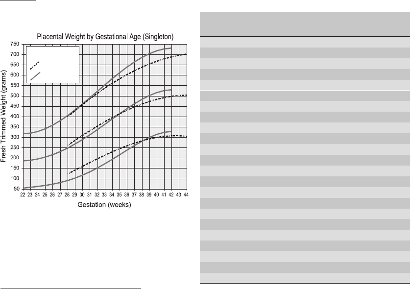
Placental Growth (Weights)
4
TABLE 5.1
Gestation Singleton Weight Twin Fetus:Placenta
in weeks Mean (b) in g Factor Ratio
19 160 [synth] 1.33 -
20 165 [synth] 1.32 -
21 175 [synth] 1.32 -
22 186.11 1.35 -
23 189.37 1.46 -
24 195.52 1.57 2.9
25 204.56 1.67 3.1
26 216.44 1.76 3.3
27 231.05 1.82 3.5
28 248.22 1.87 4.9
29 267.73 1.9 5.1
30 289.3 1.91 5.3
31 312.58 1.92 5.4
32 337.2 1.91 5.6
33 362.7 1.89 5.8
34 388.58 1.87 6.0
35 414.27 1.84 6.2
36 439.15 1.82 6.4
37 462.55 1.79 6.5
38 483.75 1.76 6.6
39 501.94 1.73 6.7
40 516.29 1.7 6.8
41 525.9 1.68 7.0
Dataset a
Dataset b
Mean ± 2SD
FIGURE 5.2 Placental weight by gestational age
(Singleton)
r
For twin placental mean weight multiply the
mean ‘Singleton Weight’ by the ‘Twin Fac-
tor’ in the table (10
th
centile is ≈ 0.75 × the
3
The renal sinus is that part of the pelvis lateral to the hilar plane
4
The sources for datasets ‘a’ and ‘b’ in these charts and table are detailed in the Bibliography (q.v.)
JWBK208-05 December 8, 2007 21:25 Char Count= 0
Paediatric and Placental 61
twin mean and 90
th
centile is ≈ 1.25 × the twin mean). [NB: The table entries marked ‘synth’ are not
true singleton weights but were synthesised for the purpose of calculating twin weights]
r
IUGR = birth weight below 2.5kg at term or below the 3
rd
centile for gestational age [‘small for
gestational age’ = birth weight below the 10
th
centile]
r
The normal fetal:placental weight ratio goes from 1:1 at 14/40 to ≈ 7:1 at 40–44/40
Large placenta (e.g. >90
th
centile)
r
Maternal conditions (e.g. DM / anaemia)
r
Fetal hypertrophy syndromes (e.g. Beckwith-Wiedemann syndrome)
r
Congenital syphilis
r
Fetal oedema (e.g. hydrops fetalis, less-severe anaemia, nephrotic syndrome)
Small placenta (e.g. <10
th
centile)
r
IUGR (from various causes)
r
PET
r
↑ Perivillous fibrin deposition
r
Villitis of unknown aetiology
r
Trisomies
Infarcts
r
May occupy upto 3–5% of a term placenta without functional consequence (but >10–30% is sig.)
r
Only diagnose on histology because the macro d/dg includes fibrin, MPVFD and chorangioma
r
Early (red infarct):
↑ maternal blood in intervillous space with central thrombus Signs of
villous vessels congested and dilated
ischaemia
syncytial trophoblast necrosis and eosinophilia
r
Later:
intervillous space collapses
perivillous fibrin deposition
villous vessels collapse → ghost-like villi
adjacent villi show ischaemia: ↑ syncytial knots, small terminal villi + big stem villi, empty
intervillous space
Syncytial Knots
r
Occur in 10–30% of villi in term placentae (! exclude d/dg thick section artefact)
r
They are a sign of placental hyperplasia / maturity and are ↑ in:
maternal DM / HT / PET / anaemia
adjacent to infarcts
r
They are uncommon <32 weeks but may be seen in mid-trimester abortions
Maternal HT incl. Pre-Eclamptic Toxaemia (PET) / Eclampsia
r
Villous BM thickening
r
Incomplete transformation of maternal vessels (i.e. they are lined by endothelium rather than tro-
phoblast) esp. in the intra-myometrial portion of the arteries
r
Maternal vessel acute atherosis (fibrinoid change/necrosis + intimal foamy lipid cells) ± thromboses
r
Haematomas (placental / retroplacental)
r
The resulting ischaemia / uteroplacental insufficiency → accelerated maturation (↑ vasculosyncytial
membranes, ↑ knots, small villi, etc.) → small-for-dates baby (placental weight may be ↑ or ↓)
Maternal Diabetes
r
Delayed maturation and big placenta (and baby)
r
Fetal ‘infarcts’ (actually villous vessel thromboses vide infra under Villous Changes Following IUD)
r
Villous BM thickening and ↑ knots
r
↑ Nucleated RBCs in villous vessels (i.e. nucleated RBCs easily found at term)
Maternal Antiphospholipid Antibody Syndrome (± SLE)
r
Clin.: a cause of habitual abortion (= recurrent miscarriage =≥3 consecutive pregnancy losses) incl.
1
st
trimester abortion; this is preventable by antithrombotic cover (see p. 351)
r
In treated patients with uncomplicated live births the placenta usu. shows no specific abnormality
JWBK208-05 December 8, 2007 21:25 Char Count= 0
Paediatric and Placental 62
r
Early POC may show incomplete vascular transformat
n
(normal endothelium in endometrial vessels)
r
Later fetal deaths may show MPVFD (but only in a minority of cases)
r
One study showed decidual small vessel thromboses (non-inflam
y
± recanalisation) and infarcts →
ischaemia (hypovascular/fibrotic villi and ↑ syncytial knots) but with ↓ vasculosyncytial membranes
Retroplacental Haematoma
r
Clin.: may be the cause of fetal distress, sudden hypoxic death or pre-term labour
r
Macro: haematomas are excavating (i.e. make a concave impression on the maternal surface) otherwise
it is probably insignificant post-partum clot
r
Overlying villi show signs of ischaemia (e.g. dilated congested vessels) or infarction
Intervillous Thrombus
r
Intervillous space expanded by laminated fibrin and blood; it is largely devoid of villi
r
There is no sig. infarction / ischaemia of the surrounding villi (unlike in d/dg haematoma)
r
Situated near centre of cotyledon and not considered clinically important
Subchorial Thrombus
r
Small: insignificant (= Langhans fibrinoid)
r
Massive: assoc
d
with fetal death, pre-term labour and 3
rd
trimester bleeding
Massive Perivillous Fibrin Deposition (MPVFD)
r
Rare but assoc
d
with IUGR and recurrent fetal loss (.
.
. important to recognise for future pregnancies)
r
Extensive pallor throughout placenta (>80%, if concentrated in basal plate = maternal floor ‘infarct’,
if with a reticular configuration = Gitter ‘infarct’)
r
Fibrinoid is composed of fibrin + extracellular matrix proteins (e.g. laminin)
r
Within abundant fibrinoid are viable villi (unlike d/dg infarction)
r
Groups of intermediate trophoblast cells within the fibrinoid (called X-cell islands)
r
d/dg infarction (see p. 61)
r
d/dg minor perivillous fibrin (<20% is usu. functionally not sig.) ≈ focal perivillous fibrin deposition
r
d/dg ‘extensive perivillous fibrin deposition’: 20–80% of the villi are surrounded by fibrin
Other Fetal Vascular Anomalies
r
Chorionic vessel thrombosis
r
Umbilical cord angioma / thrombosis
r
Chorangiosis: affected villi are large and have ↑vessels centrally (≥10 per villus). If diffuse (i.e. seen
in ≥10 fields from ≥10 different blocks) it is assoc
d
with fetal anomalies/mortality
r
Chorangioma: macro: range from several cm to microscopic (d/dg focal chorangiosis); usu. subchorial
and may be pale or red (d/dg thrombus/infarct). micro: capillary (± larger vessel) haemangioma-like
hamartoma ± infarct/thromboses/overlying hyperplastic trophoblast. ± fetal pathology: high output
CCF +/ other haemangiomas
Avascular villi
r
Fetal vessel thrombosis (maternal [e.g. DM, coagulopathy] and fetal causes)
r
Hydatidiform mole and non-molar hydropic abortion
r
Normal villi 1–2 weeks after IUD (d/dg infarct – these are ghost-like and have necrotic trophoblast)
Villous Changes Following IUD or Local Fetal Vessel Occlusion
r
Microvascular contraction and vascular apoptoses / perivascular karyorrhexis (hours)
r
Septation of large stem vessels by fibroblastic in-growth (from two days onwards)
r
Fibrous avascular villi ±Ca
2+
[with viable surface trophoblast and internal fibroblasts] (≈1–2 weeks)
Sub-involution of the Placental Site Vessels
r
Diagnosed as a cause of PPH in the absence of other RPOC
r
Dilated tortuous vessels and fully involuted vessels co-exist
r
Patent lumena / only partial occlusion by thrombus
r
Hyaline replacement of normal vessel wall structure ± mural intermediate trophoblast
r
Persistence of endovascular intermediate trophoblast (CAM5.2 +ve, hCG −ve)
JWBK208-05 December 8, 2007 21:25 Char Count= 0
Paediatric and Placental 63
Calcification
r
Extravillous: normal finding in a mature placenta
r
Intravillous: assoc
d
with IUD retained for >1 week
Meconium Staining of Membranes
r
Full changes require >6 hours of exposure (though chorionic M take up meconium after 3 hours)
and are assoc
d
with fetal distress, fetal thrombotic vasculopathy, chorioamnionitis and Group B Strep.
infection
r
Pseudostratified, club-shaped vacuolated amniocytes ± pigment; later may slough off with necrosis of
underlying decidua and peripheral layers of arterial smooth muscle in the umbilical cord after 24 hours
r
M containing green meconium pigment seen in the compact layer of the amnion after 12–24 hours
Villitis
r
May be focal or extensive; basal or non-basal, acute or chronic (chronic is the default meaning)
Acute villitis
r
Severe maternal infection ± prematurity or IUD
Chronic villitis
r
Mixed infiltrate of lymphocytes, histiocytes (±specific changes in 2% – e.g. TORCH, listeria, syphilis)
r
Associations: IUGR and may recur in successive pregnancy if extensive (?autoimmune aetiology)
Villous Oedema
r
Normal if limited and focal
r
Fetal infections: assoc
d
with ↑ nulceated RBCs in villous vessels (i.e. easily found at term)
r
Hydrops fetalis (e.g. 2
◦
to cardiac / thoracic anomalies, erythroblastosis fetalis [2
◦
to the haemolysis of
-thalassaemia or, rarely, Rhesus incompatibility], IEM or idiopathic) ±↑nulceated RBCs
r
Hydatidiform mole and non-molar hydropic abortion
Villous Fibrosis
r
Result of long-standing IUD
r
Stem vessel thrombosis / any cause of avascular villi or vascular insufficiency
r
Infection (esp. CMV)
r
Seen in IUGR
Squamous Metaplasia of the Amnion
r
Stratification, hyperkeratosis ± granular layer
r
No pathological significance
Amnion Nodosum
r
Macro: smooth pale little mounds mostly on the fetal surface
r
Micro: fragments of cells ± hair embedded in amorphous eosinophilic matrix (? vernix concretions)
r
Signifies oligohydramnios and .
.
. possible fetal anomalies
Twin Placentation
r
Monochorionic excludes dizygosity but monozygotic can be anything (from two separate placentas to
monoamniotic monochorionic)
r
Death of one twin can cause abnormalities in the other – but only if they are monochorionic
r
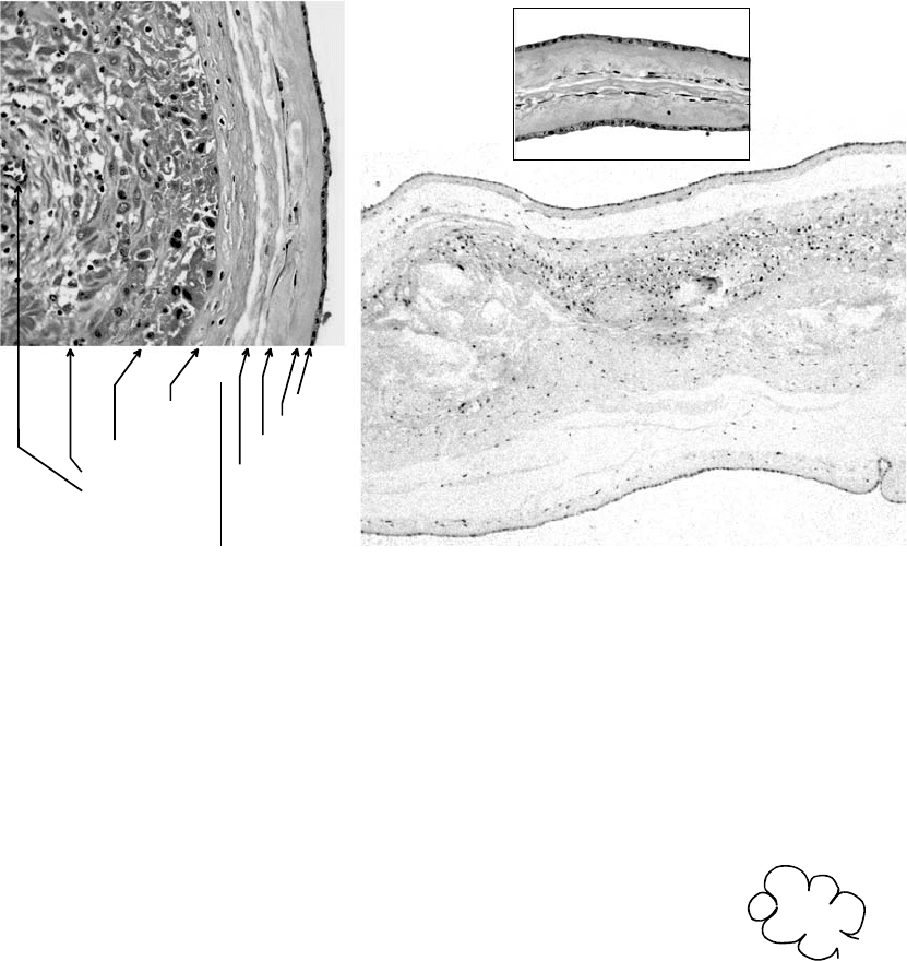
Normal (non-oedematous) amnion is upto 1/2 mm thick
r
Normal histology of amnion and chorion and intertwin membrane is shown in the figure, below
Twin-Twin Transfusion Syndrome (TTTS)
r
Is restricted to monozygotic twins
r
Donor: large poorly vascularised villi but in both the villi may
r
Recipient: smaller villi with dilated, engorged villous vessels
look normal
Complete Hydatidiform Mole (CM)
Mature / Late CM
r
Generalised hydropic change with cisterns (! d/dg: do not confuse cisterns with an amnion-deficient
gestational sac)

JWBK208-05 December 8, 2007 21:25 Char Count= 0
Paediatric and Placental 64
Layers of the Normal Chorion & Amnion Intertwin Membranes
Cellular
& Reticular
Trophoblastic
Decidual (incl.
blood vessels)
AMNION
(avascular)
Epithelial
Compact
Fibroblastic
Spongy
CHORION
DIAMNIOTIC MONOCHORIONIC
DIAMNIOTIC DICHORIONIC
Inset is x3
cf. main picture
(chorion is present between the amniotic layers)
FIGURE 5.3 Chorion, amnion and intertwin membranes
r
Avascular villi
r
No fetal tissue (‘fetal tissue’ = nucleated RBCs, amnion or fetal parts)
r
Circumferential proliferation of trophoblast (! d/dg: do not confuse with the abundant trophoblast seen
in early gestations e.g. tubal ectopics or trophoblast proliferation 2
◦
to perivillous fibrin deposition of
any cause)
Earlier CM:
r
Hydrops increases with gestational age, so at 11/40 only some villi are hydropic
r
Hydropic change is due to MPS (not fibrosis as in partial mole) and stroma is hypercellular
r
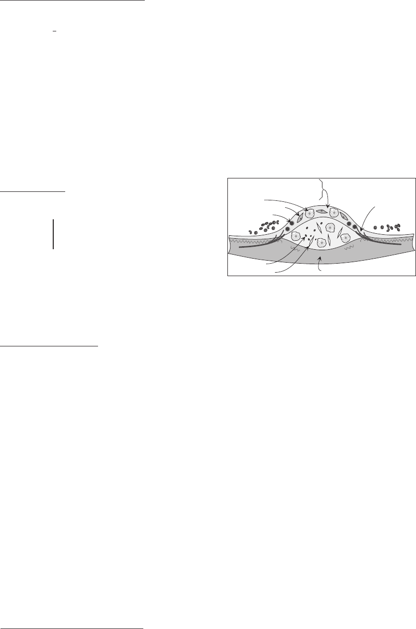
Villi may be polypoid (‘budding’ or ‘fibroadenomatoid’ – see Figure 5.4)
FIGURE 5.4 Budding
polypoid villus
r
Stromal cell karyorrhexis (not vessel) in well-preserved villi (i.e. not covered
with necrotic trophoblast)
r
Reticulin is perivascular (only)
r
>90% of CM villi are vascularised before 11/40, dropping to 30% after 20/40
r
Nucleated RBCs may be seen in some CM (an embryo begins to develop
but does not progress)
r
Trophoblast may strip off villous surface .
.
. a) circumferential excess may not be seen, b) may see
sheets of extravillous pleomorphic trophoblast (! d/dg: does not imply ChC)
r
Molar implantation site: abundant interstitial trophoblast, no endovascular plugging, ± haemorrhage
r
No dentate villi (cf. partial mole) and no rounded inclusions (may see larger irregular ovoid inclusions)
r
Immuno: Ki67 LI is high and nuclear p57 (Kip2) is ↓/lost in the villous stroma and cytotrophoblast
and intermediate trophoblasts of CM (use intervillous trophoblast islands and decidual cells as internal
+ve controls for p57) – partial mole and HA have lower Ki67 and are p57 +ve
r
CM are usu. diploid androgenones (95% monospermic XX, 5% dispermic XY)
Partial Hydatidiform Mole
r
Geographical / dentate / scalloped villi; may see a dual population of villi
r
Excess circumferential trophoblast, often vacuolated / lacy and may strip off to appear multifocal
r
Angiomatoid vessels (FVIIIRA +ve) [= ‘Branching cisterns’ esp. in late partial moles]
r
Reticulin shows ‘spider’s web’ pattern connecting one vessel to the another
r
Fibrosis ↑ with gestational age
r
Small rounded trophoblastic inclusions
r
Extensive sampling may be needed to detect these features

JWBK208-05 December 8, 2007 21:25 Char Count= 0
Paediatric and Placental 65
r
Partial moles are usu. triploid (2 of the 3 chromosome sets are from sperm) – can be useful in d/dg HA
r
Fetal tissues are common and implantation site is normal
r
! d/dg mixed curettings from a twin gestation of CM and embryo (→ dual population of villi)
r
d/dg stem villous hydrops (in PMD): no trophoblast hyperplasia, normal 3
rd
trimester fetus
r
d/dg Beckwith-Wiedemann syndrome shows sig. hydrops of some villi without other molar features
Non-molar Hydropic Abortion (HA)
FIGURE 5.5 Rounded
inclusion
r
Hydrops but of smooth round/ovoid contour and not large enough to be macroscopically visible
r
Stroma is oedematous, fibrotic and often avascular – but hypocellular cf. moles
r
Polar trophoblastic proliferation
r
Invaginated trophoblast / rounded trophoblastic inclusions (see Figure 5.5)
r
HA are usu. diploid (trisomies > monosomies) but non-molar fetal
aneuploidy may show HA with some irregular-shaped villi and inclusions
but no trophoblast proliferation (d/dg partial mole)
Invasive Hydatidiform Mole
r
Virtually all arise from CM but partial moles are monitored in case of possible diagnostic confusion
r
Molar villi seen within myometrium/vessels (may embolise to other organs e.g. lung =metastatic mole)
r
Residual trophoblast can be seen upto six months post ERPC .
.
. !beware diagnosing choriocarcinoma
in this period even if no villi are seen (i.e. sampled)
Morbidly Adherent Placentae
r
Predisposed to by any cause of decidual deficiency e.g. PMHx of Caesarian section or laser ablation of
the endometrium
r
Paucity of decidua
r
Villi and fibrinous basal plate (cytotrophoblast ++) penetrate to:
superficial myometrium = placenta accreta
deep myometrium = placenta increta
right through the myometrium = placenta percreta
Choriocarcinoma (ChC)
r
Def
n
: a tumour of villous trophoblast
r
Clin.: can occur after any kind of gestation, monitored with hCG, cured with chemoRx. Suspect ChC
if there is unexplained intracerebral haemorrhage or acute cor pulmonale
r
LN mets are unusual (consider non-gestational 1
◦
with trophoblastic differentiation – e.g. germ cell
tumour – esp. if there is no evidence of gestational origin)
r
Macro: haemorrhagic and bulky
r
Micro: defined as an admixture of syncytiotrophoblast and cytotrophoblast (±intermediate trophoblast)
that may grow in a laminar, plexiform, ‘villous’ or papillary architecture:
cytotrophoblast: usu. −ve for hCG, cells are polygonal, clear cytoplasm, well defined cell
borders, central vesicular nucleus ± prominent nucleolus / chromocentres; architecture =
cords / solid masses; ± weak staining for hPL
intermediate trophoblast: a mixture of mononuclear and multinucleated cells with
more/stronger staining for hPL than hCG and +ve for CD146 (unlike other trophoblast)
syncytiotrophoblast: usu. +ve for hCG and inhibin-, the syncytium contains smaller, denser
nuclei within more basophilic cytoplasm (cf. cytotrophoblasts) that may be vacuolated
r
No intrinsic tumour vasculature (but the tumour invades pre-existing vessels)
r
No 2
◦
or higher order chorionic villi – by definition (i.e. no villi containing stromal cores)
r
Variant: cytotrophoblastic
(cf. d/dg PSTT) is predom. mononuclear, ↑ mitoses, intravascular cf. interstitial spread
(cf. usual type ChC) is more chemoresistant
r
‘Choriocarcinoma in situ’ = intraplacental ChC
r
d/dg new pregnancy (or persistent trophoblast in CM). More likely to be ChC if abundant, very pleo-
morphic and found > 4 months after last known pregnancy.
r
d/dg PSTT: more haemorrhagic, vascular mode of spread (cf. intramuscular in PSTT), laminar structure,
cytotrophoblasts of ChC are less eosinophilic, more hCG +ve cells cf. hPL +ve

JWBK208-05 December 8, 2007 21:25 Char Count= 0
Paediatric and Placental 66
r
d/dg carcinoma with trophoblastic differentiation: CA produces little hCG for its bulk, has a vascular
stroma and is resistant to chemoRx. ChC contains paternal polymorphisms. Immuno unhelpful as ChC
is +ve for CK, EMA and some CEA.
r
d/dg invasive mole: moles have villi with stromal cores
r
d/dg pleomorphic trophoblast without villi in curettage specimens (see sections on CM and ‘Invasive
Hydatidiform Mole’ above) – do not diagnose ChC here without corroborating evidence
Placental Site Trophoblastic Tumour (PSTT)
r
Def
n
: a tumour of intermediate trophoblast (admixed mononuclear and multinucleate cells)
r
Clin.: amenorrhoea / irregular bleeding months (>4) or years after some gestation
r
Less metastatic but chemoresistant .
.
. Rx is surgery, not chemoRx
r
Eosinophilic pleomorphic cells (±cytoplasmic vacuoles / hyaline inclusions) infiltrating myometrium /
decidua (occasional scattered foci of syncytiotrophoblast may be present)
r
Hyaline and fibrin ++
r
Much necrosis (not haemorrhage) → tumour cells survive only around vessels and may show endovas-
cular growth (cling to or focally replace endothelium or are free in the lumen)
r
↑ Metastatic potential if mitoses > 5/10hpf
r
Immuno: more hPL than hCG +ve cells (i.e. the converse of ChC); +ve for inhibin-, AE1/AE3,
CAM5.2 and CD146 (= Mel-CAM)
r
d/dg exaggerated placental site reaction: mitoses, presentation > 4 months and large clusters of cells
favour PSTT while villi, decidua vera and many multinucleate cells favour exaggerated placental site
r
d/dg regressing placental site nodule: ↑↑ hyaline and central necrosis favour nodule
r
d/dg non-gestational carcinoma: PSTT presents with gynae Sx. Immuno: hPL more useful than hCG
Bibliography
Al-Adnani, M., Williams, S., Rampling, D., Ashworth, M., Malone, M. and Sebire, N.J. (2006) Histopathological reporting of paediatric cutaneous
vascular anomalies in relation to proposed multidisciplinary classification system. Journal of Clinical Pathology, 59 (12), 1278–1282.
Berenguer, B., Mulliken, J.B., Enjolras, O. et al. (2003) Rapidly involuting congenital hemangioma: clinical and histopathologic features. Pediatric
and Developmental Pathology, 6 (6), 495–510.
Boyd, T., Gang, D., Lis, G., Juozokas, A. and Pfeuger, S. (2004) Appendix 2A in Placental Pathology, fascicle 3 in Atlas of Non-Tumor Pathology
1
st
series (eds F.T. Kraus, R.W. Redline, D.J. Gersell, D.M. Nelson and J.M. Dicke), AFIP, The American Registry of Pathology, Washington D.C.
Filipe, M.I. and Lake, B.D. (1990) Histochemistry in Pathology,2
nd
edn, Churchill Livingstone, Edinburgh.
Fox, H. (1997) Trophoblastic disease, in Progress in Pathology, (eds N. Kirkham and N.R. Lemoine), Churchill Livingstone, NY, vol. 3, pp. 86–101.
Fox, H. (1997) Pathology of the Placenta,2
nd
edn, W.B. Saunders Co. Ltd., London.
Fox, H. and Wells, M. (eds) (2002) Haines & Taylor Obstetrical and gynaecological pathology,5
th
edn, Churchill Livingstone, London.
Glickman, J.N. (April 2005) Pathology of inflammatory bowel disease in children. Current Diagnostic Pathology, 11 (2), 117–124.
Gruenwald, P. and Minh, H.N.(1961) Evaluation of body and organ weights in perinatal pathology II. Weight of body and placenta in surviving and
autopsied infants. American Journal of Obstetrics and Gynecology, 82 (2), 313–319.
Guillou, L., Coquet, M., Chaubert, P. and Coindre, J.M. (1998) Skeletal muscle regeneration mimicking rhabdomyosarcoma: a potential diagnostic
pitfall. Histopathology, 33 (2), 136–144.
Hargitai, B., Marton, T. and Cox, P.M. (2004) Examination of the human placenta. Journal of Clinical Pathology, 57 (8), 785–792.
Lage, J.M., Minamiguchi, S. and Richardson, M.S. (Feb 2003) Gestational trophoblastic diseases: update on new immunohistochemical findings.
Current Diagnostic Pathology, 9 (1),1–10.
Meijer-Jorna, L.B, van der Loos, C.M., de Boer, O.J., et al. (2007) Microvascular proliferation in congenital vascular malformations of skin and soft
tissue. Journal of Clinical Pathology, 60 (7), 798–803.
Moore, I.E. and Baxendine-Jones, J. (2001) Diagnosis and molecular pathology of Hirschprung’s disease, in Recent Advances in Histopathology,
(eds D. Lowe, J.C.E. Underwood), Churchill Livingstone, Edinburgh, vol. 19, pp. 51–66.
Out, H.J., Kooijman, C.D., Bruinse, H.W. and Derksen, R.H. (1991) Histopathological findings in placentae from patients with intrauterine fetal death
and anti-phospholipid antibodies. European Journal of Obstetrics, Gynecology and Reproductive Biology, 41 (3), 179–186.
Paradinas, F.J. (June 1998) The differential diagnosis of choriocarcinoma and placental site tumour. Current Diagnostic Pathology, 5 (2), 93–101.
Pinar, H., Sung, C.J., Oyer, C.E. and Singer, D.B. (1996) Reference values for singleton and twin placental weights. Pediatric Pathology and
Laboratory Medicine, 16 (6), 901–907.
Sebire, N.J., Fox, H., Backos, M. et al. (2002) Defective endovascular trophoblast invasion in primary antiphospholipid antibody syndrome-associated
early pregnancy failure. Human Reproduction, 17 (4), 1067–1071.
Sebire, N.J., Backos, M., Gaddal, S., et al. (2003) Placental pathology,antiphospholipid antibodies, and pregnancy outcome in recurrent miscarriage
patients. Obstetric Gynecology, 101, 258–263.
Sebire, N.J. and Rees, H. (2002) Diagnosis of gestational trophoblastic disease in early pregnancy. Current Diagnostic Pathology, 8 (6), 430–440.
Skordilias, K. and Howatson, A.G. (2003) The diagnostic challengeof paediatric small round cell tumours,in Progress in Pathology, (eds N.Kirkham,
N.A. Shepherd), Greenwich Medical Media Ltd, London, vol. 6, pp. 1–27.
JWBK208-05 December 8, 2007 21:25 Char Count= 0
Paediatric and Placental 67
Smith, N.M and Keeling, J.W. (1997) Paediatric solid tumours, in Recent Advances in Histopathology, (eds P.P. Anthony, R.N.M. MacSween and
D.G. Lowe), Churchill Livingstone, Edinburgh, vol. 17, pp. 191–218.
Wainwright, H.C. (2006) My approach to performing a perinatal or neonatal autopsy. Journal of Clinical Pathology, 59 (7), 673–680.
Wigglesworth, J. and Singer, D.B. (eds) (1991) Perinatal Pathology,1
st
edn, Blackwell Medical Publishers Inc., Boston.
Wright, C. (June 2006) Congenital malformations of the lung. Current Diagnostic Pathology, 12 (3), 191–201.
Placental Weights Data Sources
Dataset ‘a’ for singleton placentas is based on the data for 1232 liveborn babies from Gruenwald and Minh (1961). (NB: Placental weights from
the autopsied infants was found to be similar to the liveborn ones in this study). I fitted this data to a 3
rd
order linear polynomial for the purpose of
plotting the chart. The fetoplacental weight ratios are also derived by fitting to data from this paper.
Dataset ‘b’ for singleton placentas is based on the data for liveborn babies as gathered by Boyd et al. (2004). I fitted this data to a 4
th
order linear
polynomial for the purpose of plotting the chart.
The twin placenta factors are based on the twin placental weights of Pinar et al., 1996.

JWBK208-06 December 8, 2007 16:1 Char Count= 0
Vascular 68
6. Vascular
Normal and Age-Related Changes
r
Elastic arteries are large Windkessel vessels (distend during systole to buffer the BP pulse) and have
the outer
2
3
of the wall supplied by vasa vasorum (e.g. aorta and main branches). Age →focal fibrosis
and fragmentat
n
of medial elastic with ↑MPS ± amyloid and some intimal fibrosis.
r
Muscular arteries are the main distributors (e.g. coronaries). The wall muscle is disposed in a regular
spiral helix angled ≈30
◦
to the transverse plane of the vessel. Age →intimal and medial fibrosis, focal
medial hyaline, IEL may show small breaks (± Ca
2+
) and focal reduplication.
r
Arterioles are defined as having: a lumen < 0.1–0.3 mm +/ ≤ 6 concentric layers of medial muscle.
r
Capillaries have no muscle, are the diameter of an RBC and have continuous, fenestrated or sinusoidal
endothelium. Age → BM thickening.
r
Post-capillary venules (PCVs) have 1–2 layers of muscle and no elastic.
r
Veins are valved capacitance vessels with a lower wall-thickness:lumen-diameter ratio and poorly
defined elastic laminae cf. arteries. The wall muscle is disposed in circular and longitudinal bundles in
larger veins. Age → intimal fibrosis and medial hypertrophy.
Atherosclerosis
Time Course
r
10 years: lipid streaks (subintimal foamy M)
fibrofatty plaques (‘atheromas’)
r
40 years:↓ Calcification, Ulceration, Thrombosis
→ clinical effects
Calcification
Cholesterol cleft
Atrophy of media & loss of elastin
Neovascularisation
Collagen
Proteoglycan
Elastin
Foam cell
Smooth muscle cell
T lymphocyte
Atheromatous Plaque
FIGURE 6.1 Atheroma
Distribution
r
Eccentric lumenal narrowing
r
Sites of turbulence (bifurcations and ostia)
r
Aorta (abdominal > thoracic), coronaries, circle of Willis, (kidney), lower limbs, small intestine
r
Arterialised veins (such as occur in a CAVG)
Systemic Hypertension
Definitions
r
HT should only be diagnosed on the average of ≥2 readings on ≥2 occasions
r
Borderline HT: diastolic 90–95 mmHg systolic 140–160 mmHg
r
Established HT: diastolic ≥ 95 or systolic ≥ 160 mmHg
r
Malignant HT: rapidly increasing BP to > 130 mmHg diastolic (die in ≈2 years from RF if no Rx)
Benign HT (= Essential HT)
r
Arteriosclerosis: medial hypertrophy and reduplication of the IEL
concentric intimal fibroplasia and patchy medial fibrosis
↑MPS + fragmented elastic (i.e. myxoid degeneration)
d/dg:
a) cystic medial necrosis (a matter of degree – see p. 73 under ‘Aortic
dissection’)
b) Marfan’s syndrome (≈ pre-mature ageing of the media, see p. 78)
c) age-related changes
Hyaline arteriolosclerosis:
r
hyaline change in the media, starts in the sub-intimal region
correlates with HT most in the kidney (least in the spleen)
diffuse, variable severity (→granular contracted kidney at end-stage)
unusual in skeletal muscle, skin, brain (± Charcot-Bouchard microa-
neurysms), heart, GIT
d/dg: a) similar changes occur in the elderly & DM (without HT) and
b) radiation injury (q.v.)
Diagnostic Criteria Handbook in Histopathology: A Surgical Pathology Vade Mecum. Paul J. Tadrous
Copyright
C
2007 by John Wiley & Sons, Ltd. ISBN: 978-0-470-51903-5
