Ramanathan Sh. (Ed.) Thin Film Metal-Oxides: Fundamentals and Applications in Electronics and Energy
Подождите немного. Документ загружается.


68 D. Ruzmetov and S. Ramanathan
R.25
ı
C; UV/
R.25
ı
C; initial/
R.100
ı
C; UV/
R.100
ı
C; initial/
and can be recast in the form (UV)/(initial), where D R.25
ı
C/=R.100
ı
C/.
tells how large the resistance drop across the MIT is and is an important parameter
characterizing the MIT strength. Then Fig. 2.13b shows how is affected by UV ir-
radiation. The MIT strength is enhanced by UV treatment in HV sample (ratio >1)
and is slightly deteriorated in the rest of the samples. It should be noted again that
UV affects only a thin layer on top of the VO
2
film (50-nm thick). So that the small
changes due to UV observed in these samples correspond to much stronger modula-
tion of the properties of the top superficial layer of the film that was exposed to UV.
2.5 The Nature of the Insulating State
The origin of the MIT in VO
2
is a subject of a debate. Two main mechanisms of
the MIT have been suggested in literature. In the Peierls model, the lattice transfor-
mation at the structural phase transition temperature .T
SPT
66
ı
C/ is accompanied
by the band structure changes that result in the opening of the band gap and, conse-
quently, the MIT [35]. In this scenario the material is referred to as band insulator.
In the Mott transition model, electron correlations alone cause the transition to the
insulating state, while the ion arrangement and lattice–electron interactions are of
secondary importance to the MIT [37,48]. If electron–electron correlations are con-
sidered to be primarily responsible for the insulating state of VO
2
, the material is
often referred to as Mott or Mott–Hubbard insulator, even though current models go
generally beyond the standard Mott–Hubbard picture [31].
The understandingof the electronic ground state was considerably improved after
the studies of Cr-doped VO
2
alloys [48]. Cr enters the V sites as 3
C
ions. The re-
sulting alloy, V
1x
Cr
x
O
2
, also exhibits a metal–insulator transition close to the T
MIT
of pure VO
2
with Cr doping in the range x D 0–0:045.HowevertheV
1x
Cr
x
O
2
alloy has three different lattice structures, labeled as M
1
,T,andM
2
,intheinsu-
lating phase depending on the temperature and Cr doping level [48]. M
1
lattice
corresponds to the pure VO
2
lattice in the insulating state described above in the
Sect. 2.3.TandM
2
are two new insulating phases. M
2
is a monoclinic lattice which
is different from M
1
in that only half of the vertical (along c-axis) V–V chains are
dimerized, i.e., V ions are displaced in the lateral (perpendicular to c-axis) direction
so that the resulting vertical V chains have a zigzag pattern (Fig. 2.6). The other half
of vertical V chains in M
2
remain straight (undistorted) as it is in the metallic tetrag-
onal phase. T is believed to be a triclinic lattice and is a transitional phase between
M
1
and M
2
[29,48]. Uniaxial pressure applied to pure VO
2
can also give rise to M
2
and T phases and leads to the phase diagram similar to the V
1x
Cr
x
O
2
alloys [32].
The study of electrical properties of M
2
and T showed that the conductivity changes
only by 25% between the phases and both phases exhibit the same activation energy
of 0.4 eV [31] that is close to the activation energy in pure VO
2
, 0.45 eV. Since the
three phases, M
1
,T,andM
2
, have very different lattice structures, the similarity
2 Metal-Insulator Transition in Thin Film Vanadium Dioxide 69
of electrical properties of the three phases (the existence of MIT, similar activation
energies and conductivity values) indicates strongly that the lattice transformation
at the phase transition is not the primary cause of the MIT. This conclusion led to
substantiating the electron correlation models, such a Mott–Hubbard model, as the
appropriate description of the MIT in VO
2
[31].
However the dispute on the primary mechanism of the MIT remained unresolved.
Convincing evidence in favor of the band-like character (Peierls insulator) of VO
2
was given by LDA calculations of Wentzcovitch et al. showing that band theory can
account for the low temperature monoclinic distorted state [35]. First principles cal-
culations of Wentzcovitch et al. employed an ab initio molecular dynamics scheme
with variable cell shape to perform unconstrained structural searches for the ground
state within the 13-dimensional parameter space of the low-T phase [35]. The result
was the stable monoclinic M
1
phase with lattice parameters in good agreement with
experimental data. The fact that the calculation failed to reproduce the band gap
opening was not considered to be discouraging, since local density approximation
was notorious for underestimation of the measured optical band gaps. These results
were corroborated by the LDA calculations of Eyert who also showed that a band
theoretical approach can account for the metal–insulator transition in M
2
phase as
well [29].
Support to the band-like character of insulating VO
2
came also from experimen-
tal studies. Cavalleri et al. applied ultrafast spectroscopy to establish time domain
hierarchy between structural and electronic effects in VO
2
[6]. In the pump-probe
reflectivity experiments conducted by Cavalleri et al. the MIT in thin films of VO
2
was induced by short optical pulses and the dynamics of the reflectivity change due
to MIT was measured with femtosecond resolution. It was shown that the transi-
tion time can be brought down to 80 fs but not less (“structural bottleneck” [6]),
even though much shorter time, 15 fs, was expected if the MIT were due to pure
electronic effects. The femtosecond time scale of the transition excluded the lat-
tice temperature effects. The existence of the structural bottleneck was explained by
the arguments that the collapse of the band gap was due to the structural motion
brought about by optical phonons [6]. Thus the atomic arrangement of the high-T
unit cell was believed to be necessary for the formation of the metallic phase of VO
2
.
During the last decade, electron energy band structure studies at synchrotron
facilities considerably advanced the understanding of the metal–insulator transition
in this specific system and showed that the band structure changes at the MIT require
an explanation that goes beyond the Peierls and standard Hubbard transition models
[49, 50]. Haverkort et al. performed X-ray absorption spectroscopy at the V L
23
edge (electron transition 2p ! 3d) to provide evidence for orbital redistribution in
the V 3d states at the MIT [49]. It was shown that the orbital occupation changes
from almost isotropic in the metallic state to almost completely one-dimensional
(along the c-axis) in the insulating state. The V ions in the chain along the c-axis
then become susceptible to the Peierls transition. However, the orbital polarization
is achieved due to the strong electron correlation and the fact that the system is
close to the Mott regime. So that the MIT in VO
2
was termed as orbital-assisted
Mott–Peierls transition [49].
70 D. Ruzmetov and S. Ramanathan
Koethe et al. performed photoemission and X-ray absorption spectroscopy of the
near-Fermi levels in VO
2
across the MIT [50]. The well-resolved spectra presented
by Koethe et al. revealed in detail the structure of the density of states near the
Fermi level that exhibited considerable changes upon MIT. The spectra of Koethe
et al. were well-reproduced by calculations of Biermann et al. based on the cluster
dynamical mean field theory (CDMFT) [51]. The lattice of the insulating phase of
VO
2
comprises zigzag vertical (along c-axis) chains of V ions that can be seen as
straight chains of V–V pairs (called also dimers or clusters). The two V ions in
each dimer are slightly tilted with respect to the vertical c-axis (see Sect. 2.3). In
the CDMFT calculation of Biermann et al. [51] the V–V dimers are taken as a key
unit and the electron correlations are taken into account only within the cluster cell
including the V–V dimer. Besides correctly reproducing the near-Fermi level density
of states measured by Koethe et al. [50], the model of Biermann et al. succeeds
in reproducing the opening of the band gap upon the structural phase transition
(SPT) which is an advantage of this work with respect to most previous published
calculations of band structure of VO
2
[29,35,52,53]. The limitations of the CDMFT
calculation [51] are that it considers only t
2g
bands of the near-Fermi V 3d levels
and the effect of the electrons in the neighboring bands is not taken into account, as
well as it includes an adjustable parameter.
Recently, Green’s function methods of band structure calculations were applied
successfully to both insulating and metallic phases of VO
2
[54, 55]. Gatti et al.
performed an ab initio GW calculation with no adjustable parameters to study the
near-Fermi DOS of VO
2
in the metallic and insulating phases [55]. The calcu-
lation described well the main features of the photoemission spectra of Koethe
et al. [50] in high- and low-T phases of VO
2
. The values of the band gap and
bonding–antibonding splitting of the d
jj
band (see also Sect. 6) obtained from the
calculation agreed with experimental results. The GW calculation of Gatti et al.
provided support also for experimental observations of Haverkort et al. [49]ofthe
orbital switching of the V 3d states responsible for the transition from the isotropic
metal to electronically more one-dimensional insulator. Later GW calculations of
Sakuma et al. confirmed the results of Gatti et al. and presented graphs of the band
structure as well [54].
Kim et al. relied on femtosecond pump-probe measurements and temperature-
dependent XRD to put forward the picture where the metal–insulator transition
and structural transformation from rutile to monoclinic lattice occur separately at
different temperatures [37]. In this picture, there exists an intermediate metallic
monoclinic phase between MIT .T
MIT
56
ı
C/ and the structural phase transi-
tion .T
SPT
65
ı
C/. The fact that there is no lattice transformation to rutile phase at
the MIT excludes the Peierls model and the driving mechanism of the MIT is con-
sidered to be the Mott transition. The origin of the metallic monoclinic phase was
explained with hole-driven MIT theory [56, 57] and Hall effect measurements of
the hole density were presented in support [37]. Evidence in favor of the Mott tran-
sition was presented also on the basis of infrared spectroscopy and nano-imaging
[58]. Calculations of band structure based on the hole-driven MIT theory [56, 57]
that reproduce measured observables as well as more experimental evidence of
2 Metal-Insulator Transition in Thin Film Vanadium Dioxide 71
monoclinic correlated metal phase and Mott transition would be desirable in order
to substantiate the new picture of MIT in VO
2
proposed by Kim et al. [37].
Summarizing, there has been achieved considerable understanding of the nature
of the insulating state of VO
2
. Theoretical descriptions were developed and exper-
imental evidence was obtained that clarify the nature and differences of insulating
and metallic phases of VO
2
. It was found that the description of the metal–insulator
transition goes beyond standard Peierls and Mott–Hubbard models and it requires
considering structural and electron correlation aspects on equal footing. At the same
time the situation right near the MIT temperature still requires clarification and more
work needs to be done to explain some new experimental results and reconcile com-
peting theoretical models.
2.6 Energy Band Structure
As discussed in the previous section, the knowledge of the density of electronic
states near the Fermi level is important in order to develop theoretical description of
insulating and metallic phases of VO
2
and understand the details of MIT.
The near-Fermi level energy band structure of VO
2
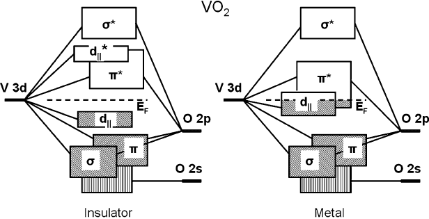
can be described using the
level diagram (Fig. 2.14) based on the molecular orbital picture proposed by Good-
enough [36]. The band structure is the result of the hybridization of V 3d and O 2p
levels and reflects the symmetries of the atomic arrangement in the crystal lattice. In
the tetragonal metallic phase the octahedral crystal field causes the splitting of V 3d
levels into e
g
and t
2g
levels ([1]: part II, section H.1). The e
g
orbitals are bridged by
the ligand (oxygen) 2p orbitals in the way that the bonding possesses -symmetry.
The corresponding levels lie further away from the Fermi level and are depicted by
antibonding
bands in Fig. 2.14 (the notations are commonly used for this system
Fig. 2.14 Band structure diagram of VO
2
near Fermi level in metallic and insulating phases as the
result of the hybridization of V and O orbitals based on Goodenough’s description [36]
72 D. Ruzmetov and S. Ramanathan
[29, 51,59]). t
2g
levels are grouped into the bands
and d
jj
that lie right near the
Fermi level. d
jj
Orbitals are aligned along the rutile c-axis and are consequently of
almost 1D character. In the low-T phase (below T
SPT
), the dimerization of the V
atoms, i.e., their pairing and tilting with respect to the c-axis as a part of the mono-
clinic distortion, causes the splitting of the d
jj
band into bonding d
jj
and antibonding
d
jj
bands and the shift of
band up and away from the Fermi level. As a result, a
band gap opening occurs between the top of d
jj
and the bottom of
. Good quanti-
tative characterization of the near-Fermi level band structure was performed by Shin
et al. using UV reflectance and photoemission spectroscopy [60]. The d
jj
splitting
was measured to be 2.5 eV, the optical band gap was 0.7 eV, and the rise of the
was 0.5 eV [60].
X-ray absorption spectroscopy (XAS) has proven to be a valuable tool to study
the unoccupied conduction bands of VO
2
crystals above and below T
MIT
and im-
prove the understanding of this system [49, 61]. The MIT characteristics vary
significantly for thin films and bulk crystals, and for thin films prepared at different
conditions [9]. Using XAS to relate this variation to the changes in the electronic
structure should provide a bridge between macroscopic observables of the MIT and
microscopic models describing the transition.
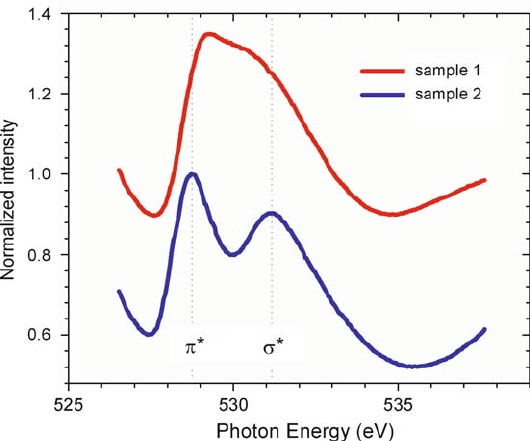
Figure 2.15 shows XAS data for two thin (57-nm) film VO
2
samples on Si
substrates [39]. Thin polycrystalline films were sputtered at different substrate
Fig. 2.15 Room-temperature XAS data for thin film VO
2
samples 1 (sputtered at 300
ı
C, grain
size 14 nm) and 2 (500
ı
C, grain size 20 nm). The spectra are normalized to the maximum of
intensity and displaced vertically for clarity. O K-edge is displayed. (From Ruzmetov et al. [39]
with permission Copyright (2007) by the American Physical Society)
2 Metal-Insulator Transition in Thin Film Vanadium Dioxide 73
temperatures which resulted in different material morphology. In particular,
sample 1 has an average crystallite size of 14 nm and sample 2–20 nm, as mea-
sured by X-ray diffraction analysis. The spectra in Fig. 2.15 display the O K-edge
of the material, i.e., the X-ray absorption enhancement is due to the electron exci-
tations from the K-shell to the unoccupied states above and near the Fermi level.
The dipole selection rules for the electron transition during photon absorption re-
quire the orbital momentum change l D˙1, so that the K-edge corresponds to
the transitions O 1s ! 2p. O 2p levels are hybridized with V 3d levels (Fig. 2.14)
and the measured spectra reflect the p-projected unoccupied density of states of
VO
2
near the Fermi level. The two peaks in Fig. 2.15 are due to the bands
and
as inscribed in the figure with the d
jj
band apparently being merged with
the
.Thed
jj
band can be sometimes separately resolved from the
band in the
polarization sensitive O K-edge XAS in single crystal VO
2
[50,61].
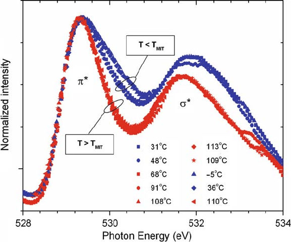
Figure 2.16 shows the evolution of the XAS O K-edge with temperature cycling
across the metal insulator transition temperature T
MIT
D 66
ı
C[39]. The spectra
were taken at consecutive temperatures: T D 31, 48, 68, 91, 108, 113, 109, 5, 36,
110
ı
C. All curves at T>T
MIT
overlap well and display narrower
width than the
Fig. 2.16 XAS O K-edge spectra for thin film VO
2
(sample 2 m) at different temperatures below
and above the T
MIT
D 66
ı
C. The spectra can be divided into two groups with wider and narrower
width. All spectra at T>T
MIT
overlap well and display narrow
band width. The discon-
tinuous linewidth broadening upon crossing the T
MIT
is ascribed to the manifestation of MIT in
the energy band structure. (From Ruzmetov et al. [39] with permission. Copyright (2007) by the
American Physical Society)
74 D. Ruzmetov and S. Ramanathan
low-T curves. The discontinuous change of the bandwidth upon crossing the MIT
temperature and constant line shape on either side of T
MIT
attest that the observed
discrete changes in the energy band structure are attributes of the phase transition.
Each spectrum in Figs. 2.15 and 2.16 can be fitted well with the sum of two
Doniach–Sunjic peaks (with Gaussian broadening) and a linear background so that
the linewidths, positions, and heights of the component peaks can be precisely ex-
tracted from the measured data [27, 39]. The results of the fitting are presented in
[39]. Sample 1 is sputtered at lower substrate temperature, has smaller grain size,
and is expected to have more disorder than sample 2 [39]. The bandwidths for this
sample appear to be larger than for sample 2 and there is also a decrease in the spac-
ing between the bands
and
. The decrease occurs mainly due to the shift of
the
peak.
Taking into account the symmetry of the orbitals in the compound can help to
obtain the microstructural information from the O K-edge XAS data extracted from
the spectra in Figs. 2.15 and 2.16, and summarized in [39]. orbitals in VO
2
point
in between the ligands (O ions) and orbitals are directed toward the ligand. There-
fore, V–V interactions affect the band more, whereas the band is influenced by
the V-ligand configuration and the indirect V–O–V interaction. Then the observed
shift in the
peak in the film with smaller grain size and increased disorder (sample
1) can be taken as evidence for the distortion of the oxygen octahedra with respect to
the V ions as compared to the samples that exhibit more single crystal VO
2
character
(samples 2 and 2m) [39]. As-grown sample 2 m is identical to sample 2 and was used
for temperature dependent measurements across T
MIT
. It was observed that neither
line position, nor spacing , change appreciably upon MIT (samples 2 and 2m)
indicating that the V–V pairing upon the MIT (as a part of the lattice transformation
from tetragonal to monoclinic) is not accompanied with the O octahedra distortion.
Finally, the widths of the peaks are the largest in the most disordered sample 1 fol-
lowed with the low-T phase of sample 2. Apparently the line widths are connected
to the amount of defects in the crystal lattice of materials. The films deposited at
low substrate temperatures have more lattice defects since the deposited atoms have
less energy to move to the most favorable thermodynamic locations. These defects
on average will cause the line broadening that is being observed in sample 1. Sim-
ilarly, the lower-symmetry monoclinic phase of VO
2
(sample 2 in Fig. 2.15) yields
broader lines than the tetragonal metallic phase (sample 2m in Fig. 2.16).
X-ray absorption spectroscopy can be used to learn what features of the band
structure are directly connected with the metal–insulator transition. Consider again
(see Sect. 2.2) the set of five vanadium oxide films with varying V–O stoichiometry
in the narrow range around the stoichiometry of VO
2
. The results of the electri-
cal characterization for this set were presented in Fig. 2.4. In order to extract the
transition temperatures .T
MIT
/ and widths .T /, the derivatives of the resistance
d.log
10
R/=dT were taken from the data in Fig. 2.4. The resulting curves had clear
minimums and could be well-fitted with Gaussians (see examples of the fitting in
Fig. 2.8b). The center and width of the fitted Gaussian were taken as T
MIT
and T .
The MIT strength was defined as the resistance ratio R.T
MIT
T /=R.T
MIT
CT /.
XAS measurements of the O K-edge at room temperature were performed on this
2 Metal-Insulator Transition in Thin Film Vanadium Dioxide 75
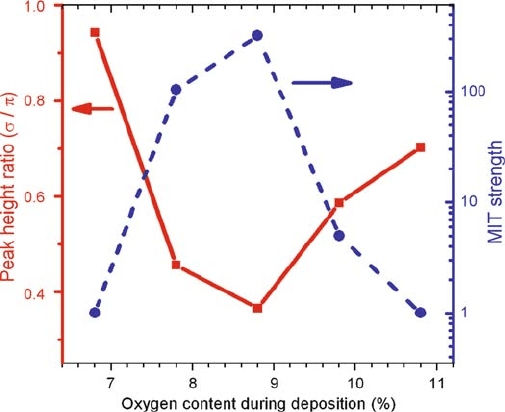
Fig. 2.17 Relative heights of
and
peaks extracted from the fitting to the XAS spectra
(presented in [27]) in comparison to the VO
2
MIT strength (i.e., ratio of resistances at temperatures
T
MIT
= CT ). (From Ruzmetov et al. [27] with permission. Copyright (2008) by the American
Physical Society)
set [27]. To extract quantitative information from the XAS data, each spectrum was
fitted with the algebraic sum of
;
peak, and linear background and corre-
sponding widths, heights, and locations of the peaks were determined. It was shown
in [27] that the
.
/ peak area was larger (smaller) in the samples with strong
MIT. The same behavior was seen in the ratio of peak intensities. To illustrate this
point, we show in Fig. 2.17 the ratio of
and
peak heights in comparison with
the MIT strength. We see a clear redistribution of the spectral weight from
to-
ward
peak in samples with strong MIT. One can conclude that the ratios of the
and
peak parameters extracted from XAS data and the absolute values of the
peak areas are strongly correlated to the MIT strength of the VO
2
films. However
the absolute values of peak heights and widths are not necessarily correlated to the
MIT properties meaning that there is a transformation of the shape of the peaks seen
in the XAS spectra in the samples with and without MIT.
The observed redistribution of the spectral weight in Fig. 2.17 from the upper
to lower band in MIT samples indicates strengthening of the
and/or d
jj
bands
with respect to the
band. Since in both orbitals responsible for the
and d
jj
bands the electron density is shifted away from the V–O bond line as compared
to the
orbitals (see the discussion of the orbital symmetries above), the noted
redistribution may be connected to the decrease of the direct V–O interaction and
increase of the V–V interactions. Therefore, one can judge that there is strengthening
of the metal bonding and weakening of the direct metal–ligand interactions as the
vanadium oxide compound approaches stoichiometric phase of pure VO
2
with a
strong MIT [27].
76 D. Ruzmetov and S. Ramanathan
Strongly differing
=
relative peak intensities have been reported in VO
2
single crystals and powders [50, 61–63]. The results from [27] presented above
are closer to the X-ray absorption data on VO
2
powders which revealed the domi-
nance of the first .
/ peak in the raw spectra [63]. The reported spectra of the O
K-edge in single crystals depend on the polarization orientation of the X-rays with
respect to the crystal axes. When the X-ray photon polarization has the electric field
vector parallel to the rutile crystal c-axis .Ejjc/, the lower band peak in Fig. 2.15
splits into two peaks assigned as
and d
jj
[50,61]. In this case the intensity of the
upper band,
, may be comparable or higher than those of
and d
jj
peaks. For
the other polarization orientation .E ?c/,thed
jj
peak is not resolved (suppressed
due to the linear orbital orientation along c-axis) and the
peak dominates over
. Then the observed increased width and area of the
peak in the samples with
strong MIT [27], may be a manifestation of the emergence of the d
jj
band in these
samples. The peak is not completely resolved because the spectra are the result of
averaging of all polarization orientations in the polycrystalline films.
Theoretical models describing the MIT within the Mott–Hubbard picture [31,53]
have received more support recently [37]. In this picture, the transition is attributed
to strong electron–electron correlations mostly in the d
jj
band. Also, a recent
model suggested by Biermann et al. based on cluster dynamical mean field theory
(CDMFT) calculation [51] shows that both correlation (Mott) and band (Peierls) ef-
fects are present. The model is well-supported by X-ray photoemission spectra of
the valence bands of VO
2
[49]. This CDMFT calculation finds a large redistribution
of the electronic occupancies in favor of the d
jj
orbital. The importance of electron
correlations is generally acknowledged when one needs to account for the transport
properties of VO
2
and the d
jj
band is considered to play a crucial role in the MIT.
The results of Ruzmetov et al. [27] presented above obtained from a set of vanadium
oxide compounds with varying anion nonstoichiometry show that the appearance of
this d
jj
band near the Fermi level in the spectra is directly related to the presence of
the metal–insulator transition.
The manifestation of MIT in the valence band structure of VO
2
films was stud-
ied using hard-UV photoemission spectroscopy at constant incident photon energy
of 150 eV [16]. The photoemission spectra displaying the binding energy of the va-
lence levels below the Fermi level in the metallic and insulating states of the film
are displayed in Fig. 2.18. The metallic state was achieved by heating the sample to
101
ı
C. Considerable electronic structure changes upon the MIT were observed in
the vicinity of the Fermi level shown in Fig. 2.18b. A clear 0.6eV shift of the peak
from the top valence band toward the Fermi level was measured. This shift is almost
as large as the band gap 0.6–0.7eV [60, 64] which implies that the Fermi level is
near the bottom of the conduction band likely due to the presence of oxygen de-
fects. This situation, i.e., coincidence of the PES threshold value and the band gap,
has also been observed in V
2
O
5
[65]. The presence of the defects creating donor-
and acceptor-like states within the energy gap which pin the position of the Fermi
level was also suggested by Berglund et al. based on the analysis of the tempera-
ture dependence of the conductivity and activation energy in VO
2
[12]. The shift
of the Fermi level toward the conduction band implies that the VO
2
semiconductor
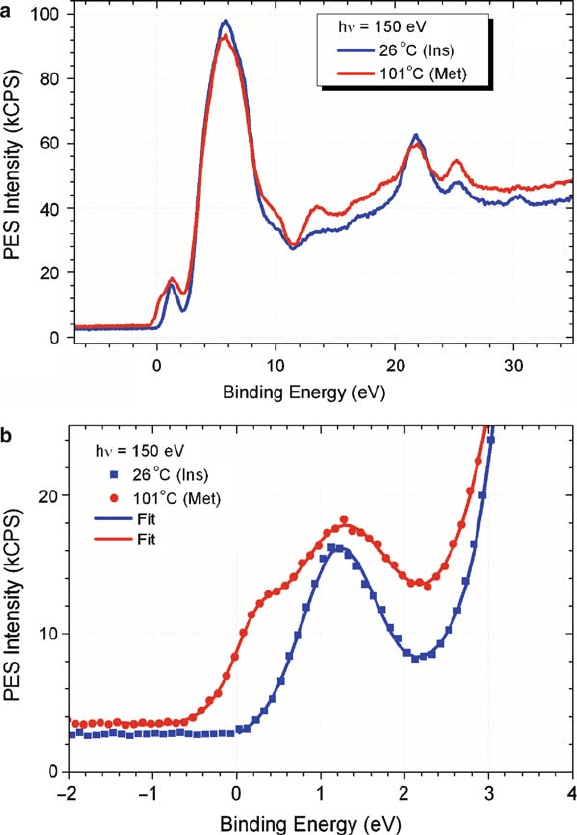
2 Metal-Insulator Transition in Thin Film Vanadium Dioxide 77
Fig. 2.18 Photoemission results on a VO
2
thin film in the metallic .101
ı
C/ and semiconducting
state .26
ı
C/.(a) Measured data. The positions of the peaks below the prominent O-2p peak do
not change upon crossing the MIT. (b) Magnified view of the near-Fermi level structure. Sym-
bols are measured data. Calculated fits are shown by solid lines. (From Ruzmetov et al. [16] with
permission. Copyright (2008) by the IOP Publishing Ltd)
is of the n-type. This is in agreement with previous studies on carrier transport in
single crystal VO
2
by Rosevear et al. indicating the carriers to be of n-type across
the transition boundary [66].
