Peterson D.R., Bronzino J.D. (Eds.) Biomechanics: Principles and Applications
Подождите немного. Документ загружается.

3-22 Biomechanics
␥
␣
cp
op
cg
tr
TEL
C-LINE
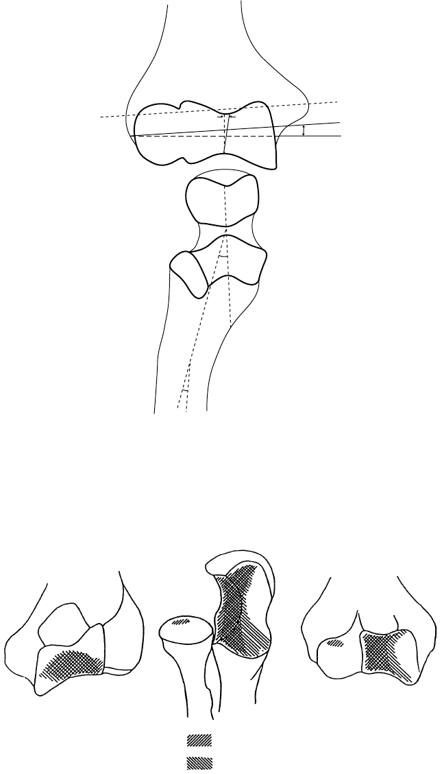
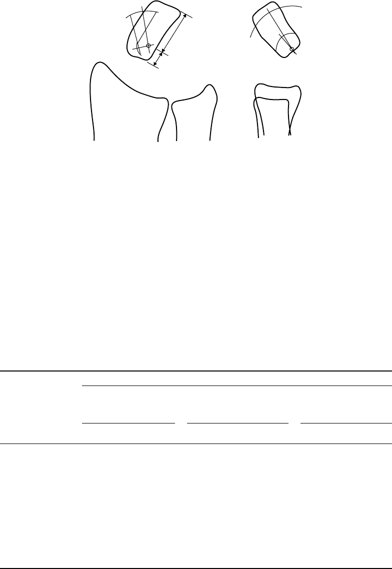
FIGURE 3.23 Components contributing to the carrying angles: α + λ + ψ.Key:α, angle between C-line and TEL;
γ , inclination of central groove (cg); λ, angle between trochlear notch (tn); ψ, reverse angulation of shaft of ulna;
TLE, transepicondylar line; C-line, line joining centers of curvature of the trochlea and capitellum; cg, central groove;
op, olecranon process; tr, trochlear ridge; cp, coronoid process. α = 2.5 ±0.0; λ = 17.5 ±5.0 (females) and 12.0 ±7.0
(males); ψ =−6.5 ± 0.7 (females) and −9.5 ± 3.5 (males). (From Shiba R., Sorbie C., Siu D.W., Bryant J.T.,
Cooke T.D.V., and Weavers H.W. 1988. J. Orthop. Res. 6: 897. With permission.)
(Posterior view)
Loads:
Valgus
Joint angle: 90°
(Anterior view)
Varus
FIGURE 3.24 Contact of the ulnohumeral joint with varus and valgus loads and the elbow at 90
◦
. Notice only
minimal radiohumeral contact in this loading condition. (From Stormont T.J., An K.N., Morrey B.F., and Chae E.Y.
1985. J. Biomech. 18: 329. Reprinted with permission of Elsevier Science Inc.)
Joint-Articulating Surface Motion 3-23
TABLE 3.15 Elbow Joint Contact Area
Total Articulating Surface
Area of Ulna and
Position Radial Head (mm
2
) Contact Area (%)
Full extension 1598 ±103 8.1 ±2.7
90
◦
flexion 1750 ±123 10.8 ± 2.0
Full flexion 1594 ±120 9.5 ±2.1
Source: Goel V.K., Singh D., and Bijlani V. 1982. J. Biomech. Eng.
104: 169.
2.5mm
90
100
80
70
60
50
40
30
20
10
120
110
0
7.8mm
Z
X
Y
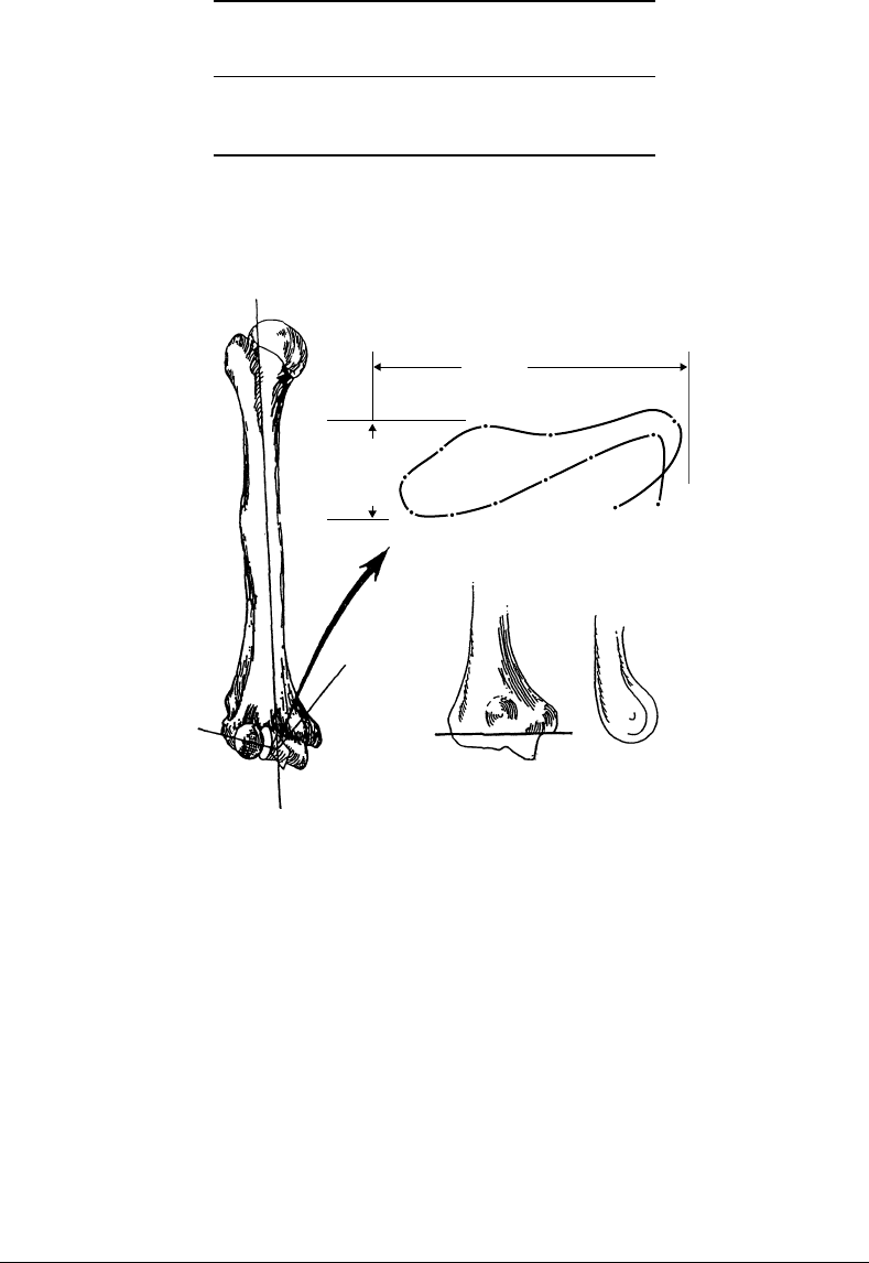
FIGURE 3.25 Very small locus of instant center of rotation for the elbow joint demonstrates that the axis may be
replicated by a single line drawn from the inferior aspect of the medial epicondyle through the center of the lateral
epicondyle, which is in the center of the lateral projected curvature of the trochlea and capitellum. (From Morrey B.F.
and Chao E.Y.S. 1976. J. Bone Joint Surg. 58A: 501. With permission.)
10
◦
; at full flexion there is a varus angulation of 8
◦
[Morrey and Chao, 1976]. More recently, the three-
dimensional kinematics of the ulno–humeral joint under simulated active elbow joint flexion–extension
was obtained by using an electromagnetic tracking device [Tanaka et al., 1998]. The optimal axis to best
represent flexion–extension motion was found to be close to the line joining the centers of the capitellum
and the trochlear groove. Furthermore, the joint laxity under valgus–varus stress was also examined. With
the weight of the forearm as the stress, a maximum of 7.6
◦
valgus–varus and 5.3
◦
of axial rotation laxity
were observed.
3.6 Wrist
The wrist functions by allowing changes of orientation of the hand relative to the forearm. The wrist joint
complex consists of multiple articulations of eight carpal bones with the distal radius, the structures of the
3-24 Biomechanics
11.0
9.75
7.5
5.25
3.0
0.75
–1.5
–3.5
–6.0
0204060
Elbow flexion (Φ°)
Carrying angle (Φ°)
80 100 120
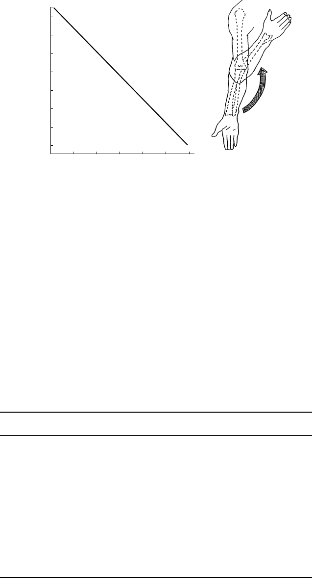
FIGURE 3.26 During elbow flexion and extension, a linear change in the carrying angle is demonstrated, typically
going from valgus in extension to varus in flexion. (From Morrey B.F. and Chao E.Y.S. 1976. J. Bone Joint Surg. 58A:
501. With permission.)
ulnocarpal space, the metacarpals, and each other. This collection of bones and soft tissues is capable of a
substantial arc of motion that augments hand and finger function.
3.6.1 Geometry of the Articulating Surfaces
The global geometry of the carpal bones has been quantified for grasp and active isometric contraction of
the elbow flexors [Schuind et al., 1992]. During grasping there is a significant proximal migration of the
radius of 0.9 mm, apparent shortening of the capitate, a decrease in the carpal height ratio, and an increase
in the lunate uncovering index (Table 3.16). There is also a trend toward increase of the distal radioulnar
TABLE 3.16 Changes of Wrist Geometry with Grasp
Analysis of
Resting Grasp Variance (p = Level)
Distal radioulnar joint space (mm) 1.6 ± 0.31.8 ±0.6 0.06
Ulnar variance (mm) −0.2 ±1.60.7 ± 1.8 0.003
Lunate, uncovered length (mm) 6.0 ±1.97.6 ± 2.6 0.0008
Capitate length (mm) 21.5 ± 2.220.8 ± 2.3 0.0002
Carpal height (mm) 33.4 ±3.431.7 ±3.4 0.0001
Carpal ulnar distance (mm) 15.8 ± 4.015.8 ± 3.0NS
Carpal radial distance (mm) 19.4 ±1.819.7 ±1.8NS
Third metacarpal length (mm) 63.8 ±5.862.
6 ±5.5NS
Carpal height ratio 52.4 ±3.350.6 ±4.1 0.02
Carpal ulnar ratio 24.9 ±5.925.4 ±5.3NS
Lunate uncovering index 36.7 ± 12.145.3 ± 14.2 0.002
Carpal radial ratio 30.6 ± 2.431.6 ± 2.3NS
Radius — third metacarpal angle (deg) −0.3 ± 9.2 −3.1 ± 12.8NS
Radius — capitate angle (deg) 0.4 ± 15.4 −3.8 ± 22.2NS
Note: 15 normal subjects with forearm in neutral position and elbow at 90
◦
flexion.
Source: Schuind F.A., Linscheid R.L., An K.N., and Chao E.Y.S. 1992. J. Hand Surg. 17A: 698.
Joint-Articulating Surface Motion 3-25
joint with grasping. The addition of elbow flexion with concomitant grasping did not significantly change
the global geometry, except for a significant decrease in the forearm interosseous space [Schuind et al.,
1992].
3.6.2 Joint Contact
Studies of the normal biomechanics of the proximal wrist joint have determined that the scaphoid and
lunate bones have separate, distinct areas of contact on the distal radius/triangular fibrocartilage complex
surface [Viegas et al., 1987] so that the contact areas were localized and accounted for a relatively small frac-
tion of the joint surface, regardless of wrist position (average of 20.6%). The contact areas shift from a more
volar location to a more dorsal location as the wrist moves from flexion to extension. Overall, the scaphoid
contact area is 1.47 times greater than that of the lunate. The scapho-lunate contact area ratio generally
increases as the wrist position is changed from radial to ulnar deviation and/or from flexion to extension.
Palmer and Werner [1984] also studied pressures in the proximal wrist joint and found that there are three
distinct areas of contact: the ulno-lunate, radio-lunate, and radio-scaphoid. They determined that the peak
articular pressure in the ulno-lunate fossa is 1.4 N/mm
2
, in the radio-ulnate fossa is 3.0 N/mm
2
, and in the
radio-scaphoid fossa is 3.3 N/mm
2
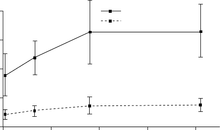
. Viegas et al. [1989] found a nonlinear relationship between increasing
load and the joint contact area (Figure 3.27). In general, the distribution of load between the scaphoid and
lunatewasconsistentwith all loadstested,with 60% ofthe total contactareainvolving the scaphoid and40%
involving the lunate. Loads greater than 46 lbs were found to not significantly increase the overall contact
area. The overall contact area, even at the highest loads tested, was not more than 40% of the available joint
surface.
Horii et al. [1990] calculated the total amount of force born by each joint with the intact wrist in the
neutral position in the coronal plane and subjected to a total load of 143 N (Table 3.17). They found that
22% of the total force in the radio-ulno–carpal joint is dissipated through the ulna (14% through the ulno-
lunate joint, and 18% through the ulno–triquetral joint) and 78% through the radius (46% through the
scaphoid fossa and 32% through the lunate fossa). At the midcarpal joint, the scapho–trapezial joint
CONT AREA (mm
2
)
CONT AREA (%)
150
100
50
10 30 50
Load (Ibs)
70 90
Contact area (mm
2
),(%)
200
0
Contact area/load (NNN)
FIGURE 3.27 The nonlinear relation between the contact area and the load at the proximal wrist joint. The contact
area was normalized as a percentage of the available joint surface. The load of 11, 23, 46, and 92 lbs was applied
at the position of neutral pronation/supination, neutral radioulnar deviation, and neutral flexion/extension. (From
ViegasS.F.,PattersonR.M., Peterson P.D.,RoefsJ.,TencerA., and Choi S.1989. J. HandSurg. 14A: 458. With permission.)

3-26 Biomechanics
TABLE 3.17 Force Transmission at the Intercarpal Joints
Joint Force (N)
Radio-ulno-carpal
Ulno-triquetral 12 ±3
Ulno-lunate 23 ±8
Radio-lunate 52 ±8
Radio-scaphoid 74 ±13
Midcarpal
Triquetral-hamate 36 ±6
Luno-capitate 51 ±6
Scapho-capitate 32 ±4
Scapho-trapezial 51 ±8
Note: A total of 143 N axial force applied across the wrist.
Source: Horii E., Garcia-Elias M., An K.N., Bishop A.T.,
Cooney W.P., Linscheid R.L., and Chao E.Y. 1990. J. Bone Joint
Surg. 15A: 393.
transmits 31% of the total applied force, the scapho–capitate joint transmits 19%, the luno-capitate joint
transmits 29%, and the triquetral-hamate joints transmits 21% of the load.
A limited amount of studies have been done to determine the contact areas in the midcarpal joint.
Viegas et al. [1990] have found four general areas of contact: the scapho-trapezial-trapezoid (STT), the
scapho-capitate (SC), the capito-lunate (CL), and the triquetral-hamate (TH). The high pressure contact
area accounted for only 8% of the available joint surface with a load of 32 lbs and increased to a maximum
of only 15% with a load of 118 lbs. The total contact area, expressed as a percentage of the total available
joint area for each fossa was: STT = 1.3%, SC = 1.8%, CL = 3.1%, and TH = 1.8%.
The correlation between the pressure loading in the wrist and the progress of degenerative osteoarthri-
tis associated with pathological conditions of the forearm was studied in a cadaveric model [Sato, 1995].
Malunion after distal radius fracture, tear of triangular fibrocartilage, and scapholunate dissociation were
all responsible for the alteration of the articulating pressure across the wrist joint. Residual articular incon-
gruity of the distal radius following intra-articular fracture has been correlated with early osteoarthritis.
In an in vitro model, step-offs of the distal radius articular incongruity were created. Mean contact stress
was significantly greater than the anatomically reduced case at only 3 mm of step-off [Anderson et al.,
1996].
3.6.3 Axes of Rotation
The complexity of joint motion at the wrist makes it difficult to calculate the instant center of motion.
However, the trajectories of the hand during radioulnar deviation and flexion/extension, when they occur
in a fixed plane, are circular, and the rotation in each plane takes place about a fixed axis. These axes are
located within the head of the capitate and are not altered by the position of the hand in the plane of
rotation [Youm et al., 1978]. During radioulnar deviation, the instant center of rotation lies at a point
in the capitate situated distal to the proximal end of this bone by a distance equivalent to approximately
one-quarter of its total length (Figure 3.28). During flexion/extension, the instant center is close to the
proximal cortex of the capitate, which is somewhat more proximal than the location for the instant center
of radioulnar deviation.
Normal carpal kinematics were studied in 22 cadaver specimens using a biplanar radiography method.
The kinematics of the trapezium, capitate, hamate, scaphoid, lunate, and triquetrum were determined
during wrist rotation in the sagittal and coronal plane [Kobagashi et al., 1997]. The results were expressed
using the concept of the screw displacement axis and covered to describe the magnitude of rotation about
and translation along three orthogonal axes. The orientation of these axes is expressed relative to the radius
Joint-Articulating Surface Motion 3-27
1
3
FIGURE 3.28 The location of the center of rotation during ulnar deviation (left) and extension (right), determined
graphically using two metal markers embedded in the capitate. Note that during radial–ulnar deviation the center lies
at a point in the capitate situated distal to the proximal end of this bone by a distance equivalent to approximately
one-quarter of its total longitudinal length. During flexion–extension, the center of rotation is close to the proximal
cortex of the capitate. (From Youm Y., McMurty R.Y., Flatt A.E., and Gillespie T.E. 1978. J. Bone Joint Surg. 60A: 423.
With permission.)
during sagittal plane motion of the wrist (Table 3.18). The scaphoid exhibited the greatest magnitude of
rotation and the lunate displayed the least rotation. The proximal carpal bones exhibited some ulnar
deviation in 60
◦
of wrist flexion. During coronal plane motion (Table 3.19), the magnitude of radial–ulnar
deviation of the distal carpal bones was mutually similar and generally of a greater magnitude than that of
the proximal carpal bones. The proximal carpal bones experienced some flexion during radial deviation
of the wrist and extension during ulnar deviation of the wrist.
TABLE 3.18 Individual Carpal Rotation Relative to the Radius (Deg) (Sagittal Plane Motion of the Wrist)
Axis of Rotation
Z
XY(+) Ulnar Deviation;
(+) Pronation; (−) Supination (+) Flexion; (−) Extension (−) Radial Deviation
Wrist Motion
a
Carpal Bone N-E60 N-E30 N-F30 N-F60 N-E60 N-E30 N-F60 N-E60 N-E60 N-E30 N-F30 N-F60
Trapezium (N = 13) −0.9 −1.3 0.9 −1.4 −59.4 −29.3 28.7 54.2 1.2 0.3 −0.4 2.5
SD 2.8 2.2 2.6 2.7 2.3 1 1.8 3 4 2.7 1.3 2.8
Capitate (N = 22) 0.9 −1 1.3 −1.6 60.3 −30.2 21.5 63.5 0 0 0.6 3.2
SD 2.7 1.8 2.5 3.5 2.5 1.1 1.2 2.8 2 1.4 1.6 3.6
Hamate (N = 9) 0.4 −1 1.3 −0.3 −59.5 −29 28.8 62.6 2.1 0.7 0.1 1.8
SD 3.4 1.7 2.5 2.4 1.4 0.8 10.2 3.6 4.4 1.8 1.2 4.1
Scaphoid (N = 22) −2.5 −0.7 1.6 2 −52.3 −26 20.6 39.7 4.5 0.8 2.1 7.8
SD 3.4 2.6 2.2 3.1 3 3.2 2.8 4.3 3.7 2.1 2.2 4.5
Lunate (N = 22) 1.2 0.5 0.3 −2.2 −29.7 −15.4 11.5 23 4.3 0.9 3.3 11.1
SD 2.8 1.8 1.7 2.8 6.6 3.9 3.9 5.9 2.6 1.5 1.9 3.4
Triquetrum (N = 22) −3.5 −2.5 2.5 −0.7 −39.3 −20.1 15.5 30.6 0 −0.3 2.4 9.8
SD 3.5 2 2.2 3.7 4.8 2.7 3.8 5.1 2.8 1.4 2.6 4.3
a
N-E60: neutral to 60
◦
of extension; N-E30: neutral to 30
◦
of extension; N-F30: neutral to 30
◦
of flexion; N-F60: neutral to
60
◦
of flexion.
SD = standard deviation.
Source: Kobayashi M., Berger R.A., Nagy L. et al. 1997. J. Biomech. 30: 8, 787.

3-28 Biomechanics
TABLE 3.19 Individual Carpal Rotation to the Radius (Deg) (Coronal Plane Motion of the Wrist)
Axis of Rotation
Z
XY(+) Ulnar Deviation;
(+) Pronation; (−) Supination (+) Flexion; (−) Extension (−) Radial Deviation
Wrist Motion
a
Carpal Bone N-RD15 N-UD15 N-UD30 N-RD15 N-UD15 N-UD30 N-RD15 N-UD15 N-UD30
Trapezium (N = 13) −4.8 9.1 16.3 0 4.9 9.9 −14.3 16.4 32.5
SD 2.4 3.6 3.6 1.5 1.3 2.1 2.3 2.8 2.6
Capitate (N = 22) −3.9 6.8 11.8 1.3 2.7 6.5 −14.6 15.9 30.7
SD 2.6 2.6 2.5 1.5 1.1 1.7 2.1 1.4 1.7
Hamate (N = 9) −4.8 6.3 10.6 1.1 3.5 6.6 −15.5 15.4 30.2
SD 1.8 2.4 3.1 3 3.2 4.1 2.4 2.6 3.6
Scaphoid (N = 22) 0.8 2.2 6.6 8.5 −12.5 −17.1 −4.2 4.3 13.6
SD 1.8 2.4 3.1 3 3.2 4.1 2.4 2.6 3.6
Lunate (N = 22) −1.2 1.4 3.9 7 −13.9 −22.5 −1.7 5.7 15
SD 1.6 0 3.3 3.1 4.3 6.9 1.7 2.8 4.3
Triquetrum (N = 22) −1.1 −1 0.8 4.1 −10.5 −17.3 −5.1 7.7 18.4
SD 1.4 2.6 4 3 3.8 6 2.4 2.2 4
a
N-RD15: neutral to 15
◦
of radial deviation; N-UD30: neutral to 30
◦
of ulnar deviation; N-UD15: neutral to 15
◦
of ulnar
deviation.
SD = standard deviation.
Source: Kobayashi M., Berger R.A., Nagy L. et al. 1997. J. Biomech. 30: 8, 787.
3.7 Hand
The hand is an extremely mobile organ that is capable of conforming to a large variety of object shapes
and coordinating an infinite variety of movements in relation to each of its components. The mobility
of this structure is possible through the unique arrangement of the bones in relation to one another, the
articular contours, and the actions of an intricate system of muscles. Theoretical and empirical evidence
suggest that limb joint surface morphology is mechanically related to joint mobility, stability, and strength
[Hamrick, 1996].
3.7.1 Geometry of the Articulating Surfaces
Three-dimensional geometric models of the articular surfaces of the hand have been constructed. The
sagittal contours of the metacarpal head and proximal phalanx grossly resemble the arc of a circle
[Tamai et al., 1988]. The radius of curvature of a circle fitted to the entire proximal phalanx surface
ranges from 11 to 13 mm, almost twice as much as that of the metacarpal head, which ranges from 6
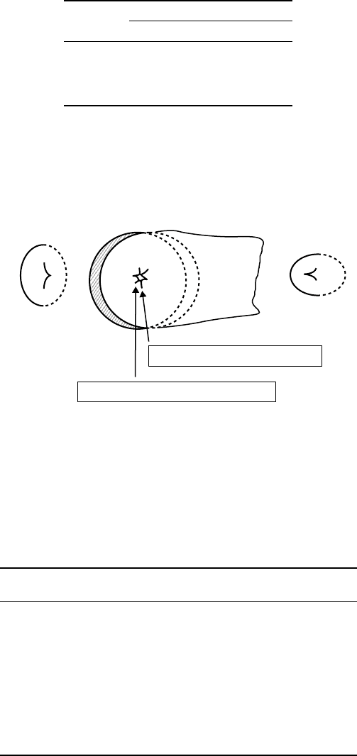
to 7 mm (Table 3.20). The local centers of curvature along the sagittal contour of the metacarpal
heads are not fixed. The locus of the center of curvature for the subchondral bony contour approx-
imates the locus of the center for the acute curve of an ellipse (Figure 3.29). However, the locus of
center of curvature for the articular cartilage contour approximates the locus of the obtuse curve of an
ellipse.
The surface geometry of the thumb carpometacarpal (CMC) joint has also been quantified [Athesian
et al., 1992].The surface area of theCMC joint is significantly greater for malesthan for females (Table 3.21).
The minimum, maximum, and mean square curvature of these joints is reported in Table 3.21. The
curvature of the surface is denoted by κ and the radius of curvature is ρ = 1/κ. The curvature is negative
when the surface is concave and positive when the surface is convex.
Joint-Articulating Surface Motion 3-29
TABLE 3.20 Radius of Curvature of the
Middle Sections of the Metacarpal Head and
Proximal Phalanx Base
Radius (mm)
Bony Contour Cartilage Contour
MCH index 6.42 ± 1.23 6.91 ± 1.03
Long 6.44 ± 1.08 6.66 ± 1.18
PPB index 13.01 ± 4.09 12.07 ±3.29
Long 11.46 ± 2.30 11.02 ±2.48
Source: Tamai K., Ryu J., An K.N., Linscheid R.L.,
Cooney W.P., and Chao E.Y.S. 1988. J. Hand Surg.
13A: 521.
A
B
C
Metacarpal head
B
C
A
C⬘
C⬘
A⬘
A⬘
B⬘B⬘
Local center of bony contour
Local center of cartilage contour
FIGURE 3.29 The loci of the local centers of curvature for subchondral bony contour of the metacarpal head
approximates the loci of the center for the acute curve of an ellipse. The loci of the local center of curvature for articular
cartilage contour of the metacarpal head approximates the loci of the bony center of the obtuse curve of an ellipse.
(From Tamai K., Ryu J., An K.N., Linscheid R.L., Cooney W.P., and Chao E.Y.S. 1988. J. Hand Surg. 13A: 521. Reprinted
with permission of Churchill Livingstone.)
TABLE 3.21 Curvature of Carpometacarpal Joint Articular Surfaces
Area
¯
κ
min
¯
κ
max
¯
κ
rms
n (cm
2
)(m
−1
)(m
−1
)(m
−1
)
Trapezium
Female 8 1.05 ± 0.21 −61 ±22 190 ±36 165 ±32
Male 5 1.63 ±0.18 −87 ±17 114 ±19 118 ±6
Total 13 1.27 ±0.35 −71 ±24 161 ±48 147 ±34
Female vs. male p ≤ 0.01 p ≤ 0.05 p ≤ 0.01 p ≤ 0.01
Metacarpal
Female 8 1.22 ± 0.36 −49 ±10 175 ±25 154 ±20
Male 5 1.74 ±0.21 −37 ±11 131 ±17 116 ±8
Total 13 1.42
± 0.40 −44 ± 12 158 ±31 140 ±25
Female vs. male p ≤ 0.01 p ≤ 0.05 p ≤ 0.01 p ≤ 0.01
Note: Radius of curvature: ρ = 1/κ.
Source: Athesian J.A., Rosenwasser M.P., and Mow V.C. 1992. J. Biomech. 25: 591.
3-30 Biomechanics
3.7.2 Joint Contact
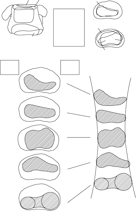
The size and location of joint contact areas of the metacarpophalangeal (MCP) joint changes as a function
of the joint flexion angle (Figure 3.30). The radioulnar width of the contact area becomes narrow in the
neutral position and expands in both the hyperextended and fully flexed positions [An and Cooney, 1991].
Neutral
45° flexion
90° flexion
Dorsal
Passive
hyperext
Active
extension
Neutral
45° Flexion
90° Flexion
Ulnar
side
Radial
side
MCH PPB
Active ext
Neutral
Passive ext
Passive ext
Active ext
Contact area
(mm
2
)
U
(a)
(b)
RRU
45° flexion
30° ext 34.5
45° flexion 33.0
90° flexion 32.8
Neutral 40.2
90° flexion
Volar
FIGURE 3.30 (a) Contact area of the MCP joint in five joint positions. (b) End on view of the contact area on each
of the proximal phalanx bases. The radioulnar width of the contact area becomes narrow in the neutral position and
expands in both the hyperextended and fully flexed positions. (From An K.N. and Cooney W.P. 1991. In B.F.
Morrey (Ed.), Joint Replacement Arthroplasty, pp. 137–146, New York, Churchill Livingstone. By permission of Mayo
Foundation.)
Joint-Articulating Surface Motion 3-31
Dorsal
Volar
Left trapezium Left metacarpal
Ulnar
16%
20%
16%
24%
14%
23%
32%
18%
Radial
Radial
Volar
Dorsal
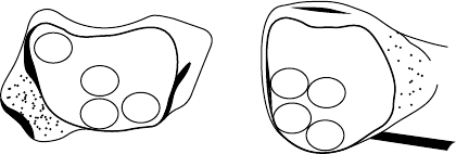
FIGURE 3.31 Summary of the contact areas for all specimens, in lateral pinch with a 25 N load. All results from
the right hand are transposed onto the schema of a carpometacarpal joint from the left thumb. (From Ateshian G.A.,
Ark J.W., Rosenwasser M.P., et al. 1995. J. Orthop. Res. 13: 450.)
In the neutral position, the contact area occurs in the center of the phalangeal base, this area being slightly
larger on the ulnar than on the radial side.
The contact areas of the thumb carpometacarpal joint under the functional position of lateral key
pinch and in the extremes of range of motion were studied using a stereophotogrammetric technique
[Ateshian et al., 1995]. The lateral pinch position produced contact predominately on the central, volar,
and volar–ulnar regions of the trapezium and the metacarpals (Figure 3.31). Pelligrini et al. [1993] noted
that the palmar compartment of the trapeziometacarpal joint was the primary contact area during flexion
adduction of the thumb in lateral pinch. Detachment of the palmar beak ligament resulted in dorsal
translation of the contact area producing a pattern similar to that of cartilage degeneration seen in the
osteoarthritic joint.
3.7.3 Axes of Rotation
Rolling and sliding actions of articulating surfaces exist during finger joint motion. The geometric shapes
of the articular surfaces of the metacarpal head and proximal phalanx, as well as the insertion location
of the collateral ligaments, significantly govern the articulating kinematics, and the center of rotation
is not fixed but rather moves as a function of the angle of flexion [Pagowski and Piekarski, 1977]. The
instant centers of rotation are within 3 mm of the center of the metacarpal head [Walker and Erhman,
1975]. Recently the axis of rotation of the MCP joint has been evaluated in vivo by Fioretti [1994]. The
instantaneous helical axis of the MCP joint tends to be more palmar and tends to be displaced distally as
flexion increases (Figure 3.32).
The axes of rotation of the CMC joint have been described as being fixed [Hollister et al., 1992],
but others believe that a polycentric center of rotation exists [Imaeda et al., 1994]. Hollister et al.
[1992] found that axes of the CMC joint are fixed and are not perpendicular to each other, or to the
bones, and do not intersect. The flexion/extension axis is located in the trapezium, and the abduc-
tion/adduction axis is on the first metacarpal. In contrast, Imaeda et al. [1994] found that there was
no single center of rotation, but rather the instantaneous motion occurred reciprocally between centers
of rotations within the trapezium and the metacarpal base of the normal thumb. In flexion/extension,
the axis of rotation was located within the trapezium, but for abduction/adduction the center of
rotation was located distally to the trapezium and within the base of the first metacarpal. The average
instantaneous center of circumduction was at approximately the center of the trapezial joint surface
(Table 3.22).
