Peterson D.R., Bronzino J.D. (Eds.) Biomechanics: Principles and Applications
Подождите немного. Документ загружается.


2-6 Biomechanics
TABLE 2.2 Skeletal Muscle Specific Tension
Specific
Species Muscle Type Preparation Tension (kPa) References
Various Synthesis 300 Josephson [1989]
Rat SO Single fiber 134 Fitts et al. [1991]
Human Slow Single fiber 133 Fitts et al. [1991]
Rat FOG Single fiber 108 Fitts et al. [1991]
Rat FG Single fiber 108 Fitts et al. [1991]
Human Fast Single fiber 166 Fitts et al. [1991]
Cat 1 Motor unit 59 Dum et al. [1982]
Cat S Motor unit 172 Bodine et al. [1987]
Cat 2A Motor unit 284 Dum et al. [1982]
Cat FR Motor unit 211 Bodine et al. [1987]
Cat 2B+2AB Motor unit 343 Dum et al. [1982]
Cat FF/FI Motor unit 249 Bodine et al. [1987]
Human Elbow Whole muscle 230–420 Edgerton et al. [1990]
Human Ankle Whole muscle 45–250 Fukunaga et al. [1996]
Rat TA Whole muscle 272 Wells [1965]
Rat Soleus Whole muscle 319 Wells [1965]
Guinea pig Hindlimb Whole muscle 225 Powell et al. [1984]
Guinea pig Soleus Whole muscle 154 Powell et al. [1984]
TABLE 2.3 Muscle Dynamic Properties
Species Muscle Type Preparation V
a
max
a/P o b/V
max
References
Rat SO Single fiber 1.49 μ/sec Fitts et al. [1991]
Human slow Single fiber 0.86 μ/sec Fitts et al. [1991]
Rat FOG Single fiber 4.91 μ/sec Fitts et al. [1991]
Rat FG Single fiber 8.05 μ/sec Fitts et al. [1991]
Human Fast Single fiber 4.85 μ/sec Fitts et al. [1991]
Mouse Soleus Whole muscle 31.7 μ/sec Close [1972]
Rat Soleus Whole muscle 18.2 μ/sec Close [1972]
Rat Soleus Whole muscle 5.4 cm/sec 0.214 0.23 Wells [1965]
Cat Soleus Whole muscle 13 μ/sec Close [1972]
Mouse EDL Whole muscle 60.5 μ/sec Close [1972]
Rat EDL Whole muscle 42.7 μ/sec Close [1972]
Cat EDL Whole muscle 31 μ/sec Close [1972]
Rat TA Whole muscle 14.4 cm/sec 0.356 0.38 Wells [1965]
a
L/sec fiber orsarcomerelengths per second, μm/secsarcomerevelocity; cm/sec whole muscle velocity.
The intrinsic velocity of shortening has been experimentally determined for a number of mus-
cle types (Table 2.3). Maximum contraction velocity of a given muscle can thus be calculated based
on a knowledge of the number of serial sarcomeres within the muscle multiplied by the maximum
contraction velocity of an individual sarcomere (Table 2.4 to Table 2.6). Sarcomere shortening
velocity varies widely among species and fiber types (Table 2.3).
2.3 Modeling
2.3.1 Cartilage
Although cartilage can be modeled as a simple elastic element, more accurate results are obtained
using a biphasic model [Mow et al., 1980], which describes the motion of the hydrating fluid relative

Musculoskeletal Soft Tissue Mechanics 2-7
TABLE 2.4 Architectural Properties of the Human Arm and Forearm
a,b
Muscle Mass Muscle Fiber Length Pennation Cross-Sectional
Muscle (g) Length (mm) (mm) Angle (◦) Area (cm
2
) FL/ML Ratio
BR (n = 8) 16.6 ±2.8 175 ±8.3 121 ±8.32.4 ± 61.33 ±22 0.69 ± 0.062
PT (n = 8) 15.9 ±1.7 130 ±4.736.4 ± 1.39.6 ±84.13 ± 52 0.28 ± 0.012
PQ (n = 8) 5.21 ±1.039.3 ±2.323.3 ±2.09.9 ± 32.07 ±33 0.58 ± 0.
021
EDCI(n = 8) 3.05 ±0.45 114 ± 3.456.9 ±3.63.1 ± 50.52 ± 0.08 0.49 ±0.024
EDCM(n = 5) 6.13 ± 1.2 112 ±4.758.8 ± 3.53.2 ±1.01.02 ± 0.20 0.50 ± 0.014
EDCR(n = 7) 4.70 ± 0.75 125 ± 10.751.2 ±1.83.2 ± 54 0.86
± 0.13 0.42 ± 0.023
EDCS(n = 6) 2.23 ±0.32 121 ± 8.052.9 ± 5.22.4 ±70.40 ±0.06 0.43 ± 0.029
EDQ (n = 7) 3.81 ± 70 152 ± 9.255.3 ± 3.72.6 ±60.64 ± 0.10 0.36 ± 0.012
EIP (n = 6) 2.86 ± 61 105 ±6.648.4 ± 2.36.3 ±
80.56 ±0.11 0.46 ± 0.023
EPL (n = 7) 4.54 ±68 138 ± 7.243.6 ± 2.65.6 ±1.30.98 ± 0.13 0.31 ± 0.020
PL (n = 6) 3.78 ±82 134 ± 11.552.3 ±3.13.5 ± 1.20.69 ±0.17 0.40 ± 0.032
FDS I(P) (n = 6) 6.01 ±1.192.5 ± 8.431
.6 ±3.05.1 ± 0.21.81 ±0.83 0.34 ± 0.022
FDS I(D) (n = 9) 6.6 ±0.8 119 ± 6.137.9 ± 3.06.7 ±0.31.63 ± 0.22 0.32 ± 0.013
FDS I(C) (n = 6) 12.4 ±2.1 207 ±10.767.6 ± 2.85.7 ±0.21.71 ± 0.28 0.33 ± 0.025
FDSM(
n = 9) 16.3 ±2.2 183 ±11.560.8 ±3.96.9 ± 0.72.53 ±0.34 0.34 ± 0.014
FDSR(n = 9) 10.2 ± 1.1 155 ± 7.760.1 ± 2.74.3 ±0.61.61 ± 0.18 0.39 ±0.023
FDSS(n = 9) 1.8 ± 0.3 103 ±6.342.4 ±2.24.9 ±0.70
.40 ±0.05 0.42 ± 0.014
FDPI(n = 9) 11.7 ± 1.2 149 ±3.861.4 ± 2.47.2 ±0.71.77 ± 0.16 0.41 ±0.018
FDPM(n = 9) 16.3 ±1.7 200 ±8.268.4 ± 2.75.7 ± 0.32.23 ±0.22 0.34 ± 0.011
FDPR(n = 9) 11.9 ±1.4 194 ±7.
064.6 ±2.66.8 ± 0.51.72 ± 0.18 0.33 ± 0.009
FDPS(n = 9) 13.7 ±1.5 150 ±4.760.7 ± 3.97.8 ±0.92.20 ± 0.30 0.40 ±0.015
FPL (n = 9) 10.0 ±1.1 168 ±10.045.1 ± 2.16.9 ±0.22.08 ± 0.22 0.24 ±0.10
a
Data from Lieber et al., 1990, 1992.
b
BR: brachioradialis; EDC I, EDC M, EDC R, and EDC S: extensor digitorum communis to the index, middle, ring, and
small fingers, respectively; EDQ: extensor digiti quinti; EIP: extensor indicis proprious; EPL: extensor pollicis longus; FDP I,
FDP M, FDP R, and FDP S: flexor digitorum profundus muscles; FDS I, FDS M, FDS R, and FDS S: flexor digitorum
superficialis muscles; FDS I(P) and FDS I(D): proximal and distal bellies of the FDS I; FDS I(C): the combined properties
of the two bellies as if they were a single muscle; FPL: flexor pollicis longus; PQ: pronator quadratus; PS: palmaris longus;
PT: pronator teres.
to the charged organic matrix. The total stress acting on the cartilage is separated into independent
solid and fluid phases:
σ
T
= σ
s
+ σ
f
where s denotes the solid phase and f the fluid phase. The relative motion of the phases defines the
equilibrium equations
∇·σ
s
=
(ν
s
− ν
f
)
k(1 + α)
2
=−∇·σ
f
where α is tissue solid content and k the tissue permeability coefficient. In addition to the equilibrium
equations, each phase is subject to separate constitutive relations:
σ
f
=−p
a
I and σ
s
=−αp
a
I + De
where p
a
is the apparent tissue stress, D is the material property tensor, and e is the strain tensor. For a
hyperelastic solid phase
De = λTr (e)I +2μe
where λ and μ are the Lam
´
e constants.

2-8 Biomechanics
TABLE 2.5 Architectural Properties of Human Lower Limb
a,b
Muscle Mass Muscle Fiber Length Pennation Cross-Sectional
Muscle (g) Length (mm) (mm) Angle (◦) Area (cm
2
) FL/ML Ratio
RF (n = 3) 84.3 ±14 316 ± 5.766.0 ±1.55.0 ±0.012.7 ±1.90.209 ±0.002
VL (n = 3) 220 ±56 324 ± 14 65.7 ±0.88 5.0 ± 0.030.6 ± 6.50.203 ± 0.007
VM (n = 3) 175 ± 41 335 ±15 70.3 ± 3.35.0 ± 0.021.1 ± 4.30.210 ± 0.005
VI (
n = 3) 160 ±59 329 ± 15 68.3 ±4.83.3 ±1.722.3 ±8.70.208 ±0.007
SM (n = 3) 108 ± 13 262 ±1.562.7 ±4.715±2.916.9 ± 1.50.239 ± 0.017
BF
L
(n = 3) 128 ± 28 342 ±14 85.3 ± 5.00.0 ± 0.012.8 ± 2.80.251 ±0.022
BF
s
(n = 3) — 271 ±11 139 ± 3.523±0.9—0.517 ±0.032
ST (n = 2) 76.9 ±7.7 317 ± 4 158 ± 2.05.0 ± 0.05.4 ± 1.00.498 ±0.0
SOL (n = 2) 215 (n = 1) 310 ± 1.519.5 ±0.525± 5.0 58.0 (n = 1) 0.063 ± 0.002
MG (n = 3) 150 ± 14 248 ±9.935.3 ±2.016.
7 ±4.432.4 ±3.10.143 ±0.010
LG (n = 3) — 217 ±11 50.7 ±5.68.3 ±1.7—0.233 ±0.016
PLT (n = 3) 5.30 ±14 85.0 ± 15 39.3 ± 6.73.3 ± 1.71.2 ±0.40.467 ±0.031
FHL (n = 3) 21.5 ± 3.3 222 ± 5.034.0 ±1.510.0 ± 2
.95.3 ±0.60.154 ±0.010
FDL (n = 3) 16.3 ±2.8 260 ±15 27.0 ±0.58 6.7 ± 1.75.1 ± 0.70.104 ± 0.004
PL (n = 3) 41.5 ±8.5 286 ± 17 38.7 ±3.210.0 ± 0.012.3 ± 2.90.136 ± 0.010
PB (n = 3) 17.3 ±2.5 230 ±13 39.3 ±
3.55.0 ± 0.05.7 ±1.0 0170 ± 0.006
TP (n = 3) 53.5 ± 7.3 254 ± 26 24.0 ±4.011.7 ± 1.720.8 ±30.095 ±0.015
TA (n = 3) 65.7 ± 10 298 ±12 77.3 ±7.85.0 ±0.09.9 ±1.50.258 ±0.015
EDL (n = 3) 35.2 ± 3.6 355 ± 13 80.3
± 8.48.3 ± 1.75.6 ±0.60.226 ±0.024
EHL (n = 3) 12.9 ±1.6 273 ± 2.487.0 ± 8.06.0 ± 1.01.8 ± 0.20.319 ± 0.030
SAR (n = 3) 61.7 ±14 503 ± 27 455 ±19 0.0 ±0.01.7 ±0.30.906 ± 0.017
GR (n = 3) 35.3 ± 7.4 335 ±
20 277 ± 12 3.3 ± 1.71.8 ±0.30.828 ±0.017
AM (n = 3) 229 ±32 305 ± 12 115 ±7.90.0 ± 0.018.2 ±2.30.378 ± 0.013
AL (n = 3) 63.5 ± 16 229 ± 12 108 ± 2.06.0 ± 1.06.8 ± 1.90.475 ±0.023
AB (n = 3) 43.8 ± 8.4 156 ± 12 103 ± 6.40.0
± 0.04.7 ± 1.00.663 ±0.036
PEC (n = 3) 26.4 ± 6.0 123 ± 4.5 104 ± 1.20.0 ± 0.02.9 ± 0.60.851 ± 0.040
POP (n = 2) 20.1 ±2.4 108 ± 7.029.0 ± 7.00.0 ± 0.07.9 ±1.40.265 ±0.048
a
Data from Wickiewicz et al., 1982.
b
AB, adductor brevis; AL, adductor longus; AM, adductor magnus; BF
L
, biceps femoris, long head; BF
S
, biceps femoris,
short head; EDL, extensor digitorum longus; EHL, extensor hallucis longus; FDL, flexor digitorum longus; GR, gracilis;
FHL, flexor hallucis longus; LG, lateral gastrocnemius; MG, medical gastrocnemius; PEC, pectineus; PB, peroneus
brevis; PL, peronius longus; PLT, plantaris; POP, popliteus; RF, rectus femoris; SAR, sartorius; SM, semimembranosus;
SOL, soleus; ST, semitendinosus; TA, tibialis anterior; TP, tibialis posterior; VI, vastus intermedius; VL, vastus lateralis;
VM, vastus medialis.
These equations can be solved analytically for the special case of confined compression against a porous
platen [Mow et al., 1980]. The surface displacement during creep under an applied load f
0
is
u
h
=
f
0
H
A
1 −
2
π
2
2
n=0
n +
1
2
−2
exp
−π
2
n +
1
2
2
H
A
kf
(1 +2a
0
)h
2
where h is the tissue thickness, and H
A
is the aggregate modulus (λ + 2μ). Those authors estimate k as
7.6 ± 3.0 × 10
−13
m
4
/Nsec and H
A
as 0.70 ± 0.09 MPa for bovine articular cartilage. Chen et al. [2001]
report strongly depth-dependent values for H
A
ranging between 1.16 ±0.20 MPa in the superficial zone to
7.75 ±1.45 MPa in the deep zone in human articular cartilage. The biphasic approach has been extended
to finite element modeling, resulting in the u–p class of models [Wayne et al., 1991].
2.3.2 Tendon and Ligament
The composition and structure of the tensile soft tissues is quite similar to that of cartilage, and the
biphasic theory can be applied to them as well. Fluid pressure serves a smaller role in tissues loaded in

Musculoskeletal Soft Tissue Mechanics 2-9
TABLE 2.6 Architectural Properties of Human Foot
a,b
Muscle Volume Muscle Length Fiber Length Cross-Sectional
Muscle (cm
3
) (mm) (mm) Area (cm
2
)
ABDH 15.2 ± 5.3 115.8 ±4.923.0 ±5.56.68 ±2.07
ABDM 8.8 ± 4.7 112.8 ± 19.023.9 ±7.43.79 ± 1.83
ADHT 1.1 ±0.624.8 ± 4.218.7 ± 5.20.62 ±0.26
ADHO 9.1 ± 3.167.4 ± 4.618.6 ±5.34.94 ± 1.36
EDB2 2
.1 ±1.269.8 ± 16.828.0 ± 6.50.79 ± 0.43
EDB3 1.3 ± 0.782.2 ±20.726.4 ± 5.10.51 ±0.30
EDB4 1.0 ± 0.770.4 ±21.123.1 ± 3.80.44 ±0.29
EHB 3.6 ±1.565.7 ± 8.527.9 ± 5.71.34 ±0.66
FDB2 4.
5 ±2.392.9 ± 15.025.4 ± 4.51.78 ± 0.79
FDB2 3.2 ±1.598.8 ±18.122.8 ±4.01.49 ± 0.71
FDB4 2.6 ±1.0 103.0 ± 9.220.8 ± 4.51.26 ±0.47
FDB5 0.7 ±0.383.2 ±3.018.2 ± 2.20.35 ± 0.16
FDMB 3.4
± 1.751.0 ±5.317.7 ± 3.82.00 ± 1.02
FHBM 3.1 ± 1.376.0 ± 19.817.5 ±4.81.80 ± 0.75
FHBL 3.4 ± 1.465.3 ±7.116.5 ± 3.42.12 ± 0.84
DI1 2.7 ± 1.451.0 ± 4.916.1 ±4.41.70 ± 0.64
DI2 2.5 ±
1.449.9 ±5.115.3 ±4.01.68 ±0.80
DI3 2.5 ± 1.244.3 ± 5.615.6 ±5.41.64 ± 0.58
DI4 4.2 ± 2.061.4 ± 4.516.0 ±4.82.72 ± 1.33
LB2 0.6 ± 0.453.9 ± 11.822.4 ± 6.50.28 ± 0.17
LB3 0.5 ± 0
.445.2 ±8.722.3 ±6.70.28 ±0.09
LB4 0.6 ± 0.437.3 ± 19.921.1 ± 9.30.30 ± 0.32
LB5 0.4 ± 0.441.0 + 12.116.2 ± 7.00.18 ± 0.13
PI1 1.5 ± 0.546.2 ±4.013.6 ±3.71.23 ±0.65
PI2 1.9 ± 0.
756.6 ±6.613.9 ±3.51.41 ±0.48
PI3 1.8 ± 0.648.8 ±9.914.2 ±5.91.38 +0.55
QPM 5.6 ±3.481.3 ± 20.127.5 ±7.01.96 ± 0.94
QPL 2.4 ±1.255.3 ± 3.923.4 + 7.11.00 ±0.41
a
Data from Kura et al., 1997.
b
ABDH, abductor hallucis; FHBM, flexor hallucis brevis medialis; FHBL, flexor
hallucis brevis lateralis; ADHT, adductor hallucis transverse; ADHO, adductor
hallucis oblique; ABDM, abductor digiti minimi; FDMB, flexor digiti minimi
brevis; DI, dorsal interosseous; PI, plantar interosseous; FDB, flexor digitorum
brevis; LB, lumbrical; QPM, quadratus plantaris medialis; QPL, quadratus plan-
taris lateralis; EHB, extensor hallucis brevis; EDB, extensor digitorum brevis.
tension, and the complication of the biphasic model is generally unnecessary. For modeling of segmental
mechanics, it is frequently sufficient to treat these structures according to a one-dimensional
approximation.
While considering tendons and ligaments as simple nonlinear elastic elements (Table 2.6) is often suffi-
cient, additional accuracy can be obtained by incorporating viscous damping. The quasi-linear viscoelastic
approach [Fung, 1981] introduces a stress relaxation function, G(t), that depends only on time, is convo-
luted with the elastic response, T
e
(λ), that depends only on the stretch ratio, to yield the complete stress
response, K (λ, t). To obtain the stress at any point in time requires that the contribution of all preceding
deformations be assessed:
T(t) =
f
−∞
G(t − τ )
∂T
e
(λ)
∂λ
∂λ
∂τ
dτ

2-10 Biomechanics
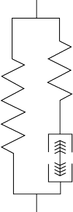
Parallel
elastic element
Series
elastic element
Contractile
element
FIGURE 2.3 The Hill model of muscle separates the active properties of muscle into a contractile element, in series
with a purely elastic element. The properties of the passive muscle are represented by the parallel elastic element.
Both the elastic response and the relaxation function are empirically determined. The common form
for the relaxation function is a sum of exponentials
G(t) = A +
i
B
i
e
−t/τ
i
The form ofthe elastic responsevaries, but usually includes apoweror exponential termtoaccommodate
the toe region.
2.3.3 Muscle
2.3.3.1 Types of Muscle Models
There are three general classes of models for predicting muscle force: biochemical, or crossbridge, models;
constitutive models; and phenomenological, or Hill, models. Crossbridge models [Huxley, 1957; Huxley
and Simmons, 1971] attempt to determine force from the chemical reactions of the crossbridge cycle.
Though accurate at the cross-bridge cycle, it is generally computationally prohibitive to model a whole
muscle in this manner. Constitutive models, such as that described by Zahalak and Ma [1990], generally
attempt to determine muscle behavior by describing populations of cross-bridges. A potentially powerful
approach, this technique has not yet been widely adopted. The vanguard of muscle modeling remains the
phenomenological model first described by Hill [1939], that describes the viscoelastic behavior of skeletal
muscle using a framework analogous to the standard linear solid (Figure 2.3). Although the series elastic
element represents primarily tendon, some series elasticity is found even in muscles lacking any external
tendon or in segments of single fibers. The parallel elastic element represents the passive properties of
the muscle, currently thought to reside primarily in titin. The contractile component is described by
independent isometric force–length (Figure 2.4) and isotonic force–velocity relations (Figure 2.5) and an
activation function [Zajac, 1989].
2.3.3.2 Muscle Force----Length Relationship
Under conditions of constant length, muscle force generated is proportional to the magnitude of the
interaction between the actin and myosin contractile filaments. Myosin filament length in all species is ap-
proximately 1.6 μm, but actin filament length varies (Table 2.7). Optimal sarcomere length and maximum
sarcomere length can be calculated using these filament lengths. For optimal force generation, each half

Musculoskeletal Soft Tissue Mechanics 2-11
100
80
60
40
20
1.0 1.5 2.0 2.5 3.0 3.5
Sarcomere length (µm)
4.5
Passive
Active
200
Percent maximum tension
0
FIGURE 2.4 The force-generating capacity of a sarcomere depends strongly on the degree of overlap of myosin and
actin filaments.
Isometric
length (L
o
)
Maximum isometric
tension (P
o
)
150
100
50
–75 –50 –25 0 25 50 75
200
Muscle force (%P
o
)
0
–100
Contractile velocity (%V
max
)
100
FIGURE 2.5 Active force generation depends strongly on shortening velocity.
myosin filament must completely overlap an actin filament, without opposing actin filaments overlapping.
No active forceis produced at sarcomerespacings shorter than themyosinfilament length or longerthan the
sum of the myosin and the pair of actin filament lengths. The range of operating sarcomere lengths varies
among muscles, but generally covers a range of ±15% of optimal length [Burkholder and Lieber, 2003].

2-12 Biomechanics
TABLE 2.7 Actin Filament Lengths
Actin Filament Optimal Length
Species Length (μm) (μm) References
Cat 1.12 2.24 Herzog et al. [1992]
Rat 1.09 2.18 Herzog et al. [1992]
Rabbit 1.09 2.18 Herzog et al. [1992]
Frog 0.98 1.96 Page and Huxley [1963]
Monkey 1.16 2.32 Walker and Schrodt [1973]
Human 1.27 2.54 Walker and Schrodt [1973]
Hummingbird 1.75 3.50 Mathieu-Costello et al. [1992]
Chicken 0.95 1.90 Page [1969]
Wild rabbit 1.12 2.24 Dimery [1985]
Carp 0.98 1.92 Sosnicki et al. [1991]
2.3.3.3 Muscle Force----Velocity Relationship
Under conditions of constant load the relationship between force and velocity is nearly hyperbolic [Hill,
1938]. The shortening force–velocity relation can be described by:
(P +a)v = b(P
0
− P)
while the lengthening relation can be described by:
F = 1.8 − 0.8
v
max
+ v
V
max
− 7.6 V
The dynamic parameters (a, b, and V
max
) vary across species and fiber types (Table 2.3).
It should be noted that this formulation omits several potentially important force-generating phenom-
ena. Notable among these are the persistent extra tension obtained following stretch [Edman et al., 1982],
the exaggerated short range stiffness [Rack and Westbury, 1974], and changes in the force–length relation
associated with activation level [Rack and Westbury, 1969]. Some of these features can be accommodated
by considering series elasticity and sarcomere length inhomogeneity [Morgan, 1990], and each represents
a nonlinearity that substantially complicates modeling and may not be necessary for first approximations
of muscle function.
Common applications of muscle modeling include forward simulation to predict output forces or
motions and inverse analysis to estimate the muscle forces that produced an observed motion. In neither
of these cases is it necessarily practical to determine muscle contractile properties empirically, and it is
frequently necessary to resort to estimation of the force–length and force–velocity relations from muscle
structure. If a muscle is considered to be a composition of uniform sarcomeres in series and in parallel, then
the deformation of single sarcomeres can be estimated from whole muscle length changes. A simplified
view of a muscle is an array of identical fibers of uniform length arranged at a common pennation angle
to the line of force. Peak isometric tension can be estimated from PCSA. Pennation angle determines the
relationship between muscle and fiber length changes:
L
m
L
m
=
L
f
L
f
cos(θ)
If sarcomere length is known at any muscle length, it is then possible to scale the sarcomere length–
tension and velocity–tension relations to the whole muscle. When reporting architectural data (Tables),
muscle and fiber lengths should be normalized to optimal sarcomere length.
Musculoskeletal Soft Tissue Mechanics 2-13
References
Butler, D.L., Grood, E.S., Noyes, F.R., and Zernicke, R.F. (1978). Biomechanics of ligaments and tendons.
In Exercise and Sport Sciences Reviews. Vol. 6: pp. 125–181, Hutton, R.S. (Ed.). The Franklin Institute
Press.
Fung, Y.C. (1981). Biomechanics: Mechanical Properties of Living Tissues. Springer-Verlag, New York.
Gans, C. (1982). Fiber architecture and muscle function. Exerc. Sport Sci. Rev. 10: 160–207.
Hill, A.V. (1938). The heat of shortening and the dynamic constants of muscle. Proc. R. Soc. Lond. [Biol].
126: 136–195.
Huxley, A.F. (1957). Muscle structure and theories of contraction. Prog. Biophys. Mol. Biol. 7:
255–318.
Huxley, A.F. and Simmons, R.M. (1971). Proposed mechanism of force generation in striated
muscle. Nature 233: 533–538.
Kastelic, J., Galeski, A., and Baer, E. (1978). The multicomposite structure of tendon. Connect. Tissue Res.
6: 11–23.
Kura, H., Luo, Z., Kitaoka, H.B., and An, K. (1997). Quantitative analysis of the intrinsic muscles of the
foot. Anat. Rec. 249: 143–151.
Lieber, R.L., Fazeli, B.M., and Botte, M.J. (1990). Architecture of selected wrist flexor and extensor muscles.
J. Hand Surg. 15: 244–250.
Lieber, R.L., Jacobson, M.D., Fazeli, B.M., Abrams, R.A., and Botte, M.J. (1992). Architecture of selected
muscles of the arm and forearm: anatomy and implications for tendon transfer. J. Hand Surg. 17:
787–798.
McElhaney, J.H., Roberts, V.L., and Hilyard, J.F. (1976) Handbook of Human Tolerance. Japan Automobile
Research Institute, Inc. (JARI), Tokyo, Japan.
Mow, V.C., Kuei, S.C., Lai, W.M., and Armstrong, C.G. (1980). Biphasic creep and stress relaxation of
articular cartilage in compression: theory and experiments. Biomech. Eng. 102: 73–84.
Powell, P.L., Roy, R.R., Kanim, P., Bello, M.A., and Edgerton, V.R. (1984). Predictability of skeletal muscle
tension from architectural determinations in guinea pig hindlimbs. J. Appl. Physiol. 57: 1715–1721.
Wayne, J.S., Woo, S.L., and Kwan, M.K. (1991). Application of the u–p finite element method to the study
of articular cartilage. J. Biomech. Eng. 113: 397–403.
Weiss, J.A. and Gardiner, J.C. (2001). Computational modeling of ligament mechanics. Crit. Rev. Biomed.
Eng. 29: 303–371.
Wickiewicz, T.L., Roy, R.R., Powell, P.L., and Edgerton, V.R. (1983). Muscle architecture of the human
lower limb. Clin. Orthop. Rel. Res. 179: 275–283.
Zahalak, G.I. and Ma, S.P. (1990). Muscle activation and contraction: constitutive relations based directly
on cross-bridge kinetics. J. Biomech. Eng. 112: 52–62.
Zajac, F.E. (1989). Muscle and tendon: Properties, models, scaling and application to biomechanics and
motor control. Crit. Rev. Biomed. Eng. 17: 359–411.

3
Joint-Articulating
Surface Motion
Kenton R. Kaufman
Kai-Nan An
Mayo Clinic
3.1 Ankle .................................................3-2
Geometry of the Articulating Surfaces
•
Joint Contact
•
Axes of Rotation
3.2 Knee..................................................3-5
Geometry of the Articulating Surfaces
•
Joint Contact
•
Axes of Rotation
3.3 Hip ...................................................3-13
Geometry of the Articulating Surfaces
•
Joint Contact
•
Axes of Rotation
3.4 Shoulder..............................................3-16
Geometry of the Articulating Surfaces
•
Joint Contact
•
Axes of Rotation
3.5 Elbow ................................................3-19
Geometry of the Articulating Surfaces
•
Joint Contact
•
Axes of Rotation
3.6 Wrist .................................................3-23
Geometry of the Articulating Surfaces
•
Joint Contact
•
Axes of Rotation
3.7 Hand .................................................3-28
Geometry of the Articulating Surfaces
•
Joint Contact
•
Axes of Rotation
3.8 Summary .............................................3-33
Acknowledgment ...........................................
3-33
References ..................................................3-35
Knowledge of joint-articulating surface motion is essential for design of prosthetic devices to restore
function; assessment of joint wear, stability, and degeneration; and determination of proper diagnosis and
surgical treatment of joint disease. In general, kinematic analysis of human movement can be arranged into
two separate categories: (1) gross movement of the limb segments interconnected by joints, or (2) detailed
analysis of joint articulating surface motion, which is described in this chapter. Gross movement is the
relative three-dimensional joint rotation as described by adopting the Eulerian angle system. Movement
of this type is described in Chapter 5: Analysis of Gait. In general, the three-dimensional unconstrained
rotation and translation of an articulating joint can be described utilizing the concept of the screw dis-
placement axis. The most commonly used analytic method for the description of 6-degree-of-freedom
displacement of a rigid body is the screw displacement axis [Kinzel et al., 1972; Spoor and Veldpaus, 1980;
Woltring et al., 1985].
3-1
