Ortiz de Montellano Paul R.(Ed.) Cytochrome P450. Structure, Mechanism, and Biochemistry
Подождите немного. Документ загружается.

154
Thomas M. Makris et al.
dioxygen, H2O2 brings the two reducing equi-
valents and two protons necessary for the
"Compound I" generation to the ferric heme in
one step. The key step in the enzymatic cycle of
the peroxidases, therefore, involves the abstrac-
tion of a proton from the proximal (closest to the
iron center) oxygen atom as the H2O2 molecule is
ligated and the delivery of a proton which is
necessary to initiate O-O bond scission^"^'
^^.
The
formation of the "0x0" complex and water is
essentially a barrierless process once protonation
occurs^^. A feature of the peroxidase mechanism
is the fact that it proceeds with no other external
source of electrons and protons, with an imidazole
side chain from a histidine residue in the distal
pocket thought to serve as the species responsible
for this proton shift. The distal histidine thus
serves, first, as a proton acceptor from the proxi-
mal oxygen, and then, as a donor of the same pro-
ton to the distal oxygen at the second step of the
reaction.
Defining the parameters involved in the bind-
ing and proton-mediated transformation of the
peroxide-coordinated ferriheme complex is criti-
cal in defining the properties of these states in
both the peroxidase and related oxygenase mech-
anisms. The pK^ of the Fe-coordinated HOO(H)
was estimated as
3.6^.0
for horseradish per-
oxidase (HRP) and cytochrome c peroxidase
(CcP)^^'
^^.
Thus, the iron-coordinated peroxide is
in an anionic form at neutral pH. The polarity of
the peroxidase distal pocket vs the dioxygen
carrier, myoglobin, or even the rather hydrophobic
nature of the P450 cytochromes, stabilizes the
resulting charge separation upon deprotonation.
The resulting hydroperoxo-ferriheme complex
is nonetheless unstable, as the peroxide ligand
weakly ligates the heme iron^^, and the dissocia-
tion rate constant for the peroxide anion as the
sixth ligand in Fe-microperoxidase-8 (Fe-MP8)
and Mn-MP8 is in the neighborhood of 10-20 s"'
(ref [60]). Similar estimates can be made from
other kinetic studies^^'
^^
which measured the K
m
and
k^^^
values for the formation of "Compound T'
in reaction of HRP with hydrogen peroxide. The
reported millimolar K^ values and the observed
k^^^ on the order of 10^M~^s~' suggest that
the dissociation rate of HOO(H) may be as high
as lO^s-i.
In contrast, the cytochromes P450 face the
challenge of "activating" dioxygen, with the
delivery of two protons in addition to the two
redox equivalents which are consumed per one O2
molecule. While the reduction of the heme iron is
provided by the protein redox partner, it is an
essential part of the P450 enzyme to catalyze the
transfer of two protons to the distal oxygen atom
of the bound peroxide anion through a sophisti-
cated proton relay mechanism. As noted in the
introduction, this pathway was shown to include
protein-bound water molecules which are stabi-
lized at the enzyme active site through specific
hydrogen-bond interactions^^' ^^^ ^^. Through
mutagenesis of the residues involved in this
network, it was shown that the overall enzyme
kinetics and the observed efficiency of coupling
redox equivalents into product was very sensitive
to the hydrogen-bonding properties of these
sites^^.
Similarly strong effects, including drastic
changes in the formed product ratios, were found
in other enzymes of the P450 superfamily^^~^^.
Kinetics of the productive and nonproductive
turnover by human cytochromes P450 with
various substrates were extensively studied by
Guengerich and coauthors^^^^. Their results
directly show the existence of multiple steps, each
potentially rate limiting depending on the specific
reaction being catalyzed.
A linked event is the resultant chemical
processing that occurs after proton delivery.
Following second proton transfer, the 0-0 bond
order is significantly diminished, so that heteroly-
sis is ensured, with release of water and generation
of the higher valent "Compound I" state. Without
efficient delivery of this second proton, one has to
consider the possibility of the radical process of
0-0 bond homolysis. Homolytic and heterolytic
scission of 0-0 bond in alkylperoxides and in
hydrogen peroxide catalyzed by various heme
enzymes have been extensively studied^'~'^^. In the
cytochromes P450, the ratio of homolytic/
heterolytic cleavage of the O-O bond has been
addressed through numerous investigations'^^' ^^'
^^~^^. The vast majority of these studies utilized
the exogenous oxidant driven pathways, with a
variety of substituted peroxides and peroxo acids.
Significantly, the results were found to depend on
the ability of the leaving group to stabilize a radi-
cal;
the more stable this species, the greater the
percentage of homolytic 0-0 bond scission^^.
The product distribution with a cumene hydroper-
oxide driven reaction was used as a measure for
Activation of Molecular Oxygen by Cytochrome P450
155
the relative ability of particular isozyme to form
a "Compound I" intermediate''^'
'^^^
^^. Attempts
were also made to correlate the "push" electron-
donating effect^
^
from the proximal thiolate with
the spectroscopic markers of the porphyrin ring
and with the formation of "Compound I" (i.e.,
the heterolysis of O-O bond) and product^^. The
formation of hydroxyl radicals via homolytic
cleavage of the O-O bond in the reconstituted
P450 CYP2B4 system resulted in covalent
modification of the protein and degradation of
the heme^^.
The general acid catalysis of heterolytic O-O
bond scission via protonation from solvent water
was suggested by many authors^^. In the absence
of additional catalysis of heterolytic scission
through the second protonation of the distal oxy-
gen atom, heterolysis/homolysis of O-O bond is
governed by the general thermodynamic stability
of the reactant and product, as predicted by Lee
and Bruice^^ for porphyrin models, and recently
analyzed by Que and Solomon^^' ^^' ^^"^^. If the
iron-peroxide complex is predominantly in a high-
spin (HS) state, as it is very often in nonheme
metal enzymes with weak or moderate ligand
field, homolysis of the 0-0 bond is favorable, as
the HS state of the iron in both the reactant and
product forms is conserved, with the process
involving release of a hydroxyl radical.
For heterolytic O-O scission following a
second protonation, in which the departing
oxygen atom leaves as water, electron density is
withdrawn from the porphyrin moiety, and a
porphyrin pi-cation radical is formed. The het-
erolysis/homolysis ratio and overall product dis-
tributions are thus coupled in the native enzyme
systems. Various parameters such as bulk and
local pH, ligation state of the metal, structure, and
redox properties of porphyrin and peroxide
species certainly play important roles in control-
ling the spin state, O-O bond order, and proton
delivery events.
Perhaps the most important is the enzyme's
ability to control the delivery of protons to the
developing negative charge of the iron-dioxygen
complex as electrons are introduced into the
center. The protonation/deprotonation events at
the bound superoxo, peroxo, and hydroperoxo
anions were theoretically analyzed and claimed to
account for the definitive reaction sequence in the
formation of the active oxo-ferryl porphyrin
pication hydroxylating intermediate in cytochrome
P450 and peroxidases^^. Despite their common
use of intermediate states, the oxygenases and
peroxidases have distinct functions, begirming
their active cycle with different heme ligand com-
plexes^^"^^. The variability of different pathways
of oxygen activation provides one with a tool
to alter the performance of the enzyme through
different mutations, or even to directly design new
types of activity into well-known systems^^' ^^^.
However, only in the last decade have the proton
delivery mechanisms in P450 catalysis been
directly revealed^^' 39-42, 62, 92-94, ioi-io4_
j^ese
important details of P450 catalysis will be dis-
cussed in detail in the remainder of this chapter.
3. Enzymatic Cycle of
Cytochrome P450
It is humbling to note that an outline of the
common catalytic cycle for the cytochromes P450
was proposed as early as 1968"^' ^^^. The critical
features of an atmospheric dioxygen binding event
interspersed between two single-electron transfer
events remain as a generally accepted outline of
the P450 catalytic "wheel"
^3,
io6 (Figure 5.2).
The sequential two-electron reduction of cyto-
chrome P450 and the existence of multiple inter-
mediates of substrate binding and reduction
was documented in bacterial P450 CYPlOl (refs
[107]-[109]) and in microsomal systems^^^' ^^^
Substrate binding to a resting state of the low-spin
(LS) ferric enzyme [1] perturbs the water, coordi-
nated as the sixth ligand of the heme iron if there
is a close fit to the active-site pocket, and alters
the spin state to favor the HS substrate-bound
complex [2]. In P450 CYPlOl, the HS Fe^^ form
of
the
protein has a more positive redox potential,
is a more efficient electron acceptor from its
redox partner, and accompanies a correspondingly
tighter association of the substrate to the reduced
form of the protein
[3]^^^.
In other systems, it
was observed that this spin shift is absent or
incomplete for some combinations of P450
isozyme and substrate^
^^.
Oxygen binding leads to
an oxy-P450 complex [4], which is the last quasi-
stable and observable intermediate in the reaction
cycle. Subsequent sequential steps are reduc-
tion of the Fe02 complex, formation of the
156
Thomas M. Makris et al.
peroxo-ferric intermediate [5a], its protonation
yielding a hydroperoxo-ferric intermediate [5b],
a second protonation at the distal oxygen atom
and subsequent heterolysis of the 0-0 bond to
form the iron-oxo "Compound I" [6] and water,
and finally the oxygenation of the substrate to
form a product complex [7] and its subsequent
release. These steps have, over the years, been
addressed by many methods, but direct observa-
tion of key intermediates was difficult due to
the high inherent reactivity of these states and
their lack of accumulation in kinetic studies. For
instance, [5a], [5b], and [6] had not been unam-
biguously observed until recently, and alternative
bidentate structures of the "reduced oxyP450" had
been proposed^^' ^^.
A beautiful feature of the overall reaction
pathway for the cytochrome P450s is the rich
chemistry afforded by the various steps in the
reductive metabolism of the heme-oxygen center.
These include nucleophilic, electrophilic, and
hydrogen-abstracting species. In addition, there
are at least three branchpoints where multiple
side reactions are possible and realized in vivo
(Figure 5.2). Abortive reactions include: (a) autox-
idation of the oxy-ferrous enzyme [4] with con-
comitant production of a superoxide anion and
return of the enzyme to its resting state [2], (b) a
"peroxide shunt," where the coordinated peroxide
[5b] dissociates from the iron and forms hydrogen
peroxide, thus completing the unproductive
(in terms of substrate turnover) two-electron
reduction of oxygen, and (c) an "oxidase uncou-
pling pathway," whereby the "ferryl-oxo"
intermediate [6] is reduced to water by two
additional electrons in lieu of hydrogen abstrac-
tion and subsequent substrate oxygenation. This
results in an effective four-electron reduction of
the dioxygen molecule with a net formation
of two molecules of water, thus anointing the
P450s with a "cytochrome oxidase" activity.
The oxidase activity in P450, in comparison to
those enzymes involved exclusively in bioenerget-
ics,
proceeds at a drastically reduced rate and
without a productive proton-pumping cycle. Thus
the physiological processing of dioxygen and
substrates via reductive metabolism is a rich and
varied set of events. The control of the specific
reactivities is determined by proton delivery in the
enzyme.
3.1.
The Ferrous-Dioxygen
Complex
Oxygen binding to reduced P450 yields the
species [4], Fe^+-00 (ferrous-dioxygen) or
Pe3+_oo~ (ferric-superoxide) complex. The
chemical structure of oxy-P450 is similar to anal-
ogous complexes in oxygen-carrier-heme proteins
(hemoglobin and myoglobin) and other heme
enzymes (HRP, HO, etc.). This ferrous Fe^^-OO
complex is EPR silent, but shows Mossbauer
quadruple splitting of the heme iron center consis-
tent with a ferric state ^^'^, similar to that observed
for "Compound IE" in HRP^^^ and for oxy-Hb^i^.
The low frequency of the O-O stretch band,
observed at 1140 cm"' in the resonance Raman
spectra of P450 CYPlOl^'^ is also typical for a
superoxide complex. These features have often
prompted discussion about the correct electronic
assignments of these complexes and more sophis-
ticated models, such as a three-centered four-
electron ozone-like bond that was envisioned
almost 30 years ago' •^.
The properties of "oxy-P450" were first
characterized in P450 CYPlOl and other
isozymes using optical absorption''^"'^'', reso-
nance Raman"^' '^^' '^^, and Mossbauer""^ spec-
troscopy. The electronic structure of P450 and
chloroperoxidase (CPO) complexes with O2, CO,
and CN~, together with their magnetic circular
dichroism (MCD) spectra were also analyzed^'' '^^.
Theoretical explanation of the specific split Soret
band for the complexes of the ferrous P450
and CPO with diatomic ligands was also pro-
vided in the mid-1970s'^^' '^^ for the case of car-
bon monoxide, and later experimentally extended
to other electron-rich ligands with a strong back-
donation (dioxygen, cyanide, thiolate)^'' '^'^.
The absorption spectra and autoxidation
properties of oxygen complexes for several
cytochromes P4505^' 121, 122, no, 131^
^j^^
related
enzymes, such as NOS'^^ and CPO'^"^, have also
been reported. While all of these states are not
particularly stable, and are quickly autoxidized,
the autoxidation rates for P450 and other thiolate-
ligated enzymes (CPO and NOS) are significantly
higher than for the oxygen carriers Mb and Hb.
This can be aided by a general acid catalysis in
the heme monooxygenases, through facile proto-
nation of the coordinated oxygen'^"*. As will be
Activation of Molecular Oxygen by Cytochrome P450
157
discussed in later sections, active proton delivery
to the bound oxygen or peroxide ligand is a salient
feature of the P450 mechanism of oxygen activa-
tion, and similar mechanisms could be responsible
for faster autoxidation of the ferrous-oxygen
intermediate in these enzymes.
The recent use of cryocrystallographic tech-
niques has enabled the direct visualization of
intermediate states in the P450 cycle, and thus
the modulation of active-site moieties within the
catalytic timeframe. With regards to dioxygen
ligation, however, the X-ray structure of Fe^^-OO
complex of P450 CYPlOl determined recently'^^
demonstrates that the structure of the heme
ferrous-oxy complex of P450 appears very similar
to analogous complexes of other heme enzymes ^^^'
^^^. Oxygen is found to be coordinated in the bent
"end-on" mode (with a Fe-0-0 angle of 136°).
This structure provides a starting point for the
formation of the end-on bound peroxo-ferric
complex, which is the first of three unstable and
highly reactive intermediates in the P450 catalytic
cycle.
3.2. Reduction of Oxy-Ferrous
P450 and Formation of
Peroxo-Ferric Complexes:
Properties, Stability, and
Spectroscopy
The stability of iron-peroxo complexes is
marginal in heme systems in the presence of a
strong proximal ligand (His, Cys, or Tyr), and the
aqueous solution at a near-neutral pH. The numer-
ous attempts to isolate such complexes obtained in
reactions of hydrogen peroxide with P450 at
ambient conditions have failed because of the
inherent low stability and fast conversion to
"ferryl-oxo" species with O-O bond scis-
sion^^^. There have been successful isolations of
the ferriheme-peroxide complex in myoglobin,
however^ ^^.
Chemical models of the Fe^^-OOH~ and
Fe^^-OOR~ complexes have been prepared by
reacting metalloporph3^ins with peroxides at
low temperatures (either 200-230 K in solutions
or freeze-quenched at 120 K and below) and stud-
ied by EPR, NMR, and optical spectroscopic
methods^^^"^^^. Similar results were obtained with
heme proteins^^^, including cytochrome P450''^^.
Particularly noteworthy is the signature LS EPR
spectra with narrow g span (^ = 2.25-2.31,
2.16-2.21,
1.93-1.96)
and the red shift of Soret
band (as compared to the spectrum of oxy-ferrous
precursor) of the Fe^^-OOH" complexes in heme
systems. The direct reactions of superoxide anion
with free Fe^^-porphyrins in the absence of strong
proximal ligand usually afford the HS Fe^^-00^~
complex with a side-on bound peroxide and iron
displaced out of the porphyrin plane toward the
bound ligand^^' ^^'
^'^^.
In order to realize such a
structure in a heme protein, it would be necessary
to break the proximal ligand bond to the iron and
force a ir-bonded configuration, acting against the
steric hindrance of the heme prosthetic group. It
turns out that the presence of the strong proximal
ligand, which favors the LS state in hexacoordi-
nated Fe^^-OOH~ complexes, is an important
restriction on the chemistry of oxygen activation
in the heme enzymes. Structure and properties of
model Fe^^-OOH~ complexes in heme enzymes
have been extensively studied theoretically^^'
^^' ^^'
93,96,97,145,146 ^hesc investigations, together with
analogous studies on heme^"^^ and nonheme
enzymes ^^ have played a definitive role in estab-
lishing the currently accepted view of the impor-
tance of the second protonation step of the distal
oxygen atom of the peroxide ligand for the effi-
cient heterolytic cleavage of the O-O bond and
"Compound I" formation.
Early attempts by Estabrook and Peterson to
create and stabilize the reduced oxy-ferrous, or
"peroxo" complex in cytochrome P450 were first
made more than 30 years ago"^' ^^^ Subsequent
efforts included steady-state and stopped-flow
studies of reconstituted systems^"^^"^^^, replacing
dioxygen by superoxide^^^' ^^^ or peroxides as
oxygen donors, or the use of alternative chemical,
photochemical ^^^' ^^^, and pulse radiolytic^^^' ^^^
methods for fast and efficient reduction of the
preformed oxy-ferrous P450. However, only in
recent years have reproducible, stable, and high-
yield preparations of Fe^^-OOH~ complexes
been obtained with cytochrome P450 and other
heme systems. This was achieved using the
methods of radiolytic reduction of oxy-ferrous
precursors in frozen solutions at 77
K"^^'
^^'
1^7-159
Irradiation with high-energy photons from a ^^Co
gamma-source or using ^^P enriched phosphate as
158
Thomas M. Makris et al.
an internal source of high-energy electrons"^^' ^^^
generates radiolytic electrons, which reduce a pre-
formed oxy-ferrous complex which is stabilized
against autoxidation by low temperature. Similar
approaches have long been used by physicists and
chemists in matrix isolation chemistry of highly
reactive intermediates^^^~^^^. In the solid state, at
the temperatures below the glass transition where
translational and rotational diffusion is minimized,
the peroxo complex is stable due to the impedi-
ment of trap migration and proton transfer events.
Historically, in biological systems, cryogenic
radiolysis was first used as a tool for studies of the
nonequilibrium intermediates in heme proteins
in the early 1970s^^^. For example, radiolysis of
several ferric proteins with different ligands in
frozen aqueous-organic solutions at 77 K was
shown to produce the corresponding ferrous
species, which then could be annealed at elevated
temperatures, and the conformational and chemi-
cal relaxation processes monitored by EPR and
optical spectroscopy methods ^^'^^^^. The first
reports on cryoradiolysis of oxy-ferrous com-
plexes in heme proteins and formation of the
Fe^^-OO(H)^"^"^ "peroxo" complex in hemoglo-
bin, myoglobin, and HRP'^^' ^^^'^^ established the
characteristic EPR spectrum of Fe3+-00(H)2-(-)
complexes with a signature narrow span of
g values (2.3-2.25, 2.2-2.14, and 1.94-1.97).
Optical absorption'^^' '^'^' '^^, Mossbauer^'^, and
EPR analysis have been used to characterize the
peroxo intermediates in heme enzymes including
cytochrome P450 CYPlOl'^^' '^^
Recently, this approach was further expanded
to understand the detailed electronic structure and
stability of peroxo-ferric intermediates in heme'^^'
41,
49, 50, 53, 157, 159, 179-181 ^^d nOUhcmC SystcmS^^'
182-188 ^g ^ result, the direct spectral identifica-
tion of the intermediates [5a] and [5b] in several
heme proteins has been clearly achieved.
Importantly, the EPR spectra were found to be
sensitive to the protonation state of the peroxide
ligand, but less responsive to the nature of the
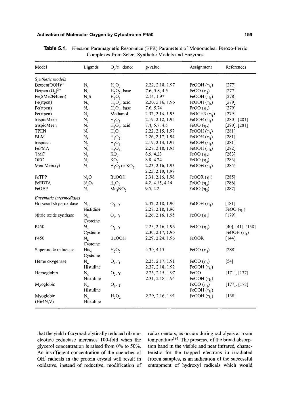
^ra«5-proximal ligand (His or Cys), Table 5.1.
UV spectroscopy shows a weak response to
the protonation state of the peroxide with the
Soret and Q bands shifting by only a few nano-
meters with iron-peroxo protonation, but high
sensitivity to the identity of the proximal ligand.
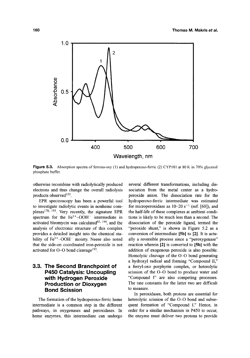
For example, the Soret band of the [5b] inter-
mediate appears at 440 nm in the thiolate-ligated
cytochrome P450, but at 420 nm in HRP and HO,
Figure 5.3.
The radiolytic reduction of
[4]
at 77 K yielded,
in most cases, an already protonated hydroperoxo-
ferric complex [5b]. In several proteins, however,
such as oxy-Mb, oxy-HRP, and the D251N variant
of cytochrome P450 CYPlOl, it was possible to
observe the unprotonated species [5a]. Interest-
ingly, the irradiation of oxy-P450 at 4 K in liquid
helium yielded the unprotonated form
[53]^*^
suggesting that an activation process of proton
transfer occurs between liquid helium and ligand
nitrogen temperatures. Most exciting is the direct
observation of the protein-catalyzed proton trans-
fer event, [5a] to form [5b], as the temperature is
raised. A second protonation and catalytic conver-
sion of the substrate to a product complex then
ensues"^ ^. Alternatively, a separate uncoupling
channel can be opened with peroxide release and
direct transition to the resting state of the enzyme,
demonstrating the subtle control of proton deliv-
ery provided by the protein matrix. The lack of
"Compound I" formation on the oxidase pathway
in HRP'^' is explained by the inability of this
enzyme to deliver this second proton to bound
dioxygen, despite the facile formation of the
"Compound I" from hydrogen peroxide.
Thus,
during the last decade, the method of
cryoradiolytic reduction has emerged as a new
tool to investigate critical intermediates of redox
systems. X-ray-induced radiation chemistry is
also increasingly recognized both as a potential
source of misinterpretations due to measurement-
induced changes of the sample, and as a new,
important tool in X-ray crystallography, where
the irradiation of protein crystals may be used to
deliberately alter the redox state of metals, flavins,
disulfides, and other cofactors'*^' i89-i9i jj^g
chemical details of
the
radiolytic process in frozen
solutions and protein crystals may, however, be
quite different. In the former, there is usually a high
concentration of glycerol or ethylene glycol present
as a cosolvent to improve the optical glass quality
of the sample at low temperature. These cosolvents
are necessary as effective quenchers of hydroxyl
radicals generated by the radiolysis of water. On
the other hand, protein crystals can sometimes
contain a much lower concentration of organic
cosolvents, thus potentially altering the processes
operating during low temperature radiolysis and
thermal annealing. For example, it was shown^^^
Activation of Molecular Oxygen by Cytochrome P450
159
Table 5.1. Electron Paramagnetic Resonance (EPR) Parameters of Mononuclear Peroxo-Ferric
Model
Synthetic models
Bztpen(OOH)2+
Bztpen (O^f^
Fe(SMe2N4tren)
Fe(rtpen)
Fe(rtpen)
Fe(rtpen)
trispicMeen
trispicMeen
TPEN
BLM
trispicen
FePMA
TMC
OEC
MemMemxyl
FeTPP
FeEDTA
FeOEP
Enzymatic intermediates
Horseradish peroxidase
Nitric oxide synthase
P450
P450
Superoxide reductase
Heme oxygenase
Hemoglobin
Myoglobin
Myoglobin
(H64N,V)
Complexes from Select
Ligands
N4
N4
N4S
N5
N5
N5
N5
N5
N5
N5
N5
N4
N4
N4
N4
N4O
N2O3
N4
N„
Histidine
N4
Cysteine
N4
Cysteine
N4
Cysteine
HiS4
Cysteine
N4
Histidine
N4
Histidine
N4
Histidine
N4
Histidine
02/e donor
H,0,
H2O2,
base
H2O2
H2O2,
acid
H2O2,
base
Methanol
up.
H2O2,
acid
H2O2
H2O2
H2O2
H2O2
KO2
KO2
H2O2 or KO2
BuOOH
H2O2
Me4N02
02,7
02,7
02,7
BuOOH
H2O2
02,7
02,7
02,7
H2O2
Synthetic Models and Enzymes
g-value
2.22,2.18, 1.97
7.6, 5.8, 4.5
2.14, 1.97
2.20,2.16, 1.96
7.6, 5.74
2.32,2.14, 1.93
2.19.2.12, 1.95
7.4, 5.7, 4.5
2.22,2.15,
1.97
2.26,2.17, 1.94
2.19,2.14, 1.97
2.27,2.18, 1.93
8.5, 4.23
8.8, 4.24
2.23,2.16, 1.93
2.25,2.10, 1.97
2.31,2.16, 1.96
4.2,4.15,4.14
9.5,
4.2
2.32,2.18, 1.90
2.27,2.18, 1.90
2.26,2.16, 1.95
2.25,2.16, 1.96
2.30,2.17, 1.96
2.29,2.24, 1.96
4.30,4.15
2.25,2.17, 1.91
2.37,2.18, 1.92
2.25,2.15,
1.97
2.31,2.18, 1.94
2.29,2.16, 1.91
Assignment
FeOOH (Til)
FeOO (TI2)
FeOOH (Til)
FeOOH
(TIJ)
FeOO (TI2)
FeOCH3 (Til)
FeOOH (Til)
FeOO (Ti2)
FeOOH (Til)
FeOOH (Til)
FeOOH (Til)
FeOOH (Til)
FeOO (TI2)
FeOO (TI2)
FeOOH (Til)
FeOOR (Til)
FeOO (TI2)
FeOO (TI2)
FeOOH (Til)
FeOO (Til)
FeOO (Til)
FeOOR
FeOO (TI2)
FeOO (Til)
FeOOH (Til)
FeOO
FeOOH (Til)
FeOO (Til)
FeOOH (Til)
FeOOH (Til)
References
[277]
[277]
[278]
[279]
[279]
[279]
[280],
[281]
[280],
[281]
[281]
[281]
[281]
[282]
[283]
[283]
[284]
[285]
[286]
[287]
[181]
FeOO (Til)
[179]
[40],
[41], [158]
FeOOH (Til)
[144]
[288]
[54]
[171],
[177]
[177],
[178]
[138]
that the yield of cryoradiolytically reduced ribonu-
cleotide reductase increases 100-fold when the
glycerol concentration is raised from 0% to 50%.
An insufficient concentration of the quencher of
OH' radicals in the protein crystal will result in
oxidative, instead of reductive, modification of
redox centers, as occurs during radiolysis at room
temperature^^^. The presence of the broad absorp-
tion band in the visible and near infrared, charac-
teristic for the trapped electrons in irradiated
frozen samples, is an indication of the successful
entrapment of hydroxyl radicals which would
160
Thomas M. Makris et al.
O
C
03
O
(0
<
600 700
Wavelength, nm
Figure 5.3. Absorption spectra of ferrous-oxy (1) and hydroperoxo-ferric (2) CYPlOl at 80 K in 70% glycerol/
phosphate buffer.
Otherwise recombine with radiolytically produced
electrons and thus change the overall radiolysis
products observed ^^^.
EPR spectroscopy has been a powerful tool
to investigate radiol)^ic events in nonheme com-
plexes ^^^' ^^^. Very recently, the signature EPR
spectrum for the Fe^^-OOH~ intermediate in
activated bleomycin was calculated^^' '^'^, and the
analysis of electronic structure of this complex
provides a detailed insight into the chemical sta-
bility of Fe^^-OOH~ moiety. Neese also noted
that the side-on coordinated iron-peroxide is not
activated for O-O bond cleavage ^^^.
3.3. The Second Branchpoint of
P450 Catalysis: Uncoupling
with Hydrogen Peroxide
Production or Dioxygen
Bond Scission
The formation of
the
hydroperoxo-ferric heme
intermediate is a common step in the different
pathways, in oxygenases and peroxidases. In
heme enzymes, this intermediate can undergo
several different transformations, including dis-
sociation from the metal center as a hydro-
peroxide anion. The dissociation rate for the
hydroperoxo-ferric intermediate was estimated
for microperoxidase as 10-20 s~' (ref [60]), and
the half-life of these complexes at ambient condi-
tions is likely to be much less than a second. The
dissociation of the peroxide ligand, termed the
"peroxide shunt," is shown in Figure 5.2 as a
conversion of intermediate [5b] to [2]. It is actu-
ally a reversible process since a "peroxygenase"
reaction wherein [2] is converted to [5b] with the
addition of exogenous peroxide is also possible.
Homolytic cleavage of the O-O bond generating
a hydroxyl radical and forming "Compound II,"
a ferryl-oxo porphyrin complex, or heterolytic
scission of the 0-0 bond to produce water and
"Compound I" are also competing processes.
The rate constants for the latter two are difficult
to measure.
In peroxidases, both protons are essential for
heterolytic scission of the 0-0 bond and subse-
quent formation of "Compound I." Hence, in
order for a similar mechanism in P450 to occur,
the enzyme must deliver two protons to provide
Activation of Molecular Oxygen by Cytochrome P450
161
the same reaction stoichiometry. If the second
proton is not available, the hydroperoxo-ferric
complex can still undergo O-O bond scission, but
must compete with dissociation of
the
complex to
form hydrogen peroxide. The weakness of perox-
ide ligand affinity provides a straightforward
explanation for the possibility of uncoupling at
this stage. The low binding affinity for the perox-
ide to the heme presumes the shift of the equilib-
rium [5b]
< >
[2] (Figure 5.2) to the right at low
peroxide concentrations. In the case of protona-
tion of the distal oxygen, the O-O bond is cleaved
with little or no apparent barrier to the reaction^"^^.
The resultant "Compound I" [6] then reacts
with the substrate or undergoes an "oxidase
shunt" process to be discussed in a subsequent
section.
Unfortunately, the only method of observing
the intermediate [6] to date in P450 enzymes has
been through the reaction of the ferric enzyme
with exogenous oxidants such as with m-
CPBA^^^^^^. Direct evidence for the quasi-stable
existence of a "Compound I" state, through meas-
urement of both formation and breakdown kinet-
ics,
in the cytochromes P450 has been provided
only recently^^^. This work utilized hyperther-
mostable CYP119 and low concentrations of per-
oxyacid to prevent secondary reactions. The
attempts to detect "Compound I" in annealing
experiments with cryogenically generated peroxo-
and hydroperoxo-ferric complexes in P450,
wherein atmospheric dioxygen was sequentially
reduced, have not been successfiil. Possible rea-
sons include the very high rate of substrate oxy-
genation by this active intermediate, and the
subtleties of competing proton delivery pathways.
Recently, Friesner et
al.
(personal communication)
estimated the barrier height of the reaction [6] -^
[7] in a P450 model to be only a few kcal mol~^
Thus,
the catal)^ic step of hydrogen abstraction
and oxygen rebound^^^ may be too rapid to allow
intermediate buildup. It is worth noting that [6]
does not accumulate even during the annealing of
cryogenically irradiated oxy-samples at low tem-
peratures 180-200 K. However, the use of solvent
exchange and ENDOR spectroscopy^^ of the prod-
uct complexes was able to provide important indi-
rect evidence for the operation of
a
"Compound I"
like species. The attempts to detect these interme-
diates in experiments with substrate-free P450
have also proved frustrating.
4. Structural Input into
the Mechanisms of
P450-Catalyzed Dioxygen
Activation
While the purely chemical characterization of
P450 reaction intermediates has largely focused
on redox-linked changes in heme-iron systems, a
complete understanding of intermediate evolution
and reactivity must involve the role of the protein
scaffold in dioxygen scission. In the case of P450
monooxygenation chemistry, this goal has been
formulated into ascertaining the role of the distal
pocket in the control of concerted proton transfer
events to a reduced oxy-ferrous intermediate,
resulting ultimately in heterolytic cleavage to pro-
duce the high-valent oxo-ferryl, or "Compound I"
intermediate. While numerous processes in the
P450 catal5^ic scheme have been linked to both
protons and the ionization state of particular
residues, we focus specifically on those proton
transfer events that result in the transformation of
the reduced ferrous-dioxygen complex, to yield
the hydroperoxo intermediate. It is critical not
only for any structural model to explain a frilly
ftanctional "wild-type" P450 reaction chemistry,
but also to give a structural basis for the compila-
tion of kinetic and spectroscopic parameters that
result from alteration of the active site by mutage-
nesis or by utilization of a variety of alternate sub-
strate structures. This then translates into
knowledge of the inherent reactivity of peroxo-
ferric complexes on the pathway to "Compound I"
formation, the control of branchpoints and their
specific chemistries providing oxidant in nucle-
ophilic, electrophilic, and radical oxidations. The
entire P450 reactivity landscape cannot be unified
into a strict and inflexible structural model, as
some aspects of proton delivery and dioxygen
activation are "finely tuned" according to the
structural requirements of a particular P450
isozyme active site and are moderated by particu-
lar substrate recognition events. Nonetheless,
many common structural motifs of the enz3mie
distal pocket have been evidenced which link
the P450 superfamily both mechanistically and
structurally.
In order to resolve structural components
potentially involved in proton donation to a
reduced ferrous-dioxygen or peroxo-anion species.
162
Thomas M. Makris et al.
at least three potential mechanisms of proton
delivery should be addressed. The first involves
ionization in which the relay of protons involves a
change in the charge state of the participating
amino acid side chain or solvent moieties. The
assignment of participating side chains involved in
proton transfer can be inferred by their local pK^
values. In a second type of process, the two partic-
ipating moieties undergo simultaneous protonation
and deprotonation in a Grotthuss mechanism^^^,
and all moieties participating in hydrogen-bonding
interactions can be potential proton donors in the
enzyme active site. Finally, the involvement of the
peptide bond itself in proton-transfer reactions has
been implicated in the proton-transfer mechanism
of
CcO^^^
and model peptides^^^.
The identification of potential proton-transfer
conduits in P450 catalysis was aided by the first
P450 crystallographic structure of CYPlOl from
Pseudomonas
putida^^^^
^^^. While this structure
played an indispensable role in visualizing the
structural features that years of P450 spectro-
scopic studies had suggested, such as confirma-
tion of the ligands of the HS substrate bound
ferriheme
species^
^"^^
^^^, the positive identifica-
tion of proton donors in the distal pocket was elu-
sive.
In fact, the P450 CYPlOl distal pocket was
shown to be mostly hydrophobic in nature, in
sharp contrast to the polar active sites of other
heme enzymes, such as CcP^^^ and catalase^^'^. Of
particular interest was the absence of charged
residues in the immediate vicinity of the heme-
oxygen binding site, such as the well-character-
ized acid-base pairs (His-Arg and His-Asp) in
the peroxidase and catalase structures, which were
thought to mediate dioxygen scission reactions.
Readily apparent from these early structures of
CYPlOl however, was the interruption of normal
hydrogen-bonding pattern in the distal I-helix, and
hydrogen bonding of the Thr252 side chain with
the carbonyl oxygen of Gly248. The resultant
"kink" in the I-helix has proved to be a common
structural element of most P450 isozymes, and
was postulated to create a pocket in which dioxy-
gen could easily bind^^^.
4.1.
A "Conserved" Alcohol Side
Chain in the Active Site
of P450
Upon examination of aligned sequences of
P450 isozymes, it has become clear that an
acid-alcohol side-chain pair is observed in a
large majority of P450 genes. These are typically
threonine, or in some cases serine, and either
aspartate or glutamate^^^, 209 j^ P450 CYPlOl
these residues are
Asp251
and
Thr252.
Given their
placement in proximity to a heme-dioxygen bind-
ing site, a plethora of mutagenesis experiments
provided mechanistic suggestions as to their role
in stabilizing distal pocket hydrogen-bonding
networks and contributing to proton delivery to a
peroxo-anion complex. For example, upon muta-
tion of the conserved threonine in CYPlOl to a
hydrophobic residue, one observes normal kinetic
parameters of pyridine nucleotide oxidation and
dioxygen consumption, yet the enzyme is fully
uncoupled, resulting almost entirely in hydrogen
peroxide production rather than the oxidation
product hydroxycamphor^'^'
^'
^ This phenotype is
certainly not exclusive to the case of CYPlOl, as
similar mutations across the space of P450 phy-
logeny have shown similar effects^^^'
^^^.
The roles
assigned for the "conserved" threonine have
included a direct proton source for the peroxo-
anion species, or a secondary effect such as stabi-
lizing critical hydrogen bonding and water
placement which in turn serves in proton delivery
and dioxygen bond scission. Several mutagene-
sis experiments were designed to discriminate
between these two possibilities. Ishimura and
colleagues, for instance, demonstrated that the
uncoupling reaction resulting in peroxide forma-
tion could be avoided if the Thr residue was
altered to a side chain which could participate in a
hydrogen-bonding interaction'^^'
^^^.
This is par-
ticularly well evidenced in the cases of Asn252
and Ser252 mutations in P450 CYPlOl, where
both substitutions retain more than half of
the enzyme's activity with regards to pyridine
nucleotide oxidation and hydroxylation turnover.
Furthermore, through the utilization of unnatural
amino acid mutagenesis^ •^, this avenue could be
explored without the stringency of naturally
occurring side chains. In an important experiment,
the hydroxyl side chain on the conserved alcohol
side chain (Thr252 in CYPlOl) was replaced
with a methoxy groups'^' ^'^, although the modi-
fied protein was not structurally characterized.
Interestingly, the mutant enzyme still retained
relatively normal kinetic parameters and nearly
full coupling of reducing equivalents into product.
Some caution is needed, however, since a common
activity of the C3^ochromes P450 is oxidative
Activation of Molecular Oxygen by Cytochrome P450
163
demethylation^^^' ^^^ and a single turnover could
result in restoration of a functional hydroxyl at the
enzyme active site. Nonetheless, this result has
often been interpreted to imply that the Thr252
side chain itself is not directly involved in donat-
ing protons to a reduced oxygen intermediate but
rather provides its ftinctionality by providing
hydrogen-bonding interactions that stabilize
active-site waters that are the immediate source of
catalytic protons ^^^.
High-resolution X-ray crystallographic studies
of the Thr252Ala mutant have also proved essen-
tial in dissecting its role in catalysis and peroxide
evolution^^. Despite mutating this residue to one
unable to form a hydrogen bond to the carbonyl
oxygen of Gly248, the I-helix "kink" is still
observed in the mutant enzyme. The observed new
solvent molecules in this mutant structure^^, in
particular Wat720, have been inferred as a source
of peroxide uncoupling in the mutant. This could
arise either by decreased stability of the peroxo
ligand in protic solvents or due to the delivery of
protons to the proximal oxygen, both of which
would result in the release of hydrogen peroxide^^.
The inclusion of extra waters as a potential source
of uncoupling has likewise been structurally
inferred with a series of camphor analogs bound
to CYPlOl (ref [36]) and in the substrate-free
enzyme^^^. The use of alternate substrates for the
enzyme displayed a varying degree of hydroxyla-
tion regiospecificity and uncoupling, resulting
either in the production of peroxide or water
through an "oxidase" channeP^^"^^^. The former
can often be explained through an increased sub-
strate mobility owing to perturbed hydrogen-
bonding interactions with Tyr96^^^' ^^^, but a
consistent water-mediated mechanism was diffi-
cult to understand for the latter. Nonetheless,
the localization and superposition of active-site
waters has emerged as being of critical impor-
tance in evaluating the proton-aided dioxygen
scission and uncoupling chemistries. The crystal-
lographic structure of Thr252Ala also demon-
strated that, both the mutated residue and Asp251
residue adopt different conformations^^. For
example, in the Ala252 mutant, the Co atom
moves 1.4 A away from the dioxygen-binding
pocket. In addition, the Asp251 backbone car-
bonyl has flipped (120°) toward the distal
helix. The participation of solvent in mediating
P450 catalysis, and the conformational adapta-
tion of the acid-alcohol pair in the distal
pocket, have since been shown to be important
in understanding peroxo-ferric stabilization and
reactivity.
The implication of "extra" water as a potential
source of uncoupling has also been included as a
parameter in the redesign of
the
substrate-binding
pocket to oxidize hydrocarbon substrates of
varying sizes. In the reaction of CYPlOl with
ethylbenzene, for example, the effective coupling
to monooxygenase activity is highly sensitive
to the sterics of the substrate-binding pocket^'^'
^^^.
Although a redesign in the distal pocket to
accommodate different substrates certainly dimin-
ished unwarranted uncoupling pathways, both at
the peroxide and oxidase branchpoints, a strict
mapping of potential solvation pathways leading
to peroxide uncoupling was difficult. Active-site
mutagenesis also served as a starting point for the
redesign of the active sites in other P450 isozymes
to oxidize a number of substrates of varying polar-
ity and size^^^~^^^. While the precise structural
details behind the uncoupling pathways in these
"redesigned" enzymes is not yet realized, an
important independence of substrate-binding
parameters and efficient coupling to monooxy-
genation is clear.
Even though the presence of an active-site alco-
hol ftinctionality is highly conserved throughout
the landscape of P450 isozymes, one important
exception has ftirther established the importance of
an active-site hydrogen-bonding network in dioxy-
gen catalysis. CYP107 (P450eryF), which catal-
yzes the hydroxylation of 6-deoxyerythronolide B
in the erythromycin biosynthetic pathway, has an
alanine (Ala245) instead of threonine at the con-
served position^^^'
^^^.
Despite the absence of the
I-helix threonine, a kink or cleft is still retained in
the structure and the side chains of Glu360 and
Ser246 and a series of ordered water molecules
contribute a distal pocket hydrogen-bonding net-
^Qj.]^232,233 Interestingly, the 5-hydroxyl group of
the macrolide substrate provides a key H-bonding
interaction to a water molecule (Wat564). This
water is in a nearly identical position as the threo-
nine hydroxyl moiety in P450 CYPlOl, thus sug-
gesting a similar proton delivery network via
"substrate-assisted catalysis"^^"*. Alteration of the
substrate C5 and C9 side chains, which normally
donates a hydrogen bond to crystallographically
observable active-site water (Wat563), results in
dramatic effects on catalytic efficiency. Through
reintroduction of the active-site hydroxyl in the
