Nielsen M. Atlas of Human Anatomy
Подождите немного. Документ загружается.

125
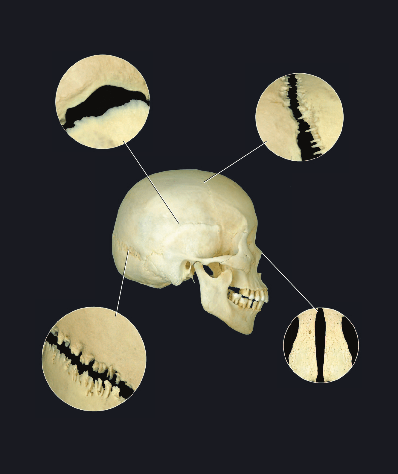
Squamous-type suture
Squamous or temporoparietal suture
Denticulate-type suture
Lamboidal or parieto-occipital suture
Serrate-type suture
Coronal or frontoparietal suture
Plane-type suture
Internasal suture
Atlas_ArticuSyst.indd Page 125 15/03/11 9:10 PM user-F391Atlas_ArticuSyst.indd Page 125 15/03/11 9:10 PM user-F391 /Users/user-F391/Desktop/Users/user-F391/Desktop
1
2
3
4
6
13
14
15
16
18
18
19
17
20
21
22
126
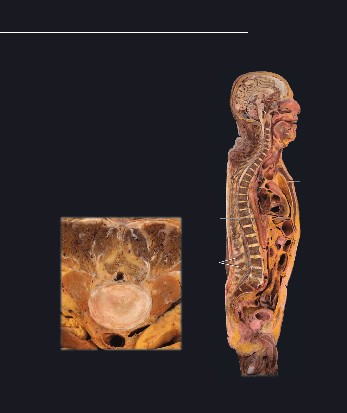
Sagittal section of head and trunk
Medial view
Synarthrosis - Cartilaginous Joints
Like the fi brous joints, the
cartilaginous joints join
neighboring skeletal ele-
1 Intervertebral disc (symphysis)
2 Nucleus pulposus of intervertebral disc
3 Anulus fibrosus of intervertebral disc
4 Pubic symphysis
5 Manubriosternal synchondrosis
6 Spheno-occipital synchondrosis
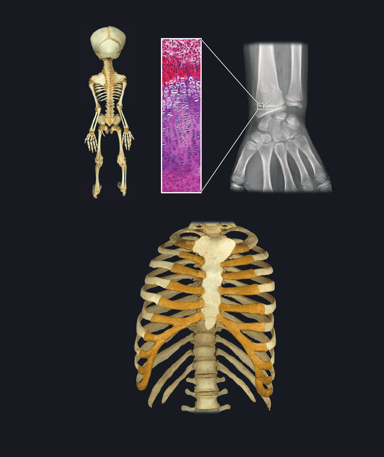
7 Epiphysial cartilage or primary cartilaginous joint
8 Sternocostal (synchondrosis)
9 Sternocostal (typically synovial but can be symphysial)
10 Interchondral (synovial)
11 Interchondral (synchondrosis)
12 Costochondral (synchondrosis)
13 Interspinous ligament (vertebral syndesmosis)
14 Nuchal ligament (vertebral syndesmosis)
15 Anterior longitudinal ligament (vertebral syndesmosis)
16 Posterior longitudinal ligament (vertebral syndesmosis)
17 Body of vertebra
18 Spinous process of vertebra
19 Lamina of vertebra
20 Psoas major muscle
21 Aorta
22 Inferior vena cava
ments with a solid mass of connective tissue, but the uniting tissue is some type of cartilage instead of collagenous connec-
tive tissue proper. The three types of cartilaginous joints are: 1) synchondroses, 2) symphyses, and 3) epiphysial cartilages
or primary cartilaginous joints. The photos on these facing pages depict the different categories of cartilaginous joints. A few
syndesmoses from the fi brous joint category are also evident.
Transverse section of lumbar intervertebral disc
Inferior view
1
2
3
4
6
13
14
15
16
18
18
19
17
20
21
22
5
Atlas_ArticuSyst.indd Page 126 15/03/11 9:10 PM user-F391Atlas_ArticuSyst.indd Page 126 15/03/11 9:10 PM user-F391 /Users/user-F391/Desktop/Users/user-F391/Desktop
1
1
7
8
10
10
11
12
9
9
9
9
9
127
Radiograph of juvenile wrist region
Anterior view
Joints of the thoracic cage
Anterior view
Epiphysial cartilage
200x
Fetal skeleton
Posterior view
1
1
7
8
10
10
11
12
9
9
9
9
9
7
Atlas_ArticuSyst.indd Page 127 15/03/11 9:10 PM user-F391Atlas_ArticuSyst.indd Page 127 15/03/11 9:10 PM user-F391 /Users/user-F391/Desktop/Users/user-F391/Desktop
1
2
5
128
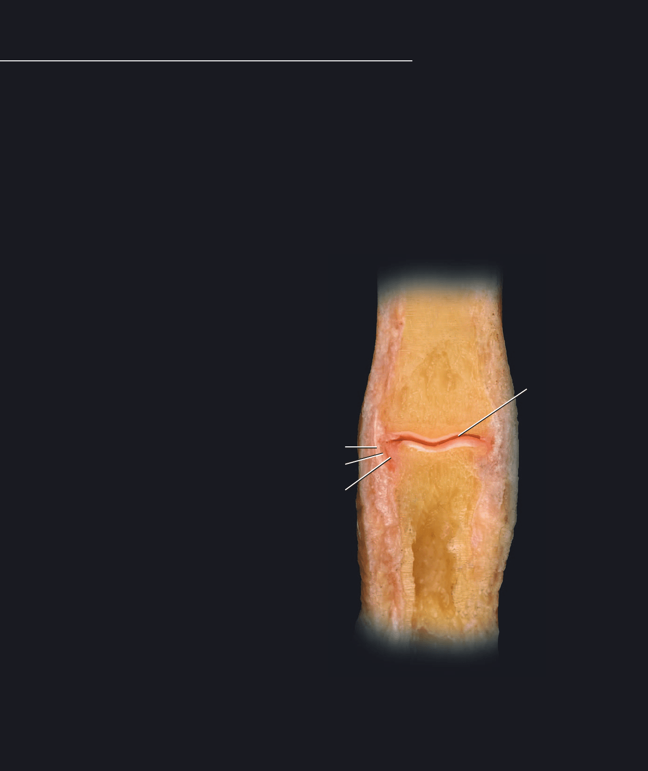
Diarthroses or Synovial Joints
Diarthroses differ from synarthroses
in one major way: instead of connect-
ing neighboring bones by a solid mass
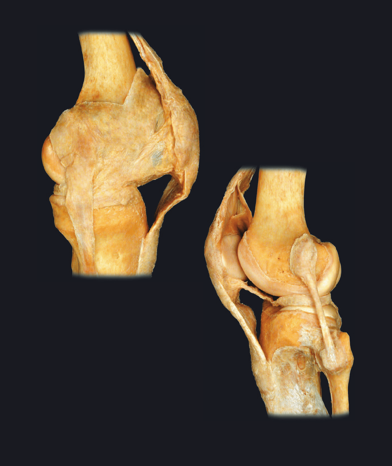
of connectve tissue, the bony connection consists of a double-layered connective tissue capsule that surrounds a lubricated
cavity between the bones. Within the capsule the ends of neighboring bony surfaces are covered by a smooth layer of hya-
line cartilage. As a result of this design there is typically a much greater range of motion present in synovial joints, and they
form the joints of the skeleton that are responsible for the major movements of the body. The outer layer of the capsule, the
fi brous membrane, is continuous with the periosteum on the adjoining bones, while the inner layer of the capsule, the syno-
vial membrane, attaches from the border of the articular cartilage on one bone to the border of the articular cartilage on the
other bone. Additionally, the synovial membrane secretes synovial fl uid, a lubricant that reduces friction between the mobile
cartilage-covered articular surfaces of the bones. The section through a fi nger joint below and the dissections of the knee
joint on the opposite page illustrate the basic features of a synovial joint. The pages that follow depict the major synovial
joints of the skeleton. One other key feature among synovial joints that is responsible for their varied range of motion is the
shape of the adjoining bone surfaces. It is this feature that anatomists use to describe the different types of synovial joints.
1 Middle phalanx of index finger
2 Proximal phalanx of index finger
3 Fibrous membrane of joint capsule
4 Synovial membrane of joint capsule
5 Articular cartilage
6 Joint cavity
7 Collateral ligament
8 Quadriceps tendon
9 Patellar ligament
10 Suprapatellar bursa
11 Synovial fold
12 Meniscus
13 Periosteum
14 Junction of periosteum (removed) with fibrous membrane
15 Junction of synovial membrane (removed) with articular cartilage
16 Femur with periosteum removed
17 Tibia with periosteum removed
18 Fibula with periosteum removed
19 Patella within quadriceps tendon
Proximal interphalangeal joint showing design of synovial joint
Frontal section, anterior view
3
7
4
6
Atlas_ArticuSyst.indd Page 128 15/03/11 9:10 PM user-F391Atlas_ArticuSyst.indd Page 128 15/03/11 9:10 PM user-F391 /Users/user-F391/Desktop/Users/user-F391/Desktop
3
7
7
4
5
5
5
8
9
10
11
12
13
14
14
15
15
16
17
18
19
129
Dissection of knee showing design of synovial joint
Medial view
Dissection of knee showing design of synovial joint
Lateral view
4
9
Atlas_ArticuSyst.indd Page 129 15/03/11 9:10 PM user-F391Atlas_ArticuSyst.indd Page 129 15/03/11 9:10 PM user-F391 /Users/user-F391/Desktop/Users/user-F391/Desktop
130
Types of Synovial Joints
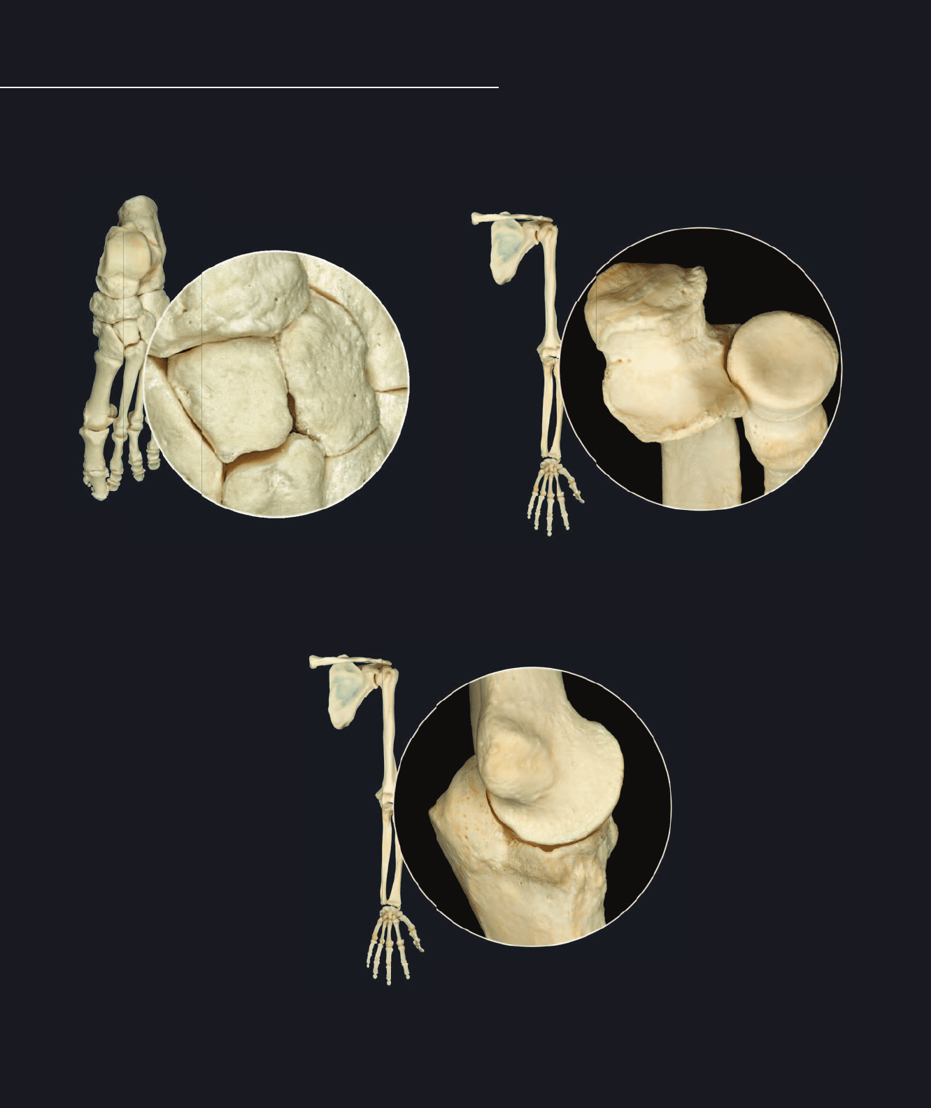
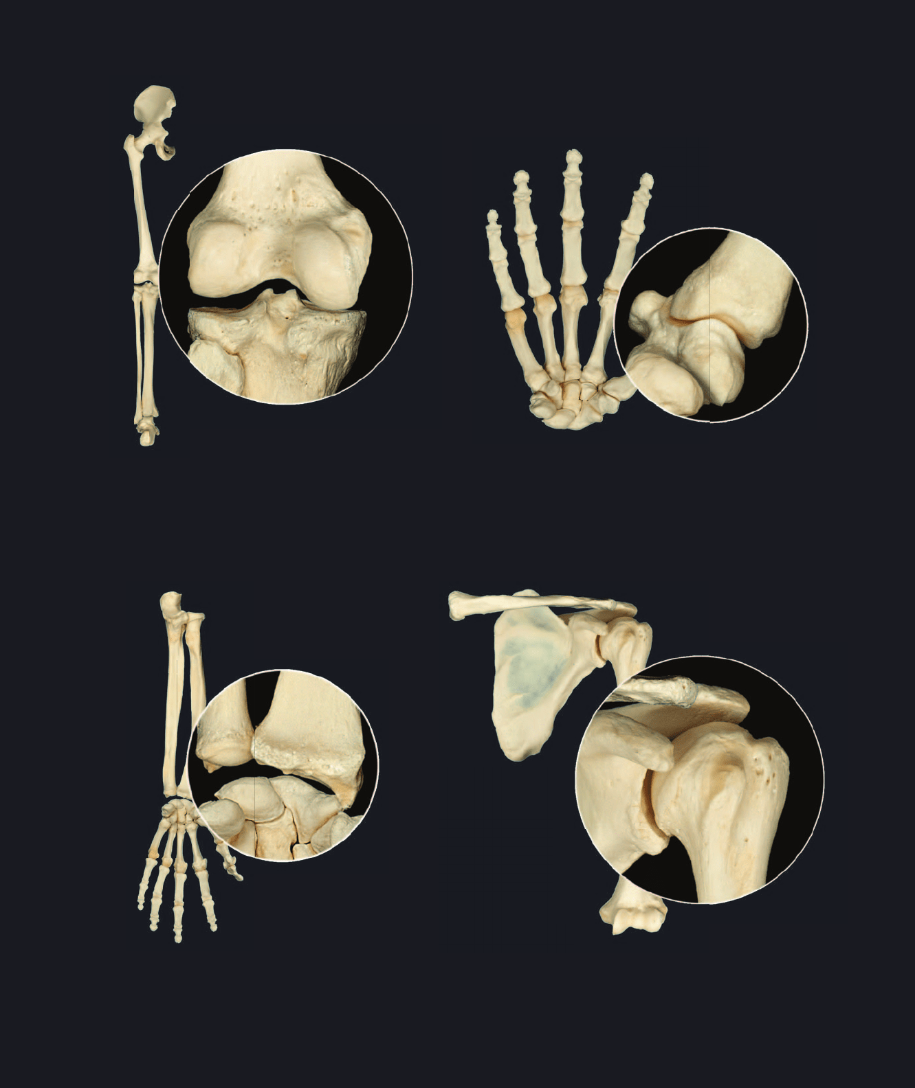
There are seven types of synovial joints in the body.
Each of the different synovial joints has the basic
structural features common to all synovial joints but is
further classifi ed based on the shape of and motion that occurs at the articular surfaces of the joint. The different types of
synovial joint are depicted below and on the opposite page. Note the shapes of the reciprocal surfaces as you study these
photos.
Hinge joint example
Humero-ulnar joint of elbow
Plane joint examples
Intertarsal joints
Pivot joint examples
Proximal radio-ulnar joint of elbow
Atlas_ArticuSyst.indd Page 130 15/03/11 9:10 PM user-F391Atlas_ArticuSyst.indd Page 130 15/03/11 9:10 PM user-F391 /Users/user-F391/Desktop/Users/user-F391/Desktop
131
Bicondylar joint example
Knee joint
Condylar joint example
Wrist joint
Saddle joint example
Metacarpal-carpal joint of thumb
Ball and socket joint example
Shoulder joint
Atlas_ArticuSyst.indd Page 131 15/03/11 9:11 PM user-F391Atlas_ArticuSyst.indd Page 131 15/03/11 9:11 PM user-F391 /Users/user-F391/Desktop/Users/user-F391/Desktop
1
2
3
4
5
6
7
8
9
10
11
12
13
14
132
Temporomandibular Joint
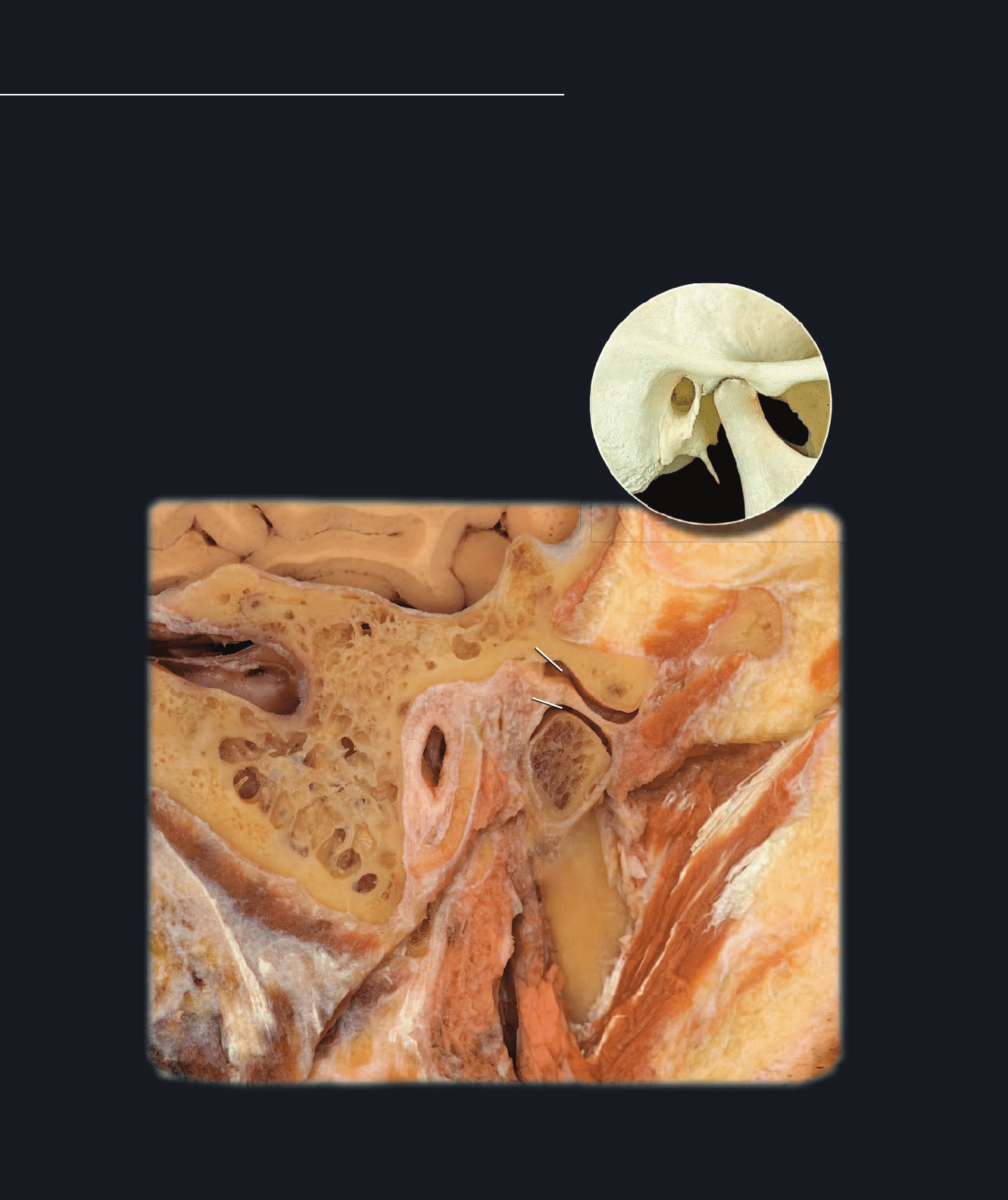
The complex temporomandibular joint differs from
other synovial joints by having an articular disc
that usually separates the joint into two separate
synovial capsules, one above and one below the disc. The articular surfaces have a covering of dense fi brocartilage rather
than the typical hyaline cartilage of most synovial joints. With its associated ligaments this joint structure accounts for the
complex series of movements that are essential during the activities of eating and speech. Each temporomandibular joint is
a condylar joint and both joints together form a bicondylar joint. The fi brous membrane of the articular capsule spans from
temporal bone to mandible only on the lateral side. Anteriorly, medially, and posteriorly the fi bers attach from mandible and
temporal bone to the articular disc. Extrinsic ligaments that help stabilize the joint are the lateral temporomandibular liga-
ment, sphenomandibular ligament, and stylomandibular ligament.
1 Mandibular condyle
2 Mandibular ramus
3 Articular tubercle of temporal bone
4 Mastoid process of temporal bone
5 Mastoid air cells
6 Superior compartment of articular cavity
7 Inferior compartment of articular cavity
8 Articular disc
9 Joint (articular) capsule
10 Masseter muscle
11 Parotid gland
12 Brain
13 External acoustic meatus
14 Sigmoid venous sinus
Section of right temporomandibular joint
Lateral view of sagittal section
Bones of temporomandibular joint
Lateral view
1
2
3
4
5
6
7
8
9
10
11
12
13
14
Atlas_ArticuSyst.indd Page 132 15/03/11 9:11 PM user-F391Atlas_ArticuSyst.indd Page 132 15/03/11 9:11 PM user-F391 /Users/user-F391/Desktop/Users/user-F391/Desktop
1
1
2
3
4
4
5
6
7
8
9
10
11
12
13
14
15
133
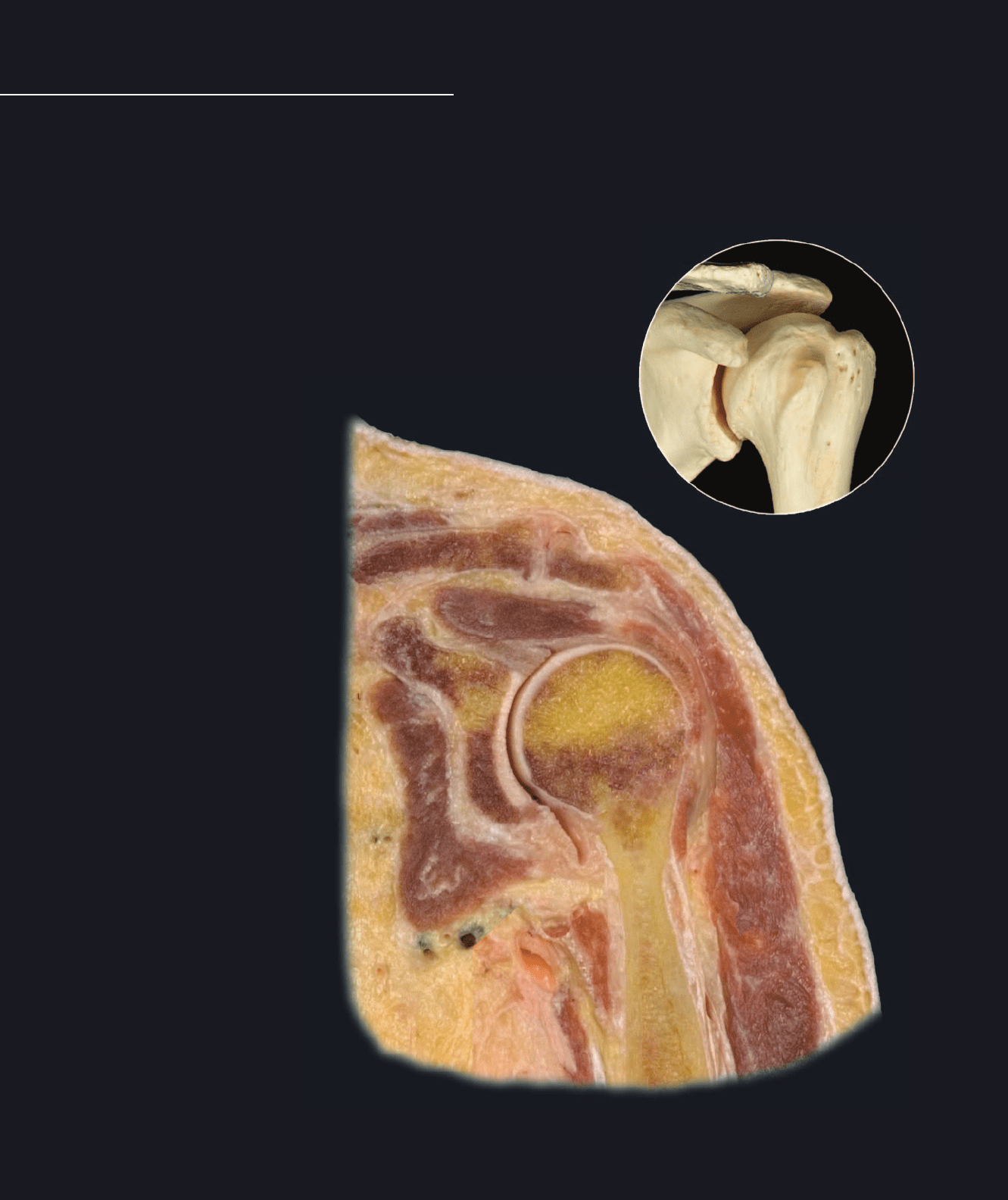
Glenohumeral Joint
The glenohumeral or shoulder joint is a ball and socket joint and is
the most mobile joint in the body. The tremendous range of motion
at this joint is the result of few external ligaments that present little
limitation to movement, and shallow, ovoid articular surfaces that make movements in all planes of space possible. In fact,
surrounding muscles and tendons play a more signifi cant role in joint support than do the joint structures. The capsular liga-
ment is extremely lax, providing limited support to the joint. Blending with the capsule are the tendons of four muscles.
Together the capsule and tendons form the rotator cuff, which is the major support structure of the joint.
1 Articular cartilage
2 Synovial membrane
3 Fibrous membrane
4 Glenoid labrum
5 Acromioclavicular ligament
6 Clavicle
7 Humerus
8 Glenoid of scapula
9 Acromion of scapula
10 Supraspinatus muscle
11 Subscapularis muscle
12 Deltoid muscle
13 Tendon of long head of biceps brachii
14 Skin
15 Subcutaneous layer
Section of left glenohumeral joint
Anterior view of frontal section
Bones of glenohumeral joint
Anterior view
1
1
2
3
4
4
5
6
7
8
9
10
11
12
13
14
15
Atlas_ArticuSyst.indd Page 133 15/03/11 9:11 PM user-F391Atlas_ArticuSyst.indd Page 133 15/03/11 9:11 PM user-F391 /Users/user-F391/Desktop/Users/user-F391/Desktop
1
1
2
3
4
5
67
8
9
10
11
134
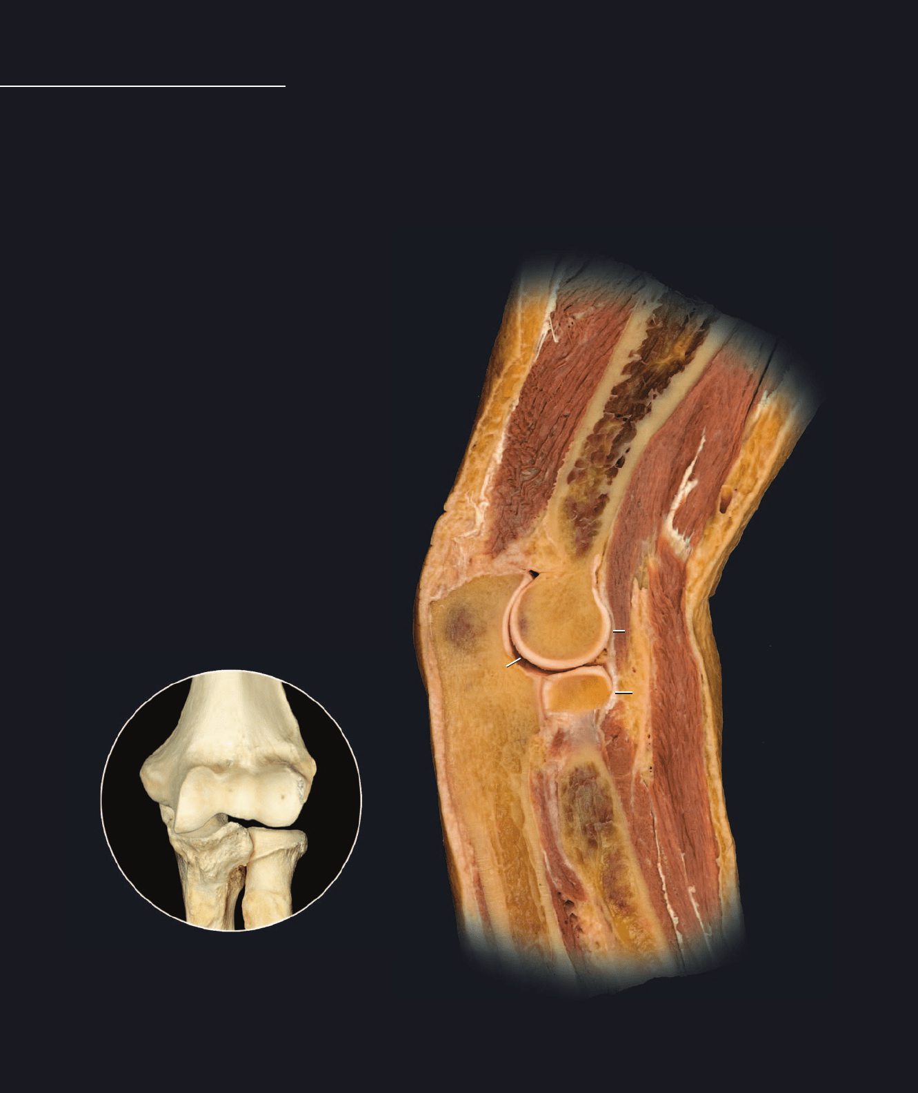
Elbow Joint
The elbow joint is a complex joint comprised of multiple articular surfaces within one articular
capsule. The elbow joint can be subdivided into three distinct articular interfaces —
the humero-ulnar joint (hinge), the humeroradial joint (combined hinge and pivot), and
1 Articular cartilage
2 Joint (articular) capsule
3 Articular (synovial) cavity
4 Capitulum of humerus
5 Olecranon of ulna
6 Head of radius
7 Anular ligament
8 Biceps brachii muscle
9 Brachialis muscle
10 Triceps brachii muscle
11 Brachioradialis muscle
Section of pronated left elbow joint
Medial view of sagittal section
Bones of elbow joint
Anterior view
the proximal radioulnar joint (pivot). Two distinct pairs of movements occur as a result of the articulations within the elbow
joint — the hinged movements of fl exion and extension, and the rotational movements of pronation and supination. Unlike
the shoulder joint, the joints fo the elbow have strong extrinsic ligaments that limit movemnts and stabilize the articulating
bones. The fi brous capsule is thin anteriorly and posteriorly, allowing for free range of motion during fl exion and extension.
On either side the capsule is reinforced by strong extrinsic ligaments, the ulnar collateral and radial collateral ligaments.
Wrapping from the back of the ulna at the base of the olecranon to the front of the ulna at the lateral surface of the coronoid
process is the semicircular anular ligament. With the radial notch of the ulna this ligament forms a fi bro-osseous ring for the
pivoting action of the radial head.
1
1
2
3
4
5
67
8
9
10
11
Atlas_ArticuSyst.indd Page 134 15/03/11 9:11 PM user-F391Atlas_ArticuSyst.indd Page 134 15/03/11 9:11 PM user-F391 /Users/user-F391/Desktop/Users/user-F391/Desktop
