Nielsen M. Atlas of Human Anatomy
Подождите немного. Документ загружается.

105
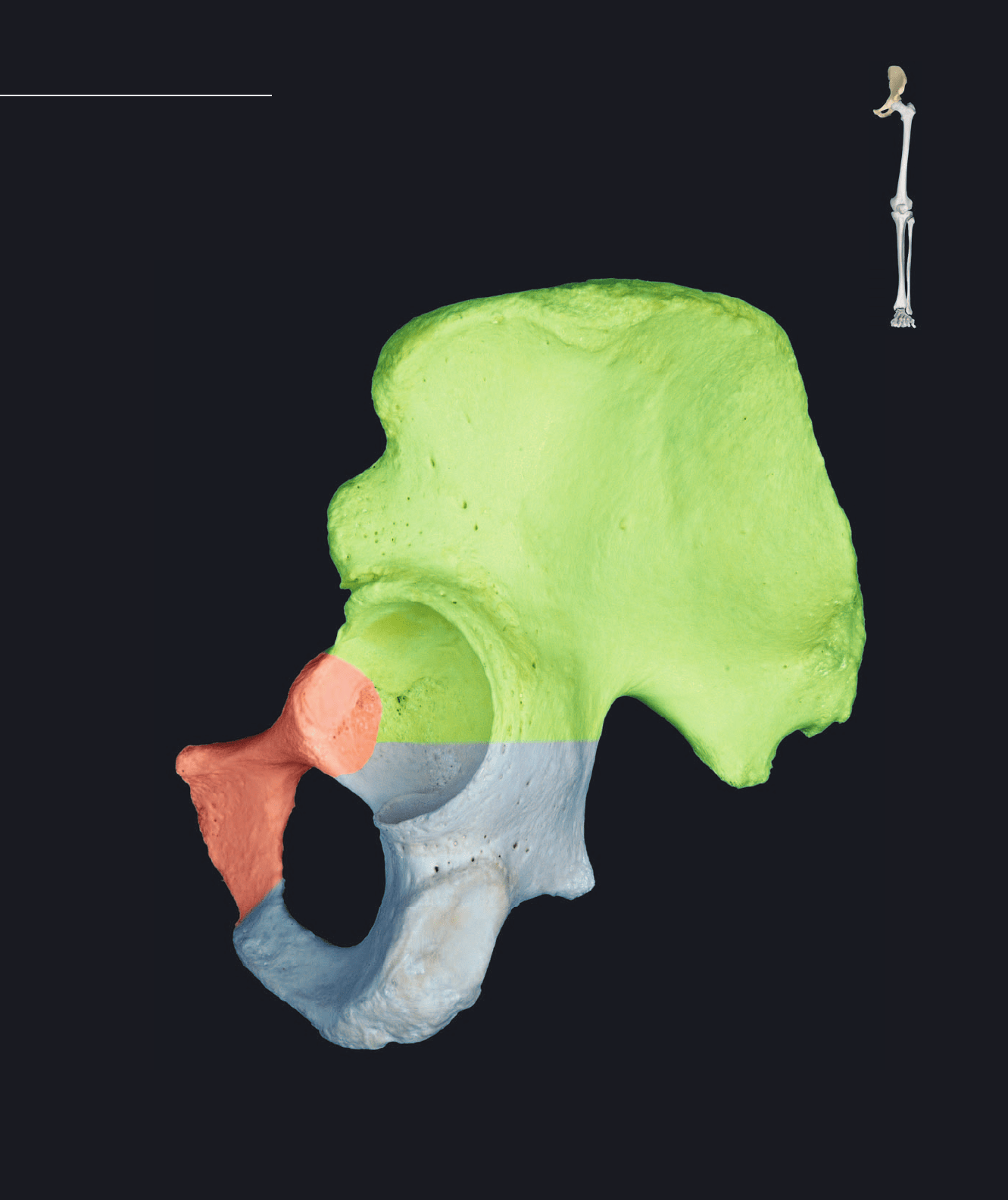
and transfers the forces of locomotion from the inferior limb to the vertebral column. Each os coxae articulates with
three bones — the femur, sacrum, and opposite os coxae. The photo on this page depicts the three bones of the os
coxae — the ilium (green), the ischium (blue), and the pubis (red). Landmarks that are shared by the bones are
depicted on this image. The following two pages show all the landmarks of the individual bones of the os coxae.
Each os coxae forms from three separate bony elements that fuse during development
at their site of union within the acetabulum. The three bony elements are the ilium, is-
chium, and pubis. This strong girdle of bone unites the inferior limb to the axial skeleton
Os Coxae
Left os coxae showing individual bones
Lateral view, anterior to left
1 Acetabulum
2 Acetabular notch
3 Lunate surface
4 Ischiopubic ramus
5 Obturator foramen
6 Greater sciatic notch
1
2
3
4
5
6
Atlas_AppendSkel.indd Page 105 15/03/11 8:57 PM user-F391Atlas_AppendSkel.indd Page 105 15/03/11 8:57 PM user-F391 /Users/user-F391/Desktop/Users/user-F391/Desktop
106
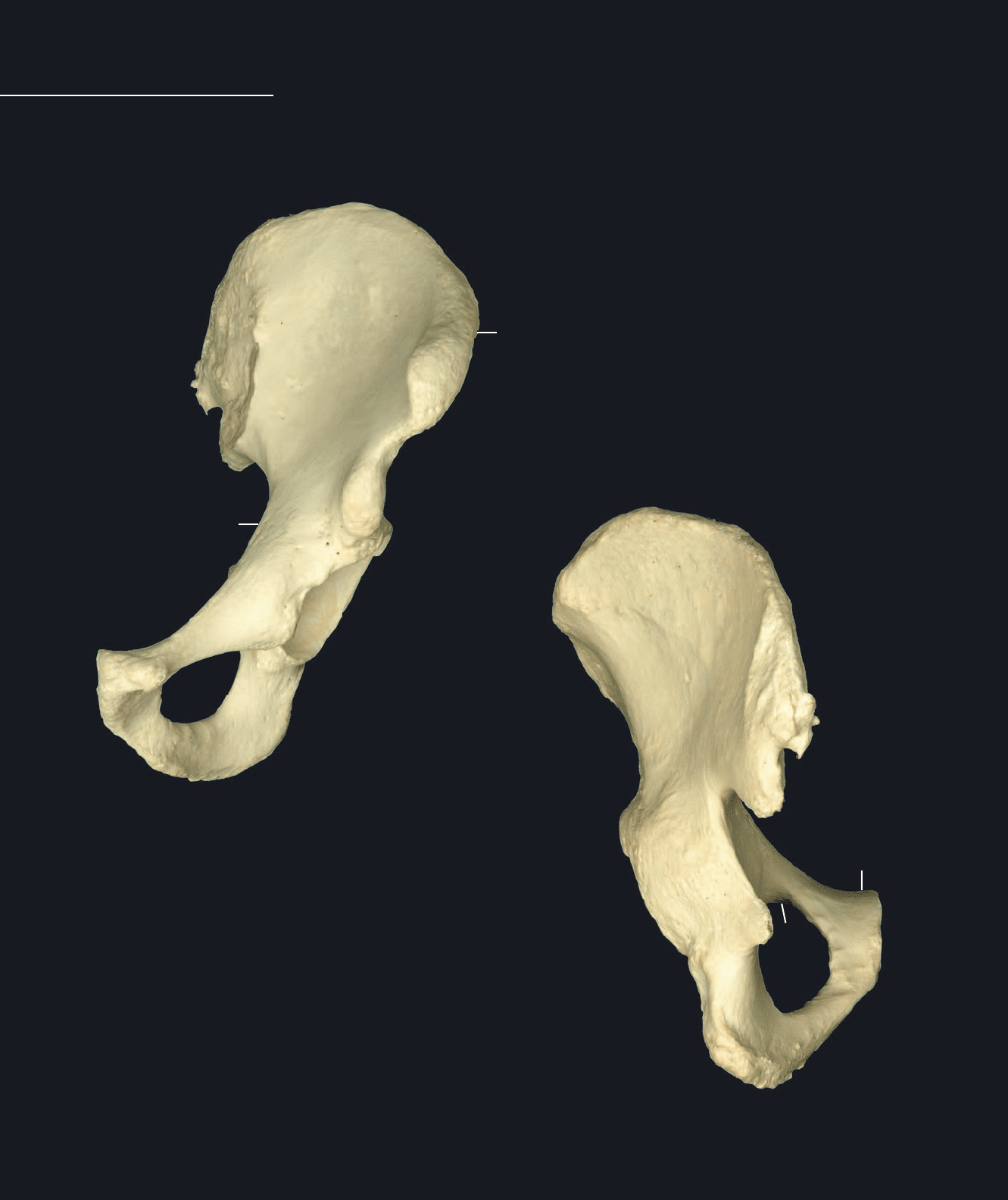
Os Coxae
Ilium
1 Body of ilium
2 Supra-acetabular groove
3 Ala or wing
4 Arcuate line
5 Iliac crest
6 Outer lip of crest
7 Intermediate zone of crest
8 Inner lip of crest
9 Tuberculum of crest
10 Anterior superior iliac spine
11 Anterior inferior iliac spine
12 Posterior superior iliac spine
13 Posterior inferior iliac spine
14 Iliac fossa
15 Anterior gluteal line
16 Posterior gluteal line
17 Inferior gluteal line
18 Auricular surface
19 Iliac tuberosity
Left os coxae
Anterior view, lateral to right
Left os coxae
Posterior view, lateral to right
1
1
3
5
5
3
78
9
10
11
12
13
14
15
16
19
21
21
22
23
25
25
26
28
29
29
32
32
4
6
33
33
28
31
Atlas_AppendSkel.indd Page 106 15/03/11 8:57 PM user-F391Atlas_AppendSkel.indd Page 106 15/03/11 8:57 PM user-F391 /Users/user-F391/Desktop/Users/user-F391/Desktop
107
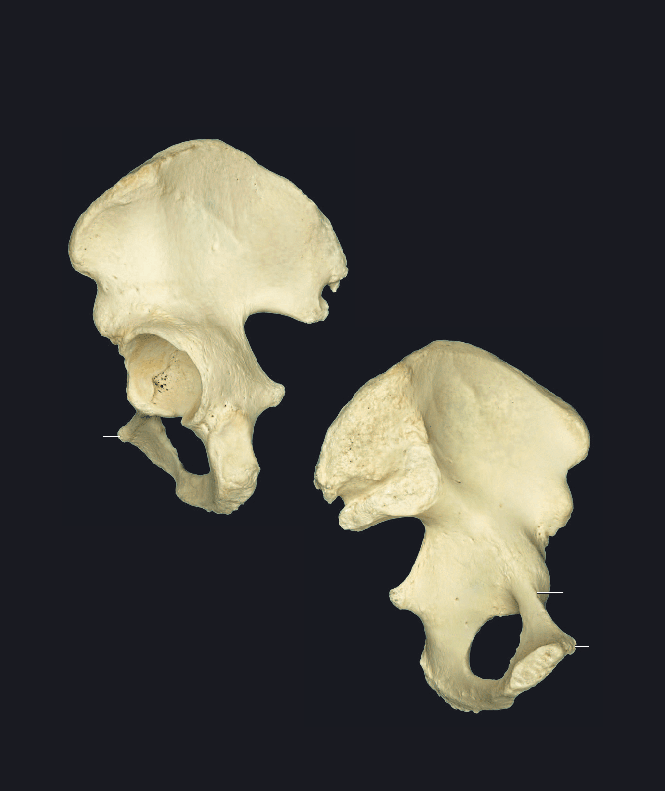
Ischium
20 Body of ischium
21 Ischial ramus
22 Ischial tuberosity
23 Ischial spine
24 Lesser sciatic notch
Pubis
25 Body of pubis
26 Pubic tubercle
27 Symphysial surface
28 Pubic crest
29 Superior pubic ramus
30 Pecten pubis or pectineal line
31 Obturator groove
32 Inferior pubic ramus
33 Obturator foramen
Left os coxae
Lateral view, anterior to left
Left os coxae
Medial view, anterior to right
1
1
5
5
3
3
4
9
2
10
10
11
12
12
13
14
15
16
17
18
19
20
20
21
21
22
23
23
25
27
29
31
32
13
33
33
24
26
24
26
30
Atlas_AppendSkel.indd Page 107 15/03/11 8:57 PM user-F391Atlas_AppendSkel.indd Page 107 15/03/11 8:57 PM user-F391 /Users/user-F391/Desktop/Users/user-F391/Desktop
108
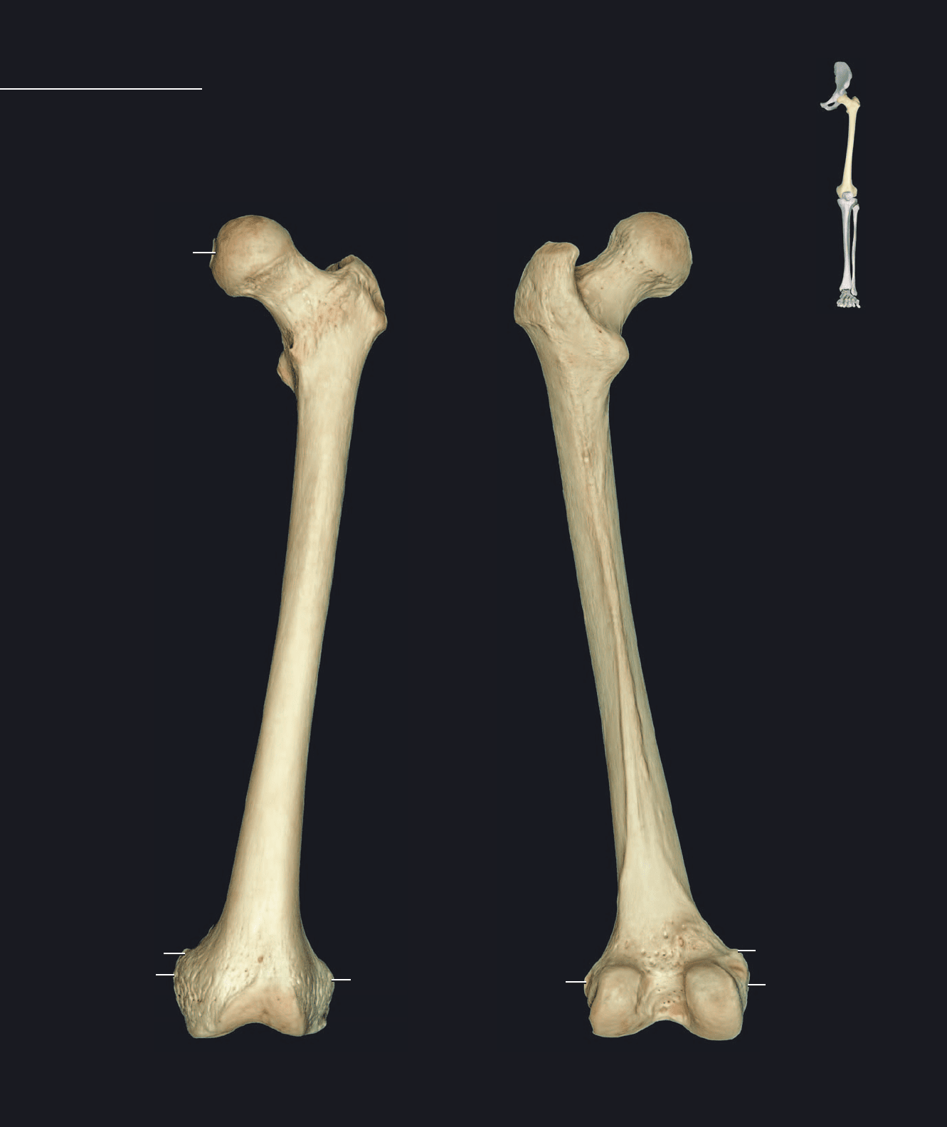
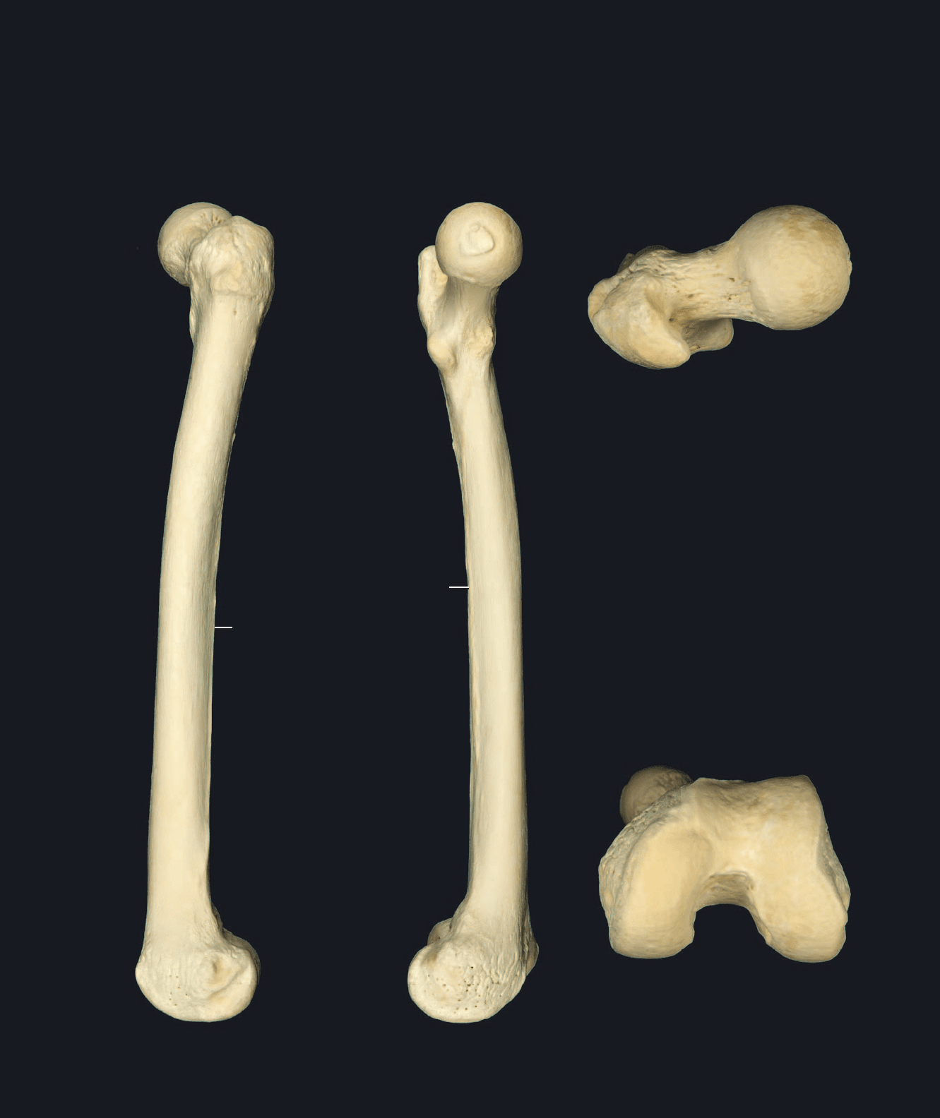
wider toward each end, the compact wall of bone becomes thinner and the medullary cavity accumulates spongy
bone. The proximal end consists of a short cantilevered neck capped by a smooth, round articular head. Projec-
tions of bone, the trochanters, form at the base of the cantilevered neck. The distal end consists of two large,
knuckle-like processes separated by an intermediate groove. The femur articulates with three bones: the os coxae,
patella, and tibia.
The femur is the longest bone of the body. The strong shaft forms a long cylindrical tube with
a slight forward bow. The strong wall of the shaft is thickest near the narrow center of the bone
where the medullary cavity is also the most spacious. As the shaft becomes progressively
Femur
Left femur
Anterior view, lateral to rigjt
Left femur
Posterior view, lateral to left
1
1
3
3
4
4
6
6
7
8
9
10 11
12
13
14
15
16
16
19
19
22
23
20
17
18
20
18
17
2
Atlas_AppendSkel.indd Page 108 15/03/11 8:57 PM user-F391Atlas_AppendSkel.indd Page 108 15/03/11 8:57 PM user-F391 /Users/user-F391/Desktop/Users/user-F391/Desktop
109
1 Head
2 Fovea for ligament of head
3 Neck
4 Greater trochanter
5 Trochanteric fossa
6 Lesser trochanter
7 Intertrochanteric line
8 Intertrochanteric crest
9 Quadrate tubercle
10 Shaft or body
11 Linea apsera
12 Pectineal or spiral line
13 Gluteal tuberosity
14 Medial supracondylar line
15 Lateral supracondylar line
16 Medial condyle
17 Medial epicondyle
18 Adductor tubercle
19 Lateral condyle
20 Lateral epicondyle
21 Groove for popliteus
22 Patellar surface
23 Intercondylar fossa
Left femur
Lateral view, anterior to left
Left femur
Medial view, anterior to right
Left femur
Inferior view, lateral to right
1
1
2
3
3
4
4
4
5
5
10
10
16 19
21
18
17
20
22
23
6
6
Left femur
Superior view, lateral to left
11
11
Atlas_AppendSkel.indd Page 109 15/03/11 8:58 PM user-F391Atlas_AppendSkel.indd Page 109 15/03/11 8:58 PM user-F391 /Users/user-F391/Desktop/Users/user-F391/Desktop
110
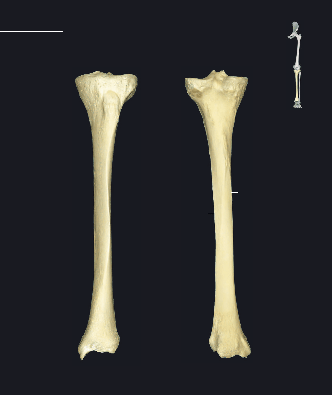
with a fl at plateau-like superior surface largely covered with articular cartilage. The smaller distal end is more knob-
like with a pronounced medial projection, the malleolus. The shaft has a strong anterior crest with sloping surfaces
to either side. The bone is easily palpable throughout its length. The tibia articulates with three bones — the femur,
fi bula, and talus.
The tibia is the large, medial bone of the leg skeleton. It is the second longest bone of the body,
only exceeded in length by the femur. Its strong shaft, consisting of thick walls of compact bone, is
triangular in cross-section. The shaft expands proximally into a fl uted extremity of spongy bone
Tibia
Left tibia
Anterior view, lateral to right
Left tibia
Posterior view, lateral to left
2 233
4
7
10 10
11
12
14
16 16
19
17
13
15
Atlas_AppendSkel.indd Page 110 15/03/11 8:58 PM user-F391Atlas_AppendSkel.indd Page 110 15/03/11 8:58 PM user-F391 /Users/user-F391/Desktop/Users/user-F391/Desktop
111
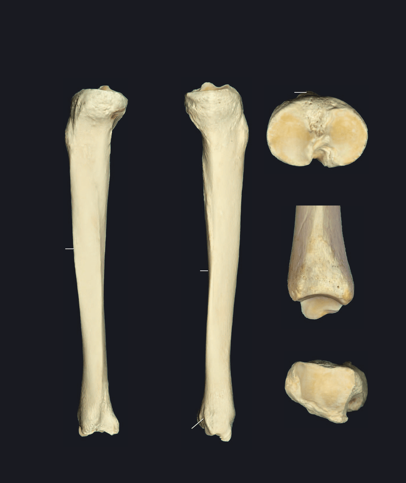
1 Superior articular surface
2 Medial condyle
3 Lateral condyle
4 Fibular articular facet
5 Anterior intercondylar area
6 Posterior intercondylar area
7 Intercondylar eminence
8 Medial intercondylar tubercle
9 Lateral intercondylar tubercle
10 Shaft or body
11 Tibial tuberosity
12 Soleal line
13 Interosseous border
14 Anterior border
15 Posterior border
16 Medial malleolus
17 Malleolar groove
18 Malleolar articular facet
19 Fibular notch
20 Inferior articular surface
Left tibia
Lateral view, anterior to left
Left tibia
Medial view, anterior to right
Left tibia
Superior view, lateral to left
Left tibia
Inferior view, lateral to right
Left tibia
Close-up of lateral view
11
2
3
4 5
6
7
7
7
10
10
11
11
13
16
16
19
20
18
19
8
9
14
15
17
11
Atlas_AppendSkel.indd Page 111 15/03/11 8:58 PM user-F391Atlas_AppendSkel.indd Page 111 15/03/11 8:58 PM user-F391 /Users/user-F391/Desktop/Users/user-F391/Desktop
112
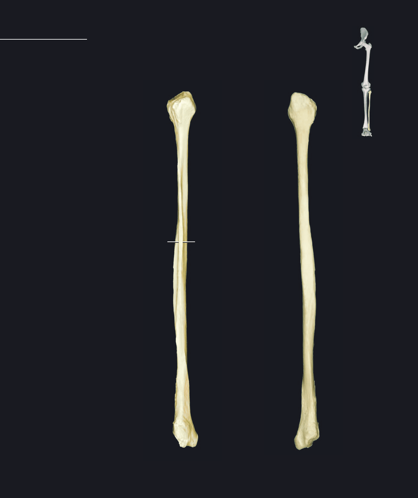
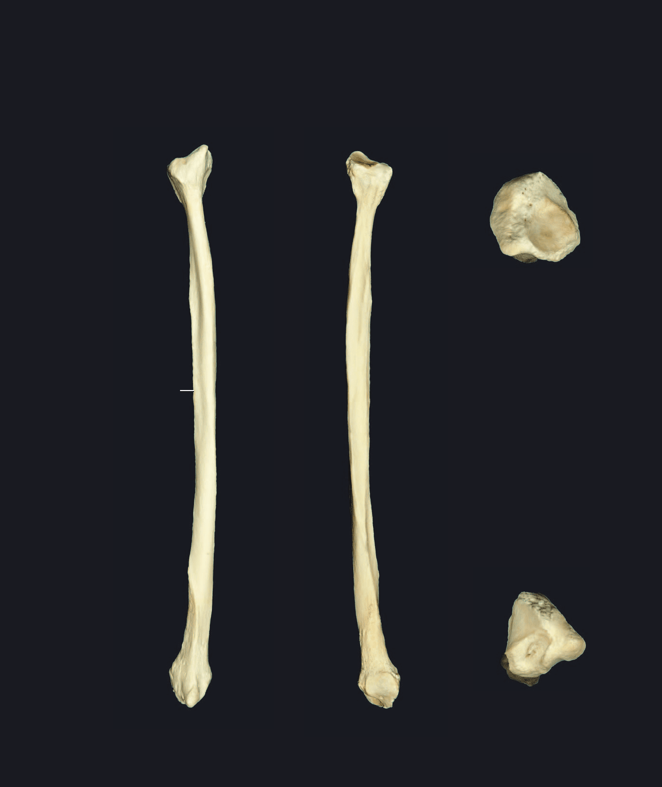
mal and distal ends, the shaft being totally surrounded with muscle. The fi bula articulates with two bones — the
tibia and talus.
The fi bula is the lateral bone of the leg skeleton. It is a slender, splint-like bone that is slightly
expanded at both ends. It plays no role in the weight-bearing function of the lower limb, but
serves as a signifi cant site of muscle attachment. It is not easily palpable except at its proxi-
Fibula
1 Head
2 Articular facet for tibia
3 Apex of head
4 Neck
5 Shaft or body
6 Interosseous border
7 Anterior border
8 Posterior border
9 Lateral malleolus
10 Articular facet for talus
11 Malleolar fossa
12 Malleolar groove
Left fi bula
Anterior view, lateral to right
Left fi bula
Posterior view, lateral to left
1
1
2
3
3
4
4
5
5
12
9
67
Atlas_AppendSkel.indd Page 112 15/03/11 8:58 PM user-F391Atlas_AppendSkel.indd Page 112 15/03/11 8:58 PM user-F391 /Users/user-F391/Desktop/Users/user-F391/Desktop
113
Left fi bula
Lateral view, anterior to left
Left fi bula
Medial view, anterior to right
Left fi bula
Inferior view, lateral to right
Left fi bula
Superior view, lateral to left
1
1
2
2
3
3
44
5
8
8
11
8
9
9
11
7
12
Atlas_AppendSkel.indd Page 113 15/03/11 8:58 PM user-F391Atlas_AppendSkel.indd Page 113 15/03/11 8:58 PM user-F391 /Users/user-F391/Desktop/Users/user-F391/Desktop
114
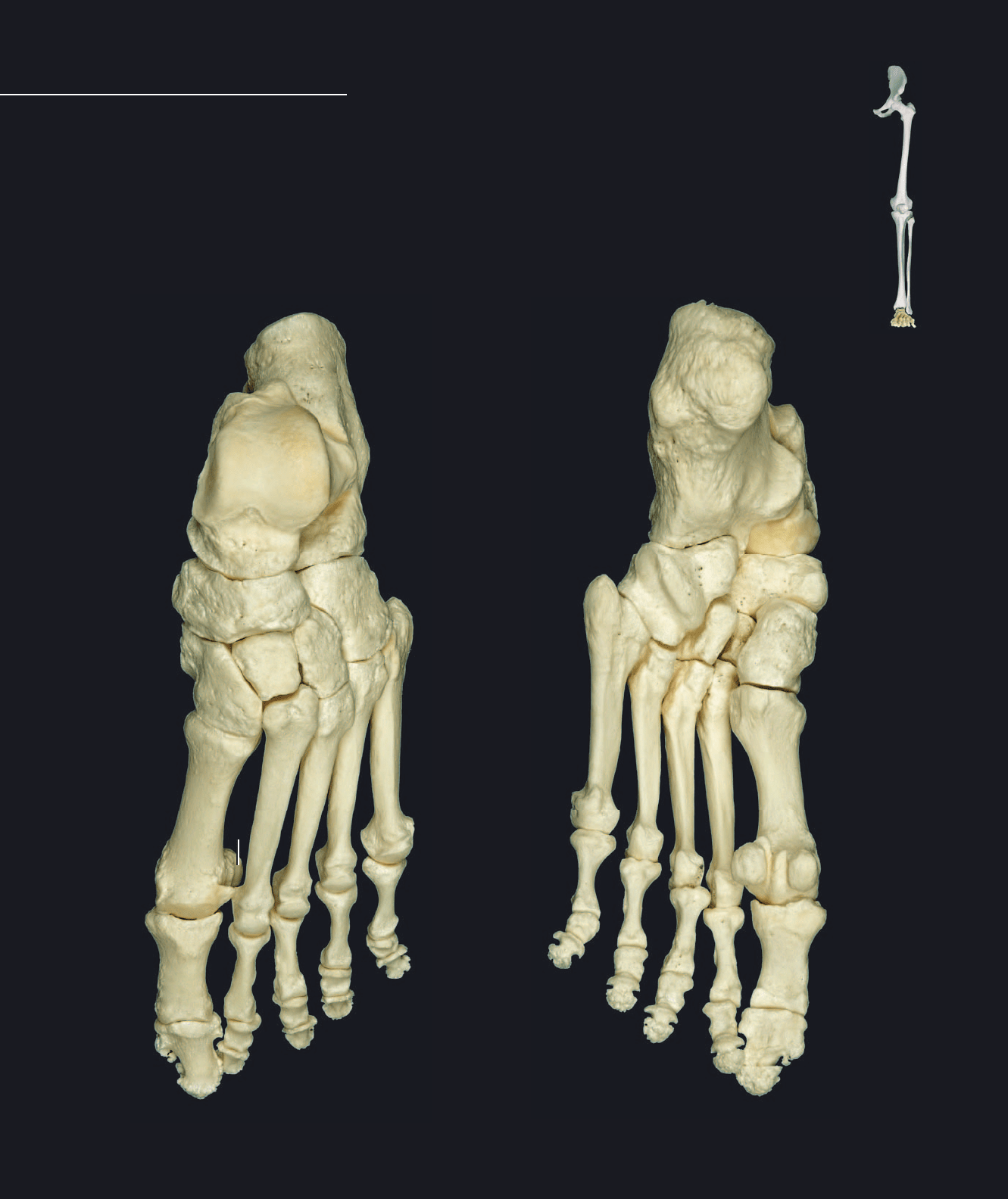
that show a greater range in size and shape than their carpal counterparts in the hand. Distal to the tarsals are the
fi ve digital rays. The four lateral digits consist of a metatarsal bone and three phalanges. The large medial digit, the
hallux or great toe, has a metatarsal bone and only two phalanges. Two prominent sesamoid bones (bones that form
in tendons) are present on the plantar surface at the head end of the fi rst metatarsal.
Like the hand, the foot is a composite structure comprised of 26 bones, not
counting the small sesamoid bones that are found in certain tendons. The
proximal end of the foot is the tarsus or ankle. There are seven tarsal bones
Foot Skeleton
1 Talus
2 Calcaneus
3 Navicular
4 Medial cuneiform
5 Intermediate cuneiform
6 Lateral cuneiform
7 Cuboid
8 Metatarsal I
9 Metatarsal II
10 Metatarsal III
11 Metatarsal IV
12 Metatarsal V
13 Proximal phalanx
14 Middle phalanx
15 Distal phalanx
16 Sesamoid bones
Left foot
Dorsal view, lateral to right
Left foot
Plantar view, lateral to left
1
1
2
2
3
3
4
4
5
5
6
6
7
7
7
8
8
9
9
10
10
11
11
12
12
16
16
13
13
13
13
13
13
13
13
13
13
14
14
14
14
14
14
14
14
15
15
15
15
15
15
15
15
15
15
16
Atlas_AppendSkel.indd Page 114 15/03/11 8:58 PM user-F391Atlas_AppendSkel.indd Page 114 15/03/11 8:58 PM user-F391 /Users/user-F391/Desktop/Users/user-F391/Desktop
