Liu A.L., Tien H.T. Advances in Planar Lipid Bilayer and Liposomes. V.6
Подождите немного. Документ загружается.


distribution of Thy-1 was reduced to 40%, indicating that the molecular packing in
the l
o
phase of synthetic mixtures is tighter than in cell membranes [16]. In giant
unilamellar vesicles (GUVs) composed of a similar synthetic lipid mixture, 30% of
the GPI-anchored placental alkaline phosphatase (PLAP) associates with the l
o
phase,
whereas a non-raft protein was virtually excluded therefrom [17]. Moreover, Wang
et al. [18] investigated the partition behaviour of differentially lipidated peptides and
found that peptides with two saturated fatty acids (the membrane anchor of a typical
cytoplasmic raft protein) were also specifically located at l
o
domains. It is under-
standable that for proteins which are associated with the membrane solely by lipid
anchors, these lipid modifications determine the differential affinity of the proteins
for co-existing lipid phases. But also specific transmembrane proteins have been
shown to be raft associated; as l
o
domains are known to be thicker than l
d
domains,
the length of the hydrophobic transmembrane helix seems to be an important factor
influencing the partition coefficient of the respective protein. Interestingly, the
glutamate receptor which is a G-protein coupled seven-helix transmembrane pro-
tein alters its conformation and ligand binding affinity in dependence of the sur-
rounding lipid phase [19]. Therefore, it is conceivable that in the cellular context
different mechanisms exist to modulate the affinity of signalling proteins for lipid
phases and that this is another level of regulating raft-based signalling processes.
As outlined above, it is well-established that lipid phases influence the spatial
distribution of proteins within the plane of the membrane. Conversely, there is also
evidence that protein-based processes can influence phase separation in lipid bilay-
ers. Hammond et al . [20] used mixtures of PC, SM and cholesterol in compositions
to give either a uniform l
d
or l
o
phase. These mixtures also contained a small but
constant mole fraction of the ganglioside GM1. Addition of cholera toxin subunit B
(CTB), a bacterial protein known to bind to several molecules of GM1, resulted in
a large-scale reorganization of the model membrane by causing the phase-sepa-
ration of large, co-existing l
d
and l
o
domains. Moreover, after GM1 cross-linking by
CTB a transmembrane raft peptide marker that was reconstituted in this model
membrane was specifically localized to the l
o
domains. This result demonstrates
how cross-linking of one membrane component can alter the localization of other
proteins and lipids and thereby could be a model for membrane domain formation
e.g., in immune-recognition receptor signalling in B and T cells [21]). The for-
mation of caveolae, which are cholesterol-rich, flask-shaped membrane invagina-
tions on the plasma membrane of specific cells is another prototypic example for
such a process. The major protein constituent, caveolin which associates with rafts
due to its three palmitic-acid modifications and its cholesterol-binding property, is
thought to drive this process by the formation of large homo-oligomeric complexes
[22]. Cross-linking and oligomerization of raft components should not only be
considered as events to locally increase the size of lipid rafts but may also act as
protein-driven mechanisms that have a global impact on the phase status of the
membrane. Moreover, it has been suggested that specific membrane proteins might
have an impact on the stability of small l
o
domains by localizing to the l
o
–l
d
phase
boundary and thereby decreasing the line tension between the phases [23]. Spe-
cifically, transmembrane proteins might act as ‘‘surfactants’’ at the phase boundaries
but also peripherally associated proteins have been shown to reside at the borders of
U. Salzer et al.54

membrane domains. The signalling proteins phospholipase Cg1 and phosphatidy-
linositol 3-kinase are located at the border of specific raft domains possibly due to
their conflicting localization signals of their membrane anchors, a saturated fatty
acid modification and a C-terminal unsaturated prenyl anchor [24,25]. H-ras is
involved in the formation of non-raft membrane microdomains thereby deter-
mining the final composition of the H-ras signalling complex [26]. In summary,
there seem to be various possibilities how membrane proteins influence the or-
ganization of lipids and hence membrane domains in the bilayer.
2.3. Detergent-Resistant Membranes
Detergent-resistant membranes (DRMs) are the most controversial aspect in the
context of the lipid raft hypothesis. It was originally observed by Brown and Rose
that GPI-linked proteins become resistant to extraction by Triton X-100 (TX-100)
during their passage through the Golgi complex [27]. These DRM complexes could
be separated from the bulk TX-100 soluble protein by density-gradient ultracen-
trifugation: due to their high lipid content, DRMs floated to the low-density (LD)
region of the gradient. As the DRMs were specifically enriched in cholesterol, SM
and glycolipids, it was suggested that the DRMs might be correlated with the
previously postulated lipid rafts at the cell membrane [28].However,itisobvious
that DRMs cannot be assumed as being identical with cellular raft domains in their
lipid and protein composition. First, various types of rafts that are assumed to co-exist
at the membrane will always be irreversibly mixed and fused in DRMs. Second, the
low temperature required for DRM preparations (0–41C) might influence the phase
state of the membrane for thermodynamic reasons. And third, it has been shown that
TX-100 affects the formation of l
o
domains in synthetic lipid mixtures [29,30].In
analysing DRMs, one has always to keep in mind that over- or under-representation
of raft proteins or lipids in DRMs is likely to be the case. Experimental evidence for
an over-representation of a raft protein in DRMs was shown in a model membrane
system. GPI-linked PLAP reconstituted in GUVs was nearly completely insoluble in
TX-100 although only 30% is localized to l
o
domains in the unperturbed vesicles
[17]. DRM preparation protocols should be carefully established for each cell system
to minimize additional artefacts. For example, a sufficiently high detergent/lipid
ratio is essential to avoid incomplete solubilization of non-raft components [31].
However, in spite of these caveats, the DRM methodology has been proven useful,
because most of the proteins that are typically present in DRMs have been shown to
be raft associated when tested by different methods (Section 2.4) [32].Recently,a
quantitative proteomic approach was applied to classify DRM proteins with respect
to the cholesterol sensitivity of their DRM association; thereby, this study revealed a
set of about 250 authentic lipid raft proteins [33].
2.4. Experimental Evidence for Lipid Rafts
Cross-linking of raft components can induce the formation of large raft aggregates
at the plasma membrane; however, in the resting state, lipid rafts have been proven
to be too small to be studied by normal microscopic techniques. Several different
Organization and Dynamics of Erythrocyte Lipid Rafts 55

advanced techniques have been used in order to determine the existence and size of
raft domains in the plasma membrane. Fluorescence resonance energy transfer
(FRET) studies showed a partial clustering of GPI-linked proteins in nanoscale
domains that were sensitive to cholesterol depletion [34,35]. Pralle et al. [36] applied
photonic force microscopy and found that the viscous drag of three different raft
proteins mediated by bead-coupled specific antibodies was significantly higher than
that of non-raft proteins. This difference was abolished when rafts were destroyed
by incubating the cells with MbCD. In this experiment, the size of the rafts was
estimated to be about 50 nm. A similar size (44 nm) was obtained in an immuno-
electron microscopy study that analyzed the distribution of a raft marker on the
cytoplasmic leaflet of the plasma membrane by spatial point pattern analysis [37].
MbCD treatment resulted in a random distribution of the marker. Interestingly,
cross-linking of a GPI-linked outer-leaflet raft protein influenced the distribution
of the inner-leaflet raft marker and indicates that there is a loose association between
inner and outer-leaflet rafts. Moreover, also non-raft microdomains specified by
their resistance to MbCD treatment have been identified. Wilson et al. [38] studied
the distribution of 4 different raft markers, i.e. the ganglioside GM1, the GPI-
linked Thy-1, the palmitoylated LAT protein and cross-linked IgE receptors FceRI,
at the plasma membrane of a mast cell line by immunoelectron microscopy. In
resting cells, Thy-1 and FceRI are found in small clusters (20–50 nm), whereas
GM-1 is rarely clustered at all, thereby indicating that various types of rafts re-
garding their composition and size might co-exist at the membrane. Upon cross-
linking, these three markers form independent larger clusters and LATwas found to
co-cluster with Thy-1 complexes. The large GM1 and Thy-1 clusters accumulate
components of the endocytic machinery indicating that aggregation of these spe-
cific rafts leads to a local endocytic event. In summary, these data actually support
the view of the raft hypothesis that small cholesterol-rich microdomains are present
at the plasma membrane where raft proteins are specifically enriched.
2.5. A Revised Version of the Lipid Raft Hypothesis
Hancock recently proposed a revised version of the lipid raft hypothesis suggesting a
dominant role for plasma membrane proteins in the formation and maintenance of
lipid domains [23]. Small, transiently forming l
o
domains might be captured by a
raft protein, thereby conferring stability to the lipid raft. Protein interaction and
oligomerisation might eventually increase both the size and the lifetime of these
lipid domains which might be involved in specific cellular processes. On the other
hand, it would be expected that small protein-stabilized l
o
domains coalesce in ever
larger domains to minimize the line of tension at the l
o
–l
d
phase boundary. As such,
large complexes are usually not observed in the plasma membrane, an active
mechanism seems to prevent large-scale phase separation. It is suggested that end-
ocytosis might be the cellular process that limits the raft-domain size [39]. Large
rafts might be removed from the plasma membrane by endocytosis and smaller,
disassembled raft units might recycle back to the plasma membrane by recycling
endosomes or other vesicular transport pathways. In fact, endocytosis is a raft-based
process and is known to be increased upon cross-linking rafts into larger domains
U. Salzer et al.56

[40]. The revised raft hypothesis stresses that proteins play an active role in the
organization of the plasma membrane by exploiting the intrinsic properties of lipid
mixtures to form co-existing liquid phases. This dynamic, complex and interactive
model replaces the ‘‘old’’ view of rafts as stable units within the membrane that
govern the lateral distribution of membrane proteins.
3. DRMs of the Erythrocyte Membrane
It was actually a solubilization study of erythrocyte membranes 33 years ago
that led to the first description of DRMs, although the term ‘‘DRM’’ was not used
then. Yu et al. [41] showed that upon extraction with the non-ionic detergent TX-
100 glycerolipids were solubilized, whereas sphingolipids remained associated with
the ‘‘detergent-insoluble ghost residue’’. When studied by electron microscopy,
these membrane residues appeared to be a ‘‘filamentous reticulum with adherent
lipoid sheets and vesicles’’. However, the main focus of that work was to char-
acterize the differential solubilization behaviour of non-glycosylated polypeptides
(mostly cytoskeletal components) and externally oriented glycoproteins (integral
membrane proteins). Nearly 30 years later, detergent resistance of erythrocyte
membrane components has regained vital interest as this feature is a prerequisite for
proteins and lipids to be classified as lipid raft components. The characterization of
DRMs is the main approach in the raft research of erythrocytes, because other
methods are difficult or even impossible to apply to these cells. Using washed
erythrocyte membranes (ghosts), Civenni et al. [42] showed that the GPI-linked
proteins AChE and CD59 were mainly associated with the detergent-insoluble
fraction upon TX-100 treatment. Unlike the endogenous proteins, in vitro incor-
porated GPI-linked proteins were not resistant to membrane extraction thereby
indicating that exogenously added GPI proteins do not co-distribute with the
endogenous proteins in lipid rafts. Lauer et al. [43] showed that the Duffy antigen, a
heptahelical transmembrane protein, and the signalling protein Gas also resist
membrane extraction and are partially found in the LD region of a sucrose gradient
in flotation experiments. Our flotation experiments revealed that stomatin, flotillin-
1 and flotillin-2 are the most abundant integral membrane proteins in erythrocytes
[44]. Whereas stomatin was a well-described erythrocyte membrane protein (also
termed band 7 protein, or protein 7.2b) [45,46] that has been shown to be absent in
erythrocytes of overhydrated hereditary stomatocytosis (OHSt) patients [47,48], the
flotillins have not been described as erythrocyte membrane proteins before. The
association with lipid rafts is a prominent feature of the flotillin proteins; in A498
kidney cells they have been described to be present in caveolae where they form
hetero-oligomeric complexes with caveolin [49] and in neurons they were shown
to co-cluster in membrane microdomains with activated GPI-linked cell adhesion
proteins [50,51]. Stomatin and the flotillins are distantly related proteins that share a
common structural domain, the SPFH domain named after the most prominent
representatives: Stomatin, Prohibitins, Flotillins and the bacterial HflK and C
proteins [52]. For stomatin, it has been shown that it is an untypical integral
Organization and Dynamics of Erythrocyte Lipid Rafts 57

membrane protein with both the N- and the C-terminus facing the cytoplasm and
with a long hydrophobic stretch probably being inserted into the membrane like a
hairpin loop [53]. Caveolins are the only other membrane protein class that show
such a peculiar monotopic membrane association [22]. Protease sensitivity exper-
iments in baby hamster kidney [54] cells and intact red blood cells (data not shown)
suggest that the flotillin proteins are not transmembrane proteins but are also
monotopically associated with the cytoplasmic face of the plasma membrane. As the
flotillin proteins and stomatin form large oligomeric complexes in the erythrocyte
membrane [44], it is tempting to assume that these proteins function as scaffolding
proteins at the cytoplasmic leaflet of erthrocyte lipid rafts [55].
In contrast to reports by Samuel et al. [56] and Murphy et al. [57], we found that
a fraction of the major erythrocyte cytoskeletal components, spectrin, actin, protein
4.2 and protein 4.1, is co-distributing with the integral raft proteins stomatin and
the flotillins in the LD region of the sucrose gradient [44]. In the following section,
we will present biochemical data that further suggest an association between DRMs
and the erythrocyte membrane skeleton and we will discuss possible candidate
molecules that might mediate this association. Moreover, we will try to explain the
seeming discrepancy between the data of our and Kasturi Haldar’s group.
3.1. Evidence for the Association of the Cytoskeleton with DRMs
When we routinely isolated red blood cell DRMs by our published method [44],
we often noticed donor-dependent variations regarding the degree of DRM
buoyancy and cytoskeleton association. To clarify which parameters might be re-
sponsible for these variations, we undertook a series of experiments with different
methodological variations of the established protocols for DRM preparation and
developed three different DRM preparation protocols. In protocol I, erythrocytes
are diluted in four volumes of tris-buffered saline (TBS, 150 mM NaCl, 10 mM Tris
pH ¼ 7.5) and this suspension is lysed by mixing with an equal volume of ice-cold
2% TX-100 in TBS for 20 min. This lysate is centrifuged for 10 min at 13,000 rpm
to concentrate the insoluble material consisting of the cytoskeleton and DRMs; this
pellet is resuspended in 1% TX-100 in TBS and then mixed with the appropriate
sucrose solution to give the bottom layer of the sucrose step gradient. This protocol
is preferably applied when whole erythrocytes are used as starting material, because
the high haemoglobin content in the original total lysate can be reduced this way.
In protocol II, the pelleting step is omitted and the TX-100 lysate is directly mixed
with the high-density sucrose solution for subsequent flotation experiments. This
protocol can be used when ghosts are used as starting material or in experiments
with whole erythrocyte lysates when the analysis of the high-density bottom frac-
tion can be neglected (due to its high haemoglobin content which would interfere
with Western blot analysis). This protocol was specifically designed to address the
question whether the association between the DRMs and the cytoskeleton is
already present in the lysate or only occurs after the centrifugation step due to the
dense packing of DRMs and the cytoskeleton in the pellet. Protocol III is a var-
iation of protocol I where the cytoskeleton-DRM pellet is resuspended in 0.2 M
sodium carbonate instead of TBS to dissolve the cytoskeletal network and to impair
U. Salzer et al.58

its interaction with DRMs. To generally obtain a separation of the DRMs with
higher density from the TX-100 soluble material, we increased the sucrose density
in the bottom layer and expanded the intermediate density region of the gradient
compared to the method published in [44].
The flotation experiments according to protocols I and III are compared in
Fig. 1. Using the protocol I with erythrocytes as starting material, the typical
distribution of protein and lipid markers in the density gradient of a flotation
experiment is shown in Fig. 1A. Only small amounts of the DRM marker proteins
stomatin, flotillin-2 and AChE are present in the LD fraction 1, whereas the major
pool of these markers co-distributes at intermediate densities (ID) in fractions 3–6.
Cholesterol partially co-distributes with AChE, flotillin-2 and stomatin in the LD
and the ID region. Moreover, we found that SM was nearly exclusively present
in the LD and ID region (data not shown), which is in line with results from
Koumanov et al. [58]. The silver-stained gel in Fig. 1A reveals that the overall protein
concentration is highest in the bottom layer (HD) of the gradient. For appropriate
silver staining, the bottom fractions were diluted because of the high amount of
protein (see figure legends), whereas equal volumes of the fractions were used for the
immunoblot, AChE, cholesterol and SM analyses. The silver stain in Fig. 1A shows
that LD fraction 1 contains only a small amount of the proteins stomatin and the
flotillins, whereas a large amount of these DRM markers is found in the ID fractions
3–6. A considerable amount of the cytoskeletal components spectrin and actin co-
distribute with the DRM markers in the ID region of the gradient.
These data reveal two pools of DRMs at different densities in the gradient: a
small pool at LD containing the typical DRM markers, and a large pool of DRMs
at IDs that is associated with considerable amounts of cytoskeletal proteins. The
cytoskeleton–DRM association is not a procedural artefact due to the tight packing
of DRMs and the cytoskeleton during the pelleting step of protocol I, because the
cytoskeleton–DRM complexes are also obtained by protocol II, which lacks this
pelleting step (data not shown). Moreover, we excluded the possibility that the
presence of high amounts of haemoglobin during lysis influences the result of a
DRM flotation experiment; several experiments that directly compared the DRMs
from erythrocytes and ghosts did not reveal major differences (data not shown). In
order to assess the relative amounts of the cytoskeleton-free and the cytoskeleton-
loaded DRM pools and to estimate the magnitude of the donor-specific variations,
we performed a statistical analysis of the results of six independent flotation
experiments (protocol I) with erythrocytes from different donors. We estimated the
relative amounts of each DRM and cytoskeletal marker in the fractions by densi-
tometry of silver-stained gels and/or quantitative immunoblotting and assessed their
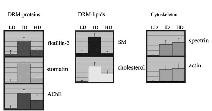
relative distribution in the LD, ID and HD regions. Fig. 2 reveals that only 5% to
maximally 20% of the DRM markers stomatin, flotillin, AChE, SM and cholesterol
are present in the cytoskeleton-free DRM pool in the LD region, whereas 60–80%
of these markers are found in the cytoskeleton-associated DRM pool at ID. More-
over, about 40% of the cytoskeletal components actin and spectrin are associated
with this DRM pool in the ID region. We want to stress here that these values of
the DRM markers give the percentage of the TX-100 insoluble fraction and that
100% does not refer to the total amount of the respective marker in the cell because
Organization and Dynamics of Erythrocyte Lipid Rafts 59
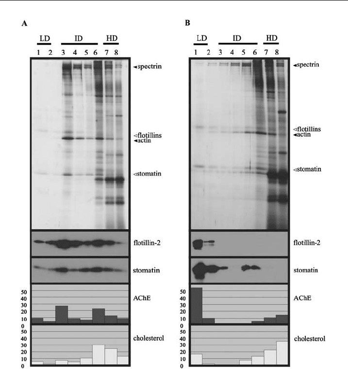
Figure 1 Comparison of cytoskeleton-loaded and cytoskeleton-free DRMs. DRMs were
prepared from erythrocytes according to protocol I (A) or protocol III (B). 150 mlpacked
erythrocytes were resuspended in 550 ml TBS, cooled, lysed with 700 ml of ice-cold 2% Triton
X-100, 10 mM EDTA, and proteinase inhibitors in TBS, incubated fo r 20 min on ice, and
centrifuged at 15000 g for 1 0 min, 41C. The pellet was resuspended in cold 1% Triton X-100
containing 5 mM EDTA and proteinase inhibitors in TBS to give 300 ml. Protocol III is identical
to protocol I except that the pellet is resuspended in 0.2 M sodium carbonate to disrupt the
cytoskeleton. These suspensions are the n mixed with 500 ml 80% sucrose in TBS, placed in a
centrifuge tube (Beckman 13 51mm),overlaidwith1ml40%,2ml35%,0.5ml20%,and
0.5 ml 5% s ucrose in TBS, and cent ri fuged in a pre-cooled SW55 rotor (Beckman) for 18 h at
235,000 g,41C. Eight fractions of 600 ml were collected from the top and equal aliquots of these
fractions were ana lysed for AChE (acetylcholinesterase) activity, and cholesterol content, and by
11% SDS-PAGE/Western blotting as indicated. For silver staining (top panels), the following
relative volumes of the fractions were applied to the gels: 1.00 (fractions 1 and 2), 0.40 (fractions
3^5) and 0.25 (fractions 6^8). LD, ID and HD indicate low-, intermediate- and high-density
regions, respectively. T he distribution of AChE and cholesterol is given in relative percentages.
Open triangles indicate the positions of lipid raft marker proteins, ¢lled triangles indicate the
positions of cytoskeletal proteins. DRM markers are preferentially found in the ID region (A),
but shift to the LD region upon disruption of the cytoskeleton (B).
U. Salzer et al.60
the TX-soluble fraction is removed in the pelleting step in protocol I. The TX-100
insoluble fraction comprises 50, 80, 90, 95 and 95% of the total cell cholesterol,
stomatin, AChE, SM and flotillin, respectively (data not shown), and 100% of the
cytoskeletal components actin and spectrin.
We have previously described that cytoskeleton-free DRMs can be prepared by
alkaline extraction of the TX-100 lysates or pellets [44] indicating that the cyto-
skeletal interactions are disrupted under alkaline conditions, whereas the integrity
of DRMs is not affected. Using protocol III, which is a modified version of the
original stripping method [44], we analysed the changes in the DRM distribution
after disruption of the cytoskeleton (Fig. 1B). The major amounts of the flotillins,
stomatin and AChE, and about 20% of cholesterol are present in the LD fractions of
the gradient. The abundance of the cytoskeletal proteins spectrin and actin in the
ID region is strongly reduced and they are mainly found in the dense fractions
(compare silver stains of Fig. 1A and 1B). The disruption of cytoskeletal interactions
strongly diminishes the ID DRM pool and leads to a large increase of DRMs at LD.
This indicates that the heavy protein load of cytoskeletal components associated
with DRMs normally prevents the migration of these DRMs to LDs.
As already mentioned above, Murphy et al. [57] did not find cytoskeletal pro-
teins associated with DRMs and suggested that differences in the detergent-to-lipid
ratio might account for this discrepancy with our previously published data [37].In
order to test whether the DRM–cytoskeleton association was due to incomplete
Figure 2 Distribution of protein a nd lipid markers wit hin the density gradient. A statistical
analysis of 20 independ ent £otation experiments using erythrocytes from six di¡erent donors
according to protocol I is shown. The amount of the indicated protein and lipid markers in the
eight fractions was assessed by densitometry of silver-stained gels, quantitative immunoblotting,
and quantitative analysis, respectively, and the relative distribution of the m arkers in the LD
(fractions 1and 2), ID (fractions 3^6), and HD region (fraction 7 and 8) of the gradient is shown
(relative percent). LD, ID, and HD indicate low-, intermediate-, and high-density regions,
respectively. Error bars indicate the standard deviation. AChE (acetylcholinesterase), SM
(sphingomyelin).
Organization and Dynamics of Erythrocyte Lipid Rafts 61

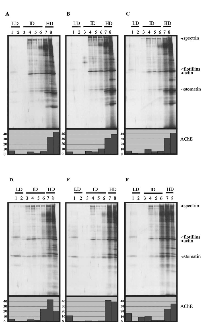
lysis, we performed flotation experiments according to protocol I with increasing
detergent-to-lipid ratios during lysis (Fig. 3). The silver stains in Fig. 3 show no
major differences in the distribution of the cytoskeletal and DRM marker proteins
within the density gradients of the three experiments, irrespective of whether
erythrocytes or ghosts are used as starting material. Therefore, this result does
not support the explanation of incomplete lysis for the cytoskeleton–DRM asso-
ciation.
To study the cytoskeleton–DRM interaction in more detail, we developed
another new method to efficiently and quickly separate cytoskeleton–DRM com-
plexes by non-equilibrium gradient centrifugation (NEGC). TX-100 lysates of
ghosts or erythrocytes are placed on top of a sucrose step gradient and subjected to
ultracentrifugation for 1 h at 38,000 g. Using this method, the TX-100-soluble
and cytosolic proteins can be recovered in the first two fractions of the gradient
comprising the loading zone and part of the 20% sucrose layer, whereas the cyto-
skeleton is present at the interphase between the 35% and the 60% sucrose layer.
Compared to the flotation method that needs at least 16 h of centrifugation, this
NEGC is a quick procedure with the respective advantages. Fig. 4A shows a typical
NEGC experiment of a ghost lysate. Most of the band 3 membrane protein and
band 6 are found in the first two fractions, and the major amounts of spectrin,
protein 4.1 and actin are present in fractions 9 and 10. A high percentage of the
DRM markers stomatin, flotillins, AChE, SM and about 50% of the membrane
cholesterol co-distribute together with the cytoskeleton in fractions 9 and 10 sup-
porting our view of an association of DRMs with the cytoskeleton. An aliquot of
fraction 9 was diluted, incubated with an alkaline buffer (pH 11) on ice to disrupt
the cytoskeleton and then subjected to a short ultracentrifugation step. The re-
sulting pellet and supernatant were analysed for the distribution of the cytoskeletal
and DRM markers. As expected, the cytoskeletal components were found in the
supernatant, whereas all the lipid and protein DRM markers were recovered in the
pellet (data not shown) indicating the presence of intact DRMs in this fraction. As
already shown in Fig. 1B, alkaline treatment disrupts the cytoskeletal interactions,
whereas DRM integrity is unaffected.
To dissolve DRMs, we performed an identical experiment as described in
Fig. 1A except that the lysis temperature was 371C. As expected, the DRM markers
are exclusively found in the TX-100-soluble fraction at the top of the gradient,
whereas the cytoskeleton is present in the bottom fraction demonstrating that the
Figure 3 DRM preparations are independent of the investigated lipid-to-detergent ratios. 50 ml
erythrocytes (A, B, and C) or ghosts (D, E, and F) are diluted with 100 ml (A, D), 250 ml(B,E)or
550 ml(C,F)andlysedwith150m l(A,D),300ml(B,E),or600ml (C, F) of ice-cold 2% Triton X-
100,10 mM EDTA, and proteinase inhibitors inTBS, incubated for 10 min on ice, and centrifuged
at 15,000xg for 20 min, 41C. The pellets were resuspended in cold 1% Triton X-100 containing
5 mM EDTA and proteinase inhibitors in TBS to give 300 ml. The sucrose gradients,
ultracentrif ugation, and gradient fractionation were performed as described in Fig. 1.The
following relative volumes of the fractions were analysed by 11% SDS-PAGE and silver
staining: 1.00 (fractions 1 and 2), 0.40 (fractions 3 ^5) and 0.25 (fractions 6^8). LD, I D and HD
indicate low-, intermediate-, and high-dens ity regions, respectively. Open triangles indicate the
positions of lipid raft marker proteins, ¢l led triangles indicate the positions of cytoskeletal
proteins.
U. Salzer et al.62
Organization and Dynamics of Erythrocyte Lipid Rafts 63