Liu A.L., Tien H.T. Advances in Planar Lipid Bilayer and Liposomes. V.6
Подождите немного. Документ загружается.

neurotransmitter release, in which its interaction with small synaptic vesicles via
synapsin was shown to play a crucial role [28,29]. Numerous interactions of spectrin
with other proteins have been inferred from using the yeast two hybrid system (for a
review see Ref. [30]).
The above-mentioned high-affinity protein–protein interactions responsible
for membrane skeleton attachment to the membrane have been the subject of
many excellent reviews (e.g., [27]) but the direct interactions of this and other
membrane-skeletal proteins with the lipid bilayer has attracted much less attention
from the researchers.
3. Interactions of Erythroid and Nonerythroid
Spectrins with Lipid Mono- and Bilayers and
other Amphipathic Ligands
3.1. Interaction of Erythroid Spectrin with Phospholipid Mono- and
Bilayers, a Historical Perspective of Seventies and Early Eighties
Spectrin was discovered and named by Marchesi and Steers [31] and its interaction
with membrane lipids or phospholipids has been studied since early seventies. The
first indication of the possibility of binding spectrin to lipids was observed in model
system in which crude low ionic strength solution extract of the erythrocyte
membranes (ghosts) which contained mostly spectrin but also actin and other
skeletal proteins were used [32].
In 1970 the interaction of spectrin with model membrane systems was studied
using liposomes, in conditions eliminating electrostatic interactions as the basis for
its binding to the bilayer in the natural membrane. Spectrin bound to negatively
charged liposomes constructed from PC, dicetyl phosphate and cholesterol, inde-
pendently of ionic strength and also to positively charged liposomes prepared by
substitution of stearyl amine for dicetyl phosphate [32].
In 1971 Juliano et al. [33] observed that the surface pressure of PS and PC
monolayers is increased by the addition of spectrin into interphase and that spectrin
interacts with sonicated PS vesicles. Spectrin bound to monolayers and liposomes at
pH values below or near its isoelectric point. At pH 7.4 spectrin failed to interact
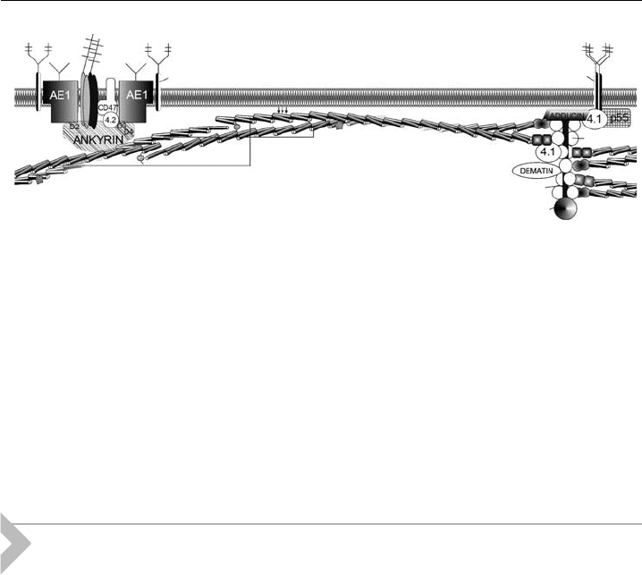
Rh AG
GLYCOPHORIN A
GLYCOPHORIN C
α spectrin
β spectrin
ACTIN
TROPOMYOSIN
TROPOMODULIN
Figure 1 Erythrocyte membrane skeleton, a schematic representation. Arrows depict lipid-
binding activity of the spectrin molecule. Other details in the text.
A.F. Sikorski et al.84

significantly with negatively charged vesicles. Ca
2+
neither increased the effect of
spectrin on PC vesicles nor altered the penetration of spectrin into PS monolayers.
The possibility of both electrostatic attractions between spectrin and phospholipids
and conformational changes in the protein-mediated penetration of spectrin into
the phospholipid bilayer, was postulated. No effect of spectrin on positively charged
stearyl amine/PC vesicles at pH 7.4 was observed [33]. Further studies showed
changes in the enthalpy of phase transition of phospholipids in the presence of
spectrin which lead to conclusion for a hydrophobic binding of spectrin to neg-
atively charged phospholipids [34]. An interaction between sonicated, well-defined
vesicles constructed from DMPC, DMPG and mixtures of these two lipids and a
spectrin–actin complex isolated from human erythrocyte membranes was demon-
strated. Spectrin was also shown to spontaneously interact with the synthetic
bilayers and these lipid–protein interactions stabilised the planar bilayer orientation
of the lipid molecules. It also protected the bilayer against the Ca
2+
and Mg
2+
and
prevented fusion of lipid vesicles which easily occurred when divalent ions were
added to pure lipid vesicles. The same laboratory performed experiments on the
interaction of PS and PS/PC vesicles with spectrin–actin from human erythrocyte
ghosts [35]. DMPS vesicles showed a decrease in enthalpy change of the lipid phase
transition upon addition of spectrin–actin. These vesicles collapsed and fused into
multilamellar structures in the presence of spectrin–actin. The data obtained from
both calorimetric and NMR techniques showed the difference between PS and PC
and showed that the negatively charged lipid polar headgroups are important for the
hydrophobic interaction of spectrin with phospholipids. The observed changes in
thermotropic properties of lipid mixtures showed that both electrostatic and as well
as hydrophobic interactions are involved in binding of spectrin to lipids. The
presence of Ca
2+
and less strongly of Mg
2+
affected the spectrin–PS interaction [5].
More detailed studies on the effect of spectrin on Ca
2+
–PS vesicles interaction
indicated that addition of this cation caused aggregation of PS vesicles and at
temperature above 251C induced the formation of an ‘‘anhydrous’’ complex of
closely apposed membranes with highly ordered crystalline acyl chains. Addition
of Mg
2+
produced much more hydrated complex with PS, without crystallisation
of the acyl chains. Both Ca
2+
and Mg
2+
evoked synergistic effect between the two
cations, which resulted in an enhancement of the ability of Ca
2+
to form its specific
complex with PS at lower concentrations. The presence of the spectrin inhibited
this synergism and interfered with the formation of the specific PS/Ca
2+
complex.
It also inhibits the fusion of PS vesicles [36].
The interactions of synthetic and natural phospholipids with spectrin purified
from human erythrocyte membranes were more precisely examined by using a
monolayer technique, at constant surface pressure [37]. High penetration of neg-
atively charged phospholipid monolayers by spectrin was observed, maximal for PG
and PS and this penetration was strongly affected by the subphase pH, surface
pressure and salt concentration. Also the lipid fatty acid composition in natural
phospholipids proved to be an important parameter. Zwitterionic or neutral lipids,
such as PC, showed only poor affinity for spectrin. Both lipids condensed upon the
addition of Ca
2+
, but only in the case of PS this was accompanied by the extrusion
of spectrin from the monolayer what led the authors to propose the model of this
Interactions of Erythroid and Nonerythroid Spectrins 85

phenomenon. Interaction of spectrin with phospholipid vesicles was stronger in the
case of mixtures of PS with another phospholipid than in the case of PS alone. Also
replacement of PC by PE improved binding [35].
The sites of lipid binding contain probably tryptophan residues, as binding of
amphipatic ligands and phospholipids were shown to quench protein-intrinsic
(tryptophan) fluorescence [38–40]. It should be noted that binding of spectrin to
membrane phospholipids was also determined on intact cells, for example it was
labelled with hydrophobic agents phenyl isothiocyanate or naphtyl isothiocyanate
[41,42]. Studies on treatment of human erythrocytes with SH–oxidizing agents,
such as tetrathionate and diamide showed that spectrin interacted with
phospholipids of the inner leaflet of membrane bilayer and this interaction was
thought essential for the maintenance of the phospholipid asymmetry in the
erythrocyte membrane [43]. Spectrin role in the maintenance of phospholipid
asymmetry was also suggested in studies on sickled, hereditary spherocytosis human
cells [44].
Also anilinonaphtyl-labelled spectrin, that exhibited a fluorescence emission
spectrum characteristic of a highly hydrophobic environment, seemed to penetrate
the lipid bilayer. The changes in the fluorescence emission spectrum obtained in the
presence of PS vesicles showed the specificity of interaction towards PS, with one
spectrin-binding site per about 750 exposed phospholipids [45].
To study the interaction of spectrin with PS vesicles, ultrastructural methods
such as an electron microscopy of rotary-shadowed platinum replicas of spectrin
dimer–PS complexes were used. PS vesicles were crosslinked by spectrin dimers
with stoichiometry that suggested the existence of multiple binding sites to PS
throughout the spectrin dimer molecule and identifying some of these in the
proximity of the tail end of spectrin. The association between spectrin dimers and
PS was demonstrated by nondenaturing gel electrophoresis. The obtained bands
corresponded with spectrin dimers connected to the
14
C-PS. The data provided
ultrastructural and biochemical evidence that spectrin binds to PS at multiple sites as
in higher concentrations the cross-linking of PS vesicles was observed [46]. Similar
results were obtained with tetrameric spectrin; the binding sites were localised
mainly at the ends of the tetramer molecule. Interesting fact is that the authors
carried out similar experiments on other phospholipids and found similar results on
vesicles prepared from PE and PS/PE vesicles but their results on PS vesicle binding
are mostly cited in the literature. Further specificity studies on the interaction of
anionic phospholipid monolayers with separated a-orb-spectrin subunits indicated
a preferential binding by the b-subunit [47].
3.2. Binding Lipid Mono- and Bilayers by Spectrins
3.2.1. Red blood cell spectrin
As was mentioned above, there are many indications coming from various studies
on cells, isolated membranes, and model systems that direct protein–lipid inter-
actions contribute to the attachment of the membrane skeleton to the membrane
hydrophobic domain.
A.F. Sikorski et al.86

In the eighties, interaction of red blood cell spectrin with membrane
phospholipids became well documented, but the main lipid partner in the inner
layer of membrane for spectrin is still a matter of debate. Some studies, such as
mentioned above, indicated PS, but there also appeared reports observing the lack
of specificity in the binding of spectrin to PS compared with PC and pointed to PE
as a main binding phospholipid [37,40,46,48]. On the other hand,
2
H and
31
P
NMR studies of bilayers constructed from DMPC and DMPS/DMPC (1:1) bound
to spectrin argued against any strong interaction of spectrin with PS. Neither the
phase transition of the DMPS/DMPC mixtures nor the spin-lattice relaxation time
(T1) of the deuterated DMPS head group was affected by spectrin [48].
Phospholipid suspensions prepared of PE, PS and their mixtures quenched the
intrinsic protein fluorescence of spectrin [14]. In the case of PE suspension up to
75% of protein fluorescence could be quenched. The pH and ionic strength values
had an effect on the interaction of phospholipid vesicles with spectrin.
Phospholipids, particularly PE, seemed to have a ‘‘stabilizing’’ effect against the
changes of protein fluorescence induced by increase of ionic strength and by ther-
mal denaturation [14]. The phospholipid suspensions influenced the proteolysis of
spectrin. In the presence of the suspension prepared from PE/PS (3:2) mixture and
of PC were observed qualitative changes in the proteolytic patterns of spectrin that
resulted probably from changes in the accessibility of some peptide bonds upon the
interaction of spectrin with phospholipids [49].
Spectrin was found to interact strongly with hydrophobic agaroses such as
Phenyl- or Octyl-Sepharose, in the presence of EDTA. The binding capacity of
spectrin depended not on the ionic strength but on its pH value. The fragments
obtained by proteolysis of spectrin also bound strongly to phenyl-agarose and
were eluted with ethylene glycol as two closely related polypeptides of 65 and
60 kDa [50].
Ray and Chakrabarti [51] studying an interaction of DMPE containing DMPC
vesicles with red blood cell spectrin dimer found that K
D
increased with an increase
of DMPE, content (from 57 nM for pure DMPC vesicles up to 720 nM for 80%
DMPE in these vesicles). However, when vesicles were prepared from pure DMPE,
the K
D
values dropped to 0.7 nM in the fluid phase and 2.6 nM in the gel phase.
These and other results led authors to the suggestion that binding site for pure
DMPE is located only at one end of the spectrin dimer.
3.2.2. Nonerythroid spectrin
Our studies revealed that nonerythroid (brain, mostly aIIbII) spectrin also binds to
membrane phospholipids [52]. Saturable binding isotherms were observed for FAT-
liposomes with K
D
values in the nanomolar range (i.e. from 16 nM at pH 7.5 for
liposomes prepared from total lipid mixture extracted from synaptic plasma mem-
brane to 500 nM for PC liposomes at pH 6.0). Purified brain spectrin induced an
increase in surface pressure in lipid monolayers composed of PE/PC, PS/PC (3:2)
and PC. The maximal effect (Dp) was observed when monolayers contained PE in
the mixture, in particular, when PE was 50–60% of the monolayer-forming lipid.
This interaction occurred optimally at pH 7.5, both in the pelleting assays and in
monolayer experiments. There was also an ionic strength optimum, corresponding
Interactions of Erythroid and Nonerythroid Spectrins 87

to 0.15 M NaCl. Monolayer experiments revealed similarly, as in the case of red
blood cell spectrin, that the major lipid-binding site is located in the b-subunit of
the brain protein [53].
3.3. Interactions of Erythroid and Nonerythroid Spectrins with
Monolayers Prepared from Anionic Phospholipids
As mentioned above many early studies indicated specificity towards anionic
phospholipids, in particular PS-containing mono- and bilayers. Our monolayer
experiments on anionic phospholipids including PI and PIP2 binding to purified
brain spectrin showed that a monolayer formed of PI or PI/PC bound brain
spectrin efficiently while red blood cell spectrin exerted much smaller effect on
these monolayers. We assumed that this event was connected to the presence of the
PH domain and had a regulatory role [54]. However, monolayers of PIP2 or PIP2/
PC were penetrated by purified brain spectrin to much lower extent than PI
monolayers [55], suggesting a larger than erythroid spectrin affinity of brain spectrin
for anionic phospholipids.
3.4. Lipidic Spectrin-Binding Sites in Natural Membranes
Our studies proved also that natural membranes contain spectrin-binding sites
which are independent of proteins. Natural, erythrocyte or neuronal membranes
were treated in such a way as to remove protein receptors, i.e., NaOH extraction
and/or treated with proteases. The affinities of these stripped membrane prepa-
rations for spectrin were similar to those found in other model systems. Moreover,
this binding is competitively inhibited by lipid vesicles [56].
3.5. Dependence of Spectrin-Binding on the Fluidity of the
Mono- and Bilayers
Interaction of spectrins with monolayers prepared from membrane phospholipids of
various fluidity seem to be dependent on the ways this parameter is regulated. Our
data [57] indicates that the presence of up to 10–20% cholesterol in the PE/PC
monolayer facilitates the penetration of the monolayer by both types of spectrin.
For monolayers constructed from mixtures of PI/PC and cholesterol, the effect of
spectrins was characterised by the presence of two maxima (at 5 and 30% cho-
lesterol) of surface pressure for erythroid spectrin, and a single maximum (at 20%
cholesterol) for brain spectrin. The binding assay results indicated a small but easily
detectable decrease in the affinity of erythrocyte spectrin for FAT-liposomes pre-
pared from a PE/PC mixture containing cholesterol, and a two- to five-fold in-
crease in maximal binding capacity (B
max
) depending on the cholesterol content.
On the other hand, the results from experiments with a monolayer constructed
from homogenous fatty acid synthetic phospholipids indicated an increase in Dp
change with the decrease in the fluidity of the phospholipids used to prepare the
monolayer. This result was further confirmed by a pelleting experiment. Adding
spectrins into the subphase of raft-like monolayers constructed from DOPC,
A.F. Sikorski et al.88

SM and cholesterol (1:1:1) induced an increase in surface pressure. The Dp change
values were, however, much smaller than those observed in the case of a natural
PE/PC (3:2) monolayer. An increased binding capacity of liposomes prepared from
a ‘‘raft-like’’ mixture of lipids for spectrins could also be concluded from the
pelleting assay [57].
3.6. Binding of Amphipatic Compounds by Spectrins
As was mentioned above, purified spectrin had the ability to bind hydrophobic and
amphipatic ligands such as 2-bromostearic acid and 9,10- and 17,18-dibromostearic
acids [38,39], fatty acids and detergents like SDS, CTAB and LDAO [40]. The
hydrophobic fluorescent probe Prodan binds to the self-associating domain of
spectrin with 1:1 stoichiometry with a K
D
of 0.4 mM. Analysis of the docking results
suggests that the binding of Prodan to the self-associating domain of spectrin in-
volves hydrophobic and hydrophilic groups of Prodan [58,59]. The same group
found several other ligands to be bound by erythroid spectrin, i.e., aureolic acid
group of antitumor antibiotics, chromomycin A3 and mithramycin, which are well
established as transcription inhibitors. The interaction leads to a change in the
tertiary structure of the protein. Also dibucaine, the quinoline-based tertiary amine
(but not benzene-based e.g., procaine, tetracaine or lidocaine) local anaesthetic was
found to bind the hydrophobic sites in red blood cell spectrin but with lower
affinity (K
D
35 mM).
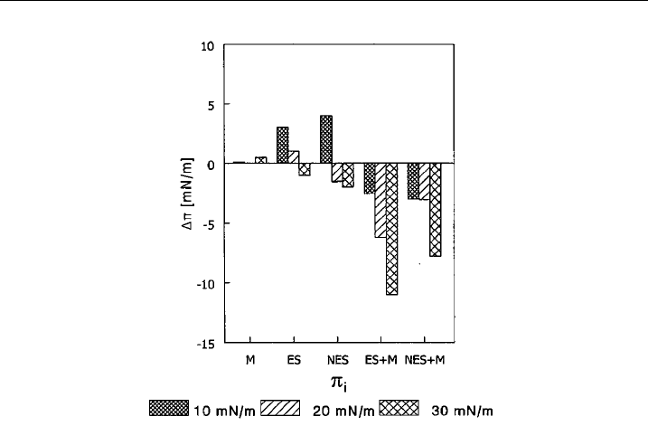
Our studies on binding antracycline cytostatic mitoxantrone to erythroid
or nonerythroid spectrin indicated strong interactions (K
D
4.6 and 1.4 mM for
erythroid and nonerythroid spectrin) and stoichiometry of 4 per erythroid spectrin
dimer and 7 molecules per nonerythroid spectrin tetramer. The K
D
values were
similar to those obtained for PE/PC (3:2) liposomes. Analysing the effect of the
presence of mitoxantrone on the interaction of both erythroid and nonerythroid
spectrins with the monolayer prepared from PE/PC (3:2) an interesting phenom-
enon was observed: the effect of spectrin on the surface pressure change increased
several-fold (see Fig. 2). The complex was also found to evoke larger change in the
fluidity of the PE/PC membrane as measured by using EPR technique with
5
0
-doxyl-stearate spin label. It was also observed that mitoxantrone possibly induces
a small change in the secondary structure of spectrins [60].
4. Spectrin–Lipid Interactions; a Molecular Approach
4.1. Lipid-Binding Sites in Spectrins
Spectrins are multidomain proteins which show specific structural features: one of
them is formation of an elongated segmental structure built of a series of repeating
units, each of about 106 amino acid residues folded in a triple-helical motif. The
other feature is the fact that outside typical segments and within them there are
domains responsible for the multiple functions of spectrin. The most important
functional domains outside typical spectrin repeats are actin-binding domain (two
Interactions of Erythroid and Nonerythroid Spectrins 89
CH motifs located at amino terminal region of b-subunit of spectrin) [13] and SH3
domain present in atypical, 10th segment of a-spectrin. Examples of other domains
are ankyrin-binding domain located in part in 14th repeat and in 15th unit of
b-spectrin [61,62] and Ca
2+
-binding EF-hand motifs located in carboxyl terminal
region of a-subunit [63].
The consequence of this multifunctionality is the existence of several lipid-
binding activities connected with the occurrence of different binding sites: (1) a
‘‘general’’ phospholipid-binding activity characteristic for the spectrin repeat motif
[64–66], (2) specific for aminophospholipids (PE and/or PS-binding sites located in
the b-subunits, (e.g., [67–70]) and (3) specific for phosphatidylinositol 4,5-bis-
phosphate, characteristic only for longer b-spectrin isoforms which poses Pleckstrin
Homology (PH) domain [54,71,72].
The first kind of the lipid-binding activity that could be attributed to the spectrin
repeat motif was actually studied by using a series of constructs containing the second
repeat of the human dystrophin rod domain [73]. DeWolf et al. [65] observed that
properly folded fragment corresponding to the second repeat unit of the rod domain
is able to modify the properties of lipid membranes (inducing a large increase in
surface shear viscosity of the monolayer) containing anionic phosphatidylserine.
Moreover, this property is dependent on the native structure as the unfolded
expressed fragment had no effect. Further studies of Le Rumeur et al. [66] showed
Figure 2 The e¡ect of 72 nM mitoxantrone on the e¡ect of spectrins on the surface pressure
change of PE/PC monolayer. ES, erythroid spectrin; NES, noner ythroid spectr in; M,
mitoxantrone. Bars represent the Dp value observed at 72 nM mitoxantrone, at 8 nM erythroid
or nonerythroid spectrin or at 8 nM spectrins and 72 nM m itoxantrone. Data obtained at three
values of initial surface pressure (10, 20 and 30 mN/m) of PE/PC (3:2) monolayer are shown.
(data taken from [60] with permission of Publisher, Taylor and Francis
R
, http://
www.informaworld.co m).
A.F. Sikorski et al.90

the engagement of tryptophan residues in this binding, as in the presence of small,
unilammellar vesicles containing PS the fluorescence characteristics changed
dramatically, indicating the tryptophan residues changed their environment into
more hydrophobic. The accessibility of these residues from the hydrophilic solvent
for quenchers was also limited. These authors conclude that dystrophin rod lies along
the membrane surface [66]. Similar experimental approach should be undertaken to
determine whether or not the same conclusion could be drawn with respect to
spectrin–phospholipids interactions.
The second class of lipid-binding sites seems to be confined to certain regions of
spectrin molecule which show some aminophospholipid and anionic phospholipids
specificity. One of the approaches to identify this class of sites was to clone and
express systematically all the fragments of both subunits of erythrocyte spectrin and
test their binding to liposomes prepared from PS and its mixture with PC. Studies
of An et al. [68,69], indicate that binding sites for PS-containing vesicles are located
in repeats 8–10 of the a-subunit, in repeats 2–4, 12–14 and in the nonhomologous
amino terminal region of the b-subunit. Their further studies [67] concerning
nonerythroid spectrin indicated that PS-binding site is located in repeats 9–11 and
in the amino terminus of a-subunit and in amino terminal region and repeat units 2
and 3 of the b-subunit. To this class of binding sites belongs, as we believe, the
ankyrin-dependent lipid-binding site which is discussed in more details below.
Pleckstrin homology domain is an example of the third kind of lipid-binding
sites specific for phosphoinositides. This kind of PIP2-binding site which is present
in nonerythroid spectrins, bIS2 and bIIS2 and also in many regulatory and cyto-
skeletal proteins [54,74] is approximately 100 amino acid residues long. Binding
of PIP2 is inhibited competitively by IP3 indicating the specificity for inositol
ring [71].
4.2. Ankyrin-Dependent Lipid-Binding Site in b-Spectrins
Our early attempt to identify the main amphipathic compound-binding site of
spectrin revealed its close proximity to the ankyrin-binding domain [50] what
suggested a functional relationship between ankyrin and lipid binding by erythroid
spectrin. Indeed, when the effect of purified ankyrin on spectrin binding to
phospholipid vesicles was tested, an inhibition of this interaction was observed [75].
The effect was greater for the vesicles containing PE (PE/PC 3:2) for which 60%
inhibition was found compared to 10–20% inhibition for PS/PC vesicles. Almost
identical results were obtained using a monolayer technique. Dixon-type [76]
analysis indicated a competitive mechanism of inhibition of PE/PC vesicles or
monolayer binding to spectrin by ankyrin. Tetrameric spectrin bound similarly to
PE/PC monolayer but inhibition with ankyrin suggests that only one of the two
possible binding sites is engaged in this interaction [77]. Moreover, when inter-
actions of nonerythroid (brain) spectrin with PE/PC monolayers in the presence of
ankyrin were analysed, a similar level of inhibition of these interactions by ankyrin
was observed [53]. Also, when isolated erythroid spectrin b-subunit was introduced
into the subphase of PE/PC monolayers in the presence of ankyrin, the inhibition
was even stronger, i.e., a three-fold lower concentration of ankyrin was needed to
Interactions of Erythroid and Nonerythroid Spectrins 91

induce the same effect. If the a-subunit was used instead of b its effect on the
monolayer surface pressure was small and entirely insensitive to incubation with
ankyrin [53,77]. It should be noted also that binding of brain spectrin to anionic
phospholipid monolayers was also competitively inhibited by purified erythrocyte
ankyrin [55].
As this event could have a regulatory role and could be engaged in the possible
mechanism of function of the membrane skeleton we decided to test whether an
ankyrin-binding domain which was identified by Kennedy et al. [61] would show
the same lipid-binding properties as spectrin molecule and to identify an ankyrin-
sensitive, lipid-binding site in erythroid and nonerythroid spectrins [70,70a]. The
results of the experiments document further the ability of cloned, expressed and
purified erythroid b-spectrin’s ankyrin-binding domain to bind PE-rich mono-
and bilayers. We found that full-length ankyrin-binding domain binds PE/PC
mono- and bilayers with affinity similar to that of native spectrin dimer and this
binding is inhibited by erythrocyte ankyrin. Binding studies were performed
by (i) pelleting assay using PE/PC liposomes and fluorescently labelled expressed
polypeptides, (ii) inhibition assay in which unlabelled, expressed polypeptides were
used to inhibit binding of purified, labelled red blood cell spectrin to PE/PC
liposomes and (iii) surface plasmon resonance technique experiments where bind-
ing of purified, expressed polypeptides to PE/PC monolayer deposited on hydro-
phobic sensor surface was observed. Results of these experiments indicate that
ankyrin-binding domain truncated mutants, that retain amino terminal part bind
the PE/PC mono- and bilayers with affinity and capacity comparable to full-length
ankyrin-binding domain. They also effectively compete for lipids with purified,
labelled spectrin. On the other hand, truncated mutants which lack 8 or 38 amino
acid residues from N-terminal region show, at least an order of magnitude lower
affinity and much higher maximal binding capacity. They also compete weakly with
spectrin for phospholipid vesicles. The expressed full-length domain induced small
decrease in order parameter of PE/PC membranes when probed with 5
0
-doxyls-
tearate similar to the effect of purified spectrin, while mutant-lacking 38 residues
from amino terminus induced small increase in order parameter which was similar
to the effect of bovine serum albumin.
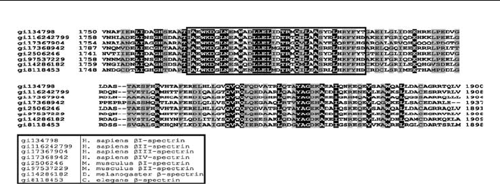
As the amino terminal part of ankyrin-binding domain of known b-spectrins
shows high degree of conservation in evolution (see Fig. 3 and also [62]), we
expected similar results in the case of nonerythroid bII spectrin. Indeed, cloned and
expressed fragments of nonerythroid (human brain) b-spectrin encompassing a
sequence corresponding to an ankyrin-binding domain was found to bind
phospholipid mono- and bilayers in a similar way to intact molecule of brain
spectrin or erythroid spectrin. Again, truncated mutant of this domain which re-
tained amino terminal region bound lipid mono- and bilayers with the affinity
similar to the full-length domain. Mutants in which this region was deleted bound
phospholipids with lower affinities and higher capacities and this binding was
insensitive to inhibition with purified ankyrin.
Transient expression of HeLa cells with GFP-conjugated constructs encoding
full-length ankyrin-binding domain of either erythroid or nonerythroid b-spectrins
induced changes in cell morphology and aggregation of membrane skeleton. These
A.F. Sikorski et al.92
changes were observed neither in cells transfected with a construct encoding GFP-
conjugated ankyrin-binding domain truncated at amino terminus by 38 amino acid
residues nor in cells transfected with a vector encoding only GFP (Bok et al.,
submitted). Also the results obtained by using site-directed mutagenesis on the
entire ankyrin-binding domain of the bI spectrin confirm that replacing hydro-
phobic with hydrophilic (serine) residues in the middle of this domain (residues
1782, 1785 and 1786) remained without effect while replacing hydrophobic
residues in the region of first 8 of them (residues 1771, 1775 and 1778) gave
substantial increase in K
D
for the interaction with PE/PC monolayers and vesicles
(Bok et al., to be published).
The above-mentioned experimental results indicate that at least in erythroid
spectrin the binding site located within or close to ankyrin-binding domain were
identified by our and Mohandas groups. It seems that there is a good chance that
these sites (segment 12–14 and amino terminal part of the ankyrin-binding domain)
are overlapping. The difference concerns nonerythroid b-spectrin; our results in-
dicate essentially the same features for both b-spectrins, while Mohandas group
could not identify this site in their constructs. In our hands binding of PS/PC (3:2)
monolayer to bacterially expressed ankyrin-binding domain and its deletion
mutants retaining N-terminal region of nonerythroid b-spectrin occurred with
K
D
’s in nanomolar range what was measured by SPR and FRET techniques [70a].
4.3. Structure of Lipid-Binding Site of Ankyrin-Binding Domain
Ankyrin-sensitive lipid-binding site of ankyrin-binding domain is located in typ-
ical triple-helical spectrin repeat rod region of the b-spectrin (helix C of the
segment 14).
We studied its structure by constructing a series of single and double spin-
labelled b -spectrin-derived peptides and analysing their spin–spin distances via
electron paramagnetic resonance spectroscopy and the Fourier deconvolution
method. The results indicate that the whole ankyrin-sensitive lipid-binding site of
b-spectrin exhibits a helical conformation revealing a distinct 3
10
-helix contribu-
tion at its N-terminus (especially the stretch 1768 IAEWKD 1773). The start of the
helix was located five residues upstream along the sequence compared to the
Figure 3 A lignme nt of known b-spectrin sequences corresponding to ankyrin-binding
domain.
Interactions of Erythroid and Nonerythroid Spectrins 93