Liu A.L., Tien H.T. Advances in Planar Lipid Bilayer and Liposomes. V.6
Подождите немного. Документ загружается.


(BaculoDirect
TM
, Invitrogen). An anti-viral is then used to select against non-
recombinant baculovirus.
Using the Bac-to-Bac
TM
baculovirus expression system, pFastBac/hSkM1-HT
was transformed into DH10-Bac E. coli and colonies were selected on the basis of
their resistance to the antibiotics: kanamycin, gentamycin, tetracycline, and by
blue/white colony screening using IPTG and X-gal. Recombinant bacmid is large
4135 kb, therefore care must be taken to purify it intact. We recommend using
DNA mini-preparation purification columns that empty by gravity flow rather than
by centrifugation. Bacmid colonies were screened by PCR analysis to identify those
containing hSkM1-HT. At this point it is also advantageous to confirm the presence
of intact bacmid DNA by gel electrophoresis. The recombinant bacmid generated
can then be transformed into the DH10-B E. coli strain which lacks a helper
plasmid as it is no longer required.
5.2. VGSC Expression in Insect Cells
The baculovirus expression system is well suited for heterologous expression of
eukaryotic proteins [57,58]. A major advantage of this system is the high-infection
efficiency of cells by the recombinant baculovirus, resulting in many cells expressing
the gene of interest. In addition, high levels of protein can be produced and this is
amenable to being scaled up for commercial production [59,60]. Different subunits
may be co-expressed producing hetero-oligomeric channels. This may be achieved
by co-infection of more than one recombinant baculovirus, or by the use of dual-
or multi-promotor baculovirus transfer vectors, for example wild type and mutated
GABA A receptor subunits have been coexpressed in this way [61–63]. Insect cells
support post-translational modifications which is particularly relevant for VGSCs
because they are heavily glycoslyated, comprising 30% carbohydrate [64]. While
glycolsyation is not essential for VGSC function it does modulate channel-opening
properties [65,66].
Insect cells grow at close to room temperature (25 1C) and do not require special
aeration conditions for volumes of up to 1 l. Baculoviruses are also safer to work
with than most mammalian viruses since they are non-infectious to vertebrates. Of
the several types of insect cell lines available, Sf 9 cells were chosen as these are
suitable for expression of non-secreted proteins. Cells were grown in Grace’s Insect
medium (Sigma) supplemented with 10% foetal bovine serum, 2% yeastolate,
3.3 mg/ml lactalbumin, pH 6.2. Cells were routinely seeded at a density of 5 10
5
cells/ml and grown to 2–3 10
6
cells/ml in 40 ml volume of media in a 50 ml
capacity spinner flask at 25 1C.
To obtain stocks of hSkM1-HT recombinant baculovirus, Sf 9 insect cells were
infected with purified recombinant bacmid DNA according to the manufacturer’s
instructions. One exception was that the resultant P1 viral supernatant was har-
vested after five days rather than only three to give a sufficiently high titre
(Z1 10
7
PFU/ml). The virus was propagated twice more by infecting cells
growing at 5 10
5
cells/ml at a multiplicity of infection (MOI) of 0.1 for five days
to obtain a 500 ml volume of recombinant baculovirus as a working stock. Virus
titre was determined by plaque assay.
Y.L. Zhang et al.34

For protein production, cells were grown to a density of 2 10
6
cells/ml, then
infected with baculovirus (1–2 10
7
PFU/ml). Protein production in Sf 9 cells
infected with hSkM1-HT recombinant baculovirus was verified by electrophoresis
and Western blot analysis using an anti-Pan Na
v
channel antibody (Alomone Labs,
Jerusalem) (1:400), a secondary peroxidase conjugated goat anti-rabbit antibody
(Amersham, England) (1:2000), and visualised using chemiluminesence (ECL
TM
,
Amersham, England). Expression of hSkM1-HT in Sf 9 insect cells was confirmed
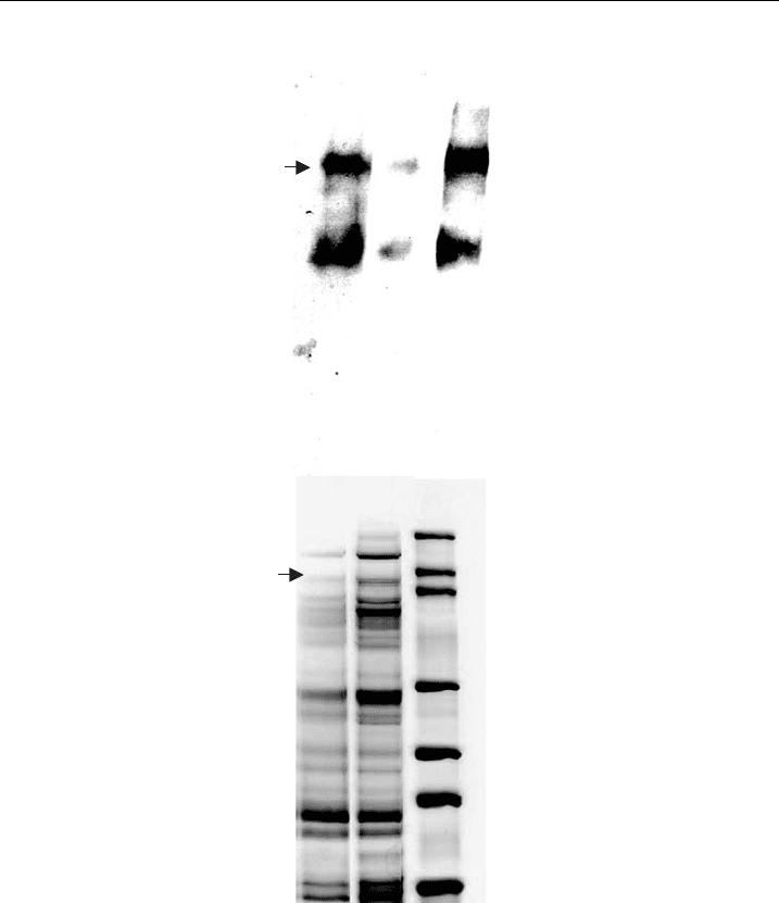
by gel electrophoresis and immunodetection (Fig. 3). Protein expression was max-
imised by establishing the MOI and post-infection time for harvest of infected Sf9
insect cells that produced the most protein. MOIs of 1, 2, 5, and 10 were tested in
cells harvested at 64 or 72 h. An MOI of 1 after 72 h of infection gave the highest
level of protein expression and was routinely used (Fig. 3). To ascertain whether
the addition of the His-tag has affected channel function, patch-clamping
cells infected with recombinant baculovirus is an ideal functional test to use prior
to purification. We have used this method previously to confirm function of
hSkM1-HT [56].
To generate protein for purification, the culture volume was scaled up to 400 ml
and grown in a 1000 ml spinner flask to provide adequate aeration. As before cells
were seeded at 5 10
5
cells/ml and cells were grown to a density of 2 10
6
cells/
ml, then infected with baculovirus (1–5 10
7
PFU/ml). After addition of baculo-
virus the total culture volume was approximately 500 ml.
5.3. Purification of Recombinant VGSC using IMAC
Purification of intact and functional VGSC protein is challenging due to its large
size (260 kDal) and its tendency to degrade rapidly during isolation. This has been
MOI 1, 72hr
MOI 2, 64hr
MOI 2, 72hr
MOI 5, 64hr
MOI 5, 72hr
MOI 10, 64hr
MOI 10, 72hr
Control
MOI 1, 64hr
Figure 3 Express ion of His-tagged voltage-gated sodium channels in insect cell s. Western
blot of Sf 9 insect cells infected with hSkM1-HT recombinant baculovirus at a multiplicity
of infection of 1, 2, 5, or 10, and harvested at 64 or 72 h post-infection. Protein expression
was detected using an anti-Pan Na
V
antibody. The control was uninfected Sf 9 cells (adapted
from Zhang et al. [ 56]).
Recombinant Voltage-Gated Sodium Channels into Planar BLMs 35

attributed to calcium-dependent proteases, therefore calcium chelators or protease
inhibitors are important ingredients throughout the purification procedure. Ethyl-
enediaminetetraacetic acid (EDTA) is often used as a calcium chelator but since it
inhibits metal ion binding, IMAC compatible protease inhibitors were used. For
successful reconstitution it is important that the protein be solubilised but not
denatured. To purify the protein in a form that would remain functional it was
solubilised under non-denaturing conditions using the non-ionic detergent, non-
idet P-40, which can be easily removed using gel filtration. Triton X-100 is another
commonly used detergent in membrane protein biochemistry, but it was not used
since it can be difficult to remove, has been shown to form channels in BLMs [67]
and can interfere with VGSC function [67,68].
In optimising the metal affinity chromatography method for the sodium chan-
nel we identified key factors that were important for improving protein yield.
Protease inhibitors reduced but did not eliminate protein degradation, therefore we
shortened the length of the procedure in an attempt to reduce this further. The
initial 16-h overnight extraction was reduced to just 30 min, as the cells were
sufficiently lysed after this time and were less prone to protein degradation during
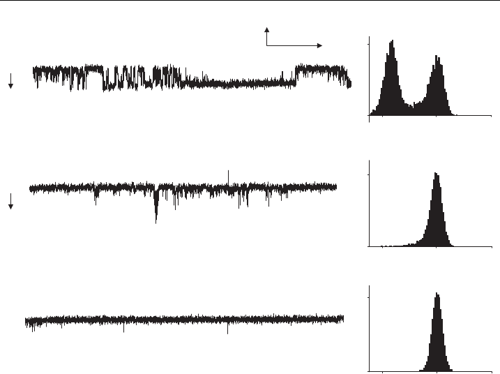
subsequent steps (Fig. 4). Thus the total time from harvest of infected cells until the
formation of proteoliposomes was less than 8 h. Another key factor in the opt-
imisation was the addition of phospholipids during the purification steps. Lipid was
added to binding, wash, and elution buffers to help maintain the conformational
integrity of the protein [69].
290kDa
240kDa
M1 2
Figure 4 Detection of IMAC-puri¢edVGSCs. Silver-stai ned polyacrlyamide gel electrophoresis
(PAGE) of puri¢ed hSkM1-HT protein fractions from a Ni
2+
a⁄nity column, following extrac-
tion for 30 min (1), or 16 h (2).
Y.L. Zhang et al.36

Virus-infected cells (500 ml) were harvested 72 h post-infection by centrifuga-
tion at 2000 g, washed three times in ice cold phosphate-buffered saline, trans-
ferred to microfuge tubes and pelleted at 13,000 rpm in a bench-top centrifuge. In
some cases cell pellets were stored at –80 1C until use. Cells were resuspended in
(20 ml) sonication buffer containing: 100 mM NaCl, 50 mM Tris-HCl, 10% glyc-
erol, and protease inhibitor cocktail tablets (complete mini EDTA-free, Roche,
Germany), pH 7.9. These protease inhibitors are compatible with IMAC, being
EDTA-free, and they inhibit serine and cysteine proteases, but not metallopro-
teases. Cells were sonicated at medium intensity (Vibracell, Sonics and Materials
Inc., Danbury, USA) in three, 40 s bursts, at 1 min intervals and kept on ice.
The addition of 2% NP-40 completed the lysis buffer and the cells were gently
agitated on a rocker for 30 min at 4 1C, then centrifuged at 100,000 g for 40 min
at 4 1C.
The supernatant was passed through an agarose matrix preloaded with Ni–NTA
(nitrilotriacetic acid) resin (Invitrogen, Carlsbad, USA). Solution flow through
the column was by gravity flow. Following equilibration, the column was loaded
with the solubilised protein extract in lysis buffer. Ten volumes of binding buffer
containing: 500 mM NaCl, 20 mM Tris-HCl, 5 mM imidazole, 10% glycerol, pH
7.9 were added, followed by 10 volumes of wash buffer (binding buffer containing
15 mM imidazole). Washing conditions were optimised by altering the imidazole
concentration. It was found that 15 mM imidazole was the highest concentration
at which the target protein was retained in the matrix column and much of the
non-specific protein was removed. The protein was eluted with five volumes
of elution buffer (binding buffer containing 250 mM imidazole, plus protease
inhibitor, pH 7.9). Because subsequent steps to remove detergent utilised a
binding matrix, all eluate fractions were collected. Samples of the eluate were
analysed by sodium dodecyl (lauryl) sulfate-polyacrylamide gel electrophoresis
(SDS-PAGE) and the different extraction times compared (Fig. 4). This showed
that more protein was present when the extraction time was 30 min compared
to 16 h.
SDS/tris-glycine gel electrophoresis indicated a molecular mass of 260 kDa, as
determined by Western blot analysis using anti-Pan Na
v
antibody (Alomone Labs,
Jerusalem, Israel) (Fig. 5A). The protein degraded in the elution buffer after only
12 h at 4 1C, even in the presence of protease inhibitor. However, if freshly purified
hSkM1-HT protein was reconstituted immediately into liposomes, it did not de-
grade (Fig. 5A and 5B). Since the protein was more stable when in liposomes,
hSkM1-HT protein was reconstituted immediately following purification. Under
these conditions, channel activity was still present after storage at –80 1C for 4
months. The presence of contaminating bands in the silver-stained gel (Fig. 5B)
indicated that the preparation would benefit from further purification, such as size
exclusion chromatography. However, since uninfected cells undergoing the same
procedure gave no channel activity when reconstituted, the level of purity was
considered sufficient for this application.
Detergent removal is an important issue for pBLM research, since its presence
will disturb the formation of liposomes and the stability of lipid bilayers. The
Recombinant Voltage-Gated Sodium Channels into Planar BLMs 37

dialysis technique is widely used to remove detergent and to buffer exchange, but is
time-consuming, usually taking more than 10 h. Affinity purification provides an
alternative efficient means of detergent removal. The detergent was, therefore,
removed using an Extracti-Gel
s
D column (Pierce, Rockford, USA). By adding
lipids to the detergent-removing gel elution buffer, the detergent could be removed
and the protein reconstituted into liposomes in a single procedure.
A
B
12 3
500KDa
290KDa
240KDa
160KDa
116KDa
97KDa
66KDa
23
Figure 5 VGSC reconstitute d into liposomes. PAGE of puri¢ed hSkM1-HT protein in eluate or
in proteoliposomes detected byWestern blot (A) and silver staining (B). Lane1 ^ freshly puri¢ed
protein eluate. Lane 2 ^ puri¢ed protein eluate stored at 4 1C overnight, Lane 3 ^ puri¢ed protein
reconstituted into liposomes and stored at 4 1C ove rnight (adapted from Zhang et al. [56]).
Y.L. Zhang et al.38

6. Functional Reconstitution
6.1. Reconstitution of Ion Channels into Liposomes
To reconstitute VGSC protein into liposomes, the composition of phospholipids
is important for subsequent fusion of proteoliposomes with pBLMs. Mixed
lipids containing acidic lipids, such as phosphatidylserine (PS) can facilitate fusion
[70,71]. We used a 10% lipid stock mixture containing a 5:3:1:1 ratio of
phosphoethanolamine (PE), phosphatidylserine (PS), phosphatidylcholine (PC)
(extracted from egg yolk [72]) and cholesterol (CH) (Avanti Polar Lipids), dispersed
by sonication in 10 mM potassium phosphate, pH 7.2. For reconstitution of
hSkM1-HT channels into liposomes, the lipid stock mix was diluted 1:20 in 10 ml
of eluate from the Ni–NTA column, giving a final lipid concentration of 0.5%.
The gel filtration column was pre-equilibrated with 0.5% lipid mixture in the
phosphate buffer. The protein was also eluted in 10 ml of the phosphate buffer.
This resulted in formation of proteoliposomes in the eluate. To concentrate the
liposomes the eluate mixture was centrifuged at 100,000 g for one hour. The
supernatant was discarded and liposomes were resuspended in a reconstitution
buffer containing: 300 mM NaCl, 15 mM HEPES (N-[2-Hydroxyethyl]-piper-
azine-N
0
-[2-ethanesulfonic acid]), 200 mM sucrose, adjusted to pH 7.4 with KOH,
and were either used immediately for pBLM experiments, or stored at 80 1C
for later use.
6.2. Incorporation of Proteoliposomes into pBLMs
Theories and methods on fusion of proteoliposomes with lipid bilayers have been
illustrated in many books and research articles [40,73,74]. Fusion is reported to
occur in two stages. First, contact occurs between the liposome and the bilayer, and
this is followed by fusion. The probability of fusion may be increased by swelling
the vesicles (proteoliposomes). Miller and Racker [32] found that successful fusion
required the presence of acidic lipids, a small amount of Ca
2+
in the vesicle-
containing (cis) side, and loading the vesicles with membrane impermeable solute
such as sucrose. Establishing an osmotic gradient across the pBLM has been re-
ported to further increase the probability of fusion [75,76]. An osmotic gradient can
be achieved by setting up asymmetric cis and trans solutions. The swelling would be
induced via water flow across the vesicle membrane. In theory, once incorporation
occurs, fusion can be stopped by addition of an osmicant to the opposite chamber
or replacement of the solution in the cis chamber with the same solution as that in
the trans chamber. We found that establishment of such conditions was not nec-
essary because fusion occurred within 15–20 min.
pBLMs were formed from a mixture of 5% PC and 2% CH in n-octane across a
200 mm diameter hole in a polystyrene cup separating two chambers. Each chamber
contained 1 ml of filter-sterile buffer containing: 300 mM NaCl, 10 mM HEPES,
pH 7.4. Single channel recordings from planar bilayer membrane experiments were
filtered at 3 kHz, sampled at 150 ms intervals and analysed using TAC 4.2.0 (Bruxton,
Seattle) single channel analysis software.
Recombinant Voltage-Gated Sodium Channels into Planar BLMs 39

STX dihydrochloride was obtained from the National Research Council
of Canada. PbTx-1 was from Calbiochem (San Diego, United States). VTD
was purchased from Sigma. TTX was purchased from Alomone Labs (Jerusalem,
Israel).
6.3. Orientation of Reconstituted hSkM1-HT Channels in pBLMs
To determine the orientation of the reconstituted channels in the pBLM we used
membrane impermeant VGSC inhibitors that inhibit by binding to the extracellular
face of the channel. TTX and STX inhibit VGSCs by binding to a common site
near the extracellular mouth of the pore. The chamber to which hSkM1-HT
proteoliposomses were added was defined as cis. Single or multiple channel events
observed in the presence of 100 mM VTD could be blocked completely by TTX or
STX, from which we inferred the directionality of the extracellular side of the
channel. Our results showed that the hSkM1-HT reconstituted channels faced
either orientation in pBLMS, but that the STX/TTX-binding site tended to face
the side to which liposomes were added (cis). This suggests that when in the
liposomes, the channels tend to be oriented in the same direction as they are in
the cell plasma membrane, with the extracellular STX/TTX-binding site facing the
outside. Reconstituted VGSC have been reported to face either the same side, the
opposite side, or both sides, from which vesicles are added [49,54,77]. Orientation
differences of protein in liposomes are likely to be related to how disruptive the
isolation/purification method is; that is how much of the original cell membrane
remains intact versus how fragmented it becomes prior to reconstitution into lipo-
somes.
6.4. Pharmacological Function of Reconstituted hSkM1-HT
Since channel insertion could occur in either orientation, proteoliposomes- (20–
40 ml) containing hSkM1-HT were added to both cis and trans chambers to increase
the probability of incorporation. We recently investigated the pharmacological in-
tegrity of the channel by examining the ability of known activators and inhibitors
that bind to different sites on the channel, to modulate its activity [56]. VTD and
PbTx-1 promote sodium channel opening by binding to receptor sites 2 and 5,
respectively, whereas the VGSC-specific inhibitors, TTX and STX, bind to re-
ceptor site 1 at the external mouth of the pore [2]. As sodium channel currents
rapidly inactivate, we used the channel activator veratridine which slows their
inactivation and allows the channels to be observed under steady-state conditions
where the membrane potential is held constant.
Control experiments using liposomes prepared from uninfected cells that un-
derwent the same purification procedure as for infected cells showed no channel
activity. This showed that the untargeted protein present (Fig. 5B) did not produce
ion channel activity. Protein purified from cells infected with hSkM1-HT recom-
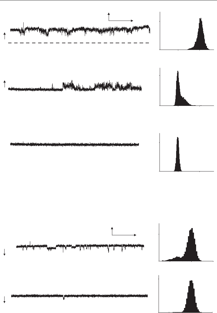
binant baculovirus showed channel activity in the presence of 100 mM VTD
(Fig. 6A). The potent and selective sodium channel blocker, TTX, at a concen-
tration of 100 nM, inhibited channel activity (Fig. 6B and 6C). This confirmed the
Y.L. Zhang et al.40

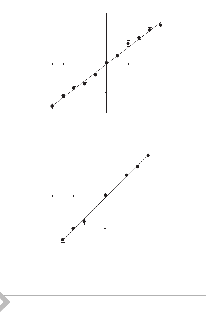
identity of the activity as that of sodium channels. VTD-activated channels were
also inhibited by 100 nM STX (Fig. 7). In the example shown in Fig. 7, there were
at least two active channels that were partially inhibited after 7 min of exposure to
STX and were completely inhibited after a further 20 min of exposure; hSkM1-HT
channels activated by the algal toxin, PbTx-1, were inhibited by STX (Fig. 8). We
also noted that channels activated by VTD or PbTx-1 differed in their maximal
conductance level (Fig. 9). VTD-activated channel openings had a slope conduct-
ance of 21 pS (Fig. 5H), whereas PbTx-1-activated channels had a slope conduct-
ance of 16 pS. hSkM1 channels activated by voltage alone have a reported slope
conductance of 25 pS [78]. Lower conductance levels for VGSCs activated by VTD
or PbTx-1 have been reported previously [79–81]. It has been suggested that VTD
stabilises the open conformation by blocking the open channel to reduce its unitary
conductance [82]. Subconductance levels were prevalent in the single channel re-
cordings in the presence of either VTD or PbTx-1, as reported for native VGSCs
(Fig. 8A) [79,80]. The results showed that the reconstituted VGSCs retained their
expected pharmacological profile.
I (pA)
-20
2
0
1000
100 µM VTD + 100 nM TTX
B
C
O
C
C
I (pA)
-20
2
0
1200
C
O
100 µM VTD
A
-20
2
Events
0
500
10 s
2 pA
100 µM VTD + 100 nM TTX
Events
Events
I (pA)
Figure 6 Reconstituted hSkM1-HT channels activated by veratridine and inhibited by TTX.
Representative channel activity and relative histograms reco rded over 1min are shown for
protein reconstitute d into liposomes and incorporated into pBLMs. P rotein was puri¢e d from
cells infected with hSkM1-HT recombinant baculovirus, recorded at ^80 mV in the presence of
100 mM veratridine. (A) control, (B) 8 min afte r addition of 100 nM TTX, and (C) 10 min after
TTX was added. The records shown were ¢ltered at 300 Hz. The arrow indicates the direction
of channel opening; C is closed and O is open.
Recombinant Voltage-Gated Sodium Channels into Planar BLMs 41

0
800
100 µM VTD + 100 nM STX
B
O
C
A
100 µM VTD
I (pA)
0
600
O
10 s
2 pA
Events
0
1200
C
C
I (pA)
I (pA)
0
3
0
3
0
3
Events Events
C
Figure 7 Reconstituted hSkM1-HT channels activated by veratridine and inhibited by STX.
Chan nel activity for hSkM1-HT at +80 mV in the presence of 100 mM veratridine. (A) control,
(B) 7 min, and (C) 27 min after 100 nM STX was presented. The baseline (0 pA) is indicated by
the broken line in A.The records shown were ¢ltered at 300 Hz.The arrow i ndicates the direction
of channel opening; C is closed and O is open (data are from a representative experiment).
10 s
2p A
C
O
60 nM PbTx-1
A
-10 1
0
1000
60 nM PbTx-1 + 50 nM STX
B
C
O
I (pA)
-10 1
Events
0
1800
I (pA)
Events
Figure 8 Reconstituted hSkM 1-HT ch annels activated by brevetoxin. Channel activity and
relative histograms fo r a re presentative experiment obtained at 80 mV in the presence of 50 nM
PbTx-1. (A) control and (B) 6 min after 60 nM STX was added.The records shown were ¢ltered
at 300 Hz.
Y.L. Zhang et al.42

7. Conclusion
This chapter describes a rapid and simplified technique for the purification
and reconstitution of VGSCs. This method could be applied to a broad range of
-2.5
2.5
-100
100
V (mV)
I (pA)
A
-1.5
1.5
-100
100
V (mV)
I (pA)
B
Figure 9 Current^voltage relationship for single hSkM 1-HT channels activated by (A)
veratridine and (B) PbTx-1 in symmetrical 300 mM NaCl solutions (data are from three or more
experiments and Fig. 9A is from Zhang et al. [56]).
Recombinant Voltage-Gated Sodium Channels into Planar BLMs 43
