Lennarz W.J., Lane M.D. (eds.) Encyclopedia of Biological Chemistry. Four-Volume Set . V. 4
Подождите немного. Документ загружается.

BMP-ligands, carry a cluster of L45 loop sequences that
are distinct from the Alk3 group of receptors but are
compatible with the BMP-activated Smads 1, 5, and 8.
As different ligands can recruit different type I receptors
into the signaling complexes, differential activation of
Smad substrates by Alk1 and 5 groups of receptors may
account for the activation of distinct downstream signals
by the same ligand under different conditions.
Endocytic trafficking of activated receptors plays an
important role in regulating receptor-dependent signal-
ing. This may result from the activation of signaling by
promoting association between activated receptors and
various signaling intermediates in the endosomal
compartments, or by dampening receptor-dependent
signaling through degradation of the activated receptor
complexes. Recent data suggests both mechanisms can
regulate TGF-
b
receptor-mediated signaling. In the
presence of R-Smads, the FYVE domain protein SARA
promotes uptake of the SARA/Smad/receptor complex
to early endosomal membranes, promoting clathrin-
dependent endocytosis, receptor-mediated phosphoryl-
ation and activation of the R-Smad. In contrast,
Calveolin-dependent endocytosis and proteosomal
degradation of the activated receptor complex is
mediated by recruitment of the HECT domain E3
ligase SMURF to the activated type I receptor by the
inhibitory Smad7. This results in the uptake of the
SMURF/Smad7/receptor complex to Calveolin positive
endosomes, and promotes SMURF-mediated ubiquiti-
nation and proteosomal degradation of the signaling
complex.
In addition to the canonical Smad-signaling path-
ways, there is considerable evidence of cell-type-
dependent regulation of alternative signaling pathways
by different members of the TGF-
b
superfamily. These
include activation of MAPK signaling pathways ERK,
JNK and p38 MAPK, PI3 kinase, PKC, and inhibition
of p70
S6K
signaling. These signals may be required to
mediate and/or augment maximal Smad-dependent
responses, or they may exert distinct downstream
responses in different cell types. However, the precise
mechanisms linking these noncanonical signaling path-
ways with the activated receptor complexes are
incompletely understood. Some of these effects may
be indirect, resulting from the induction of Smad-
dependent target genes that themselves regulate these
responses. For example, while JNK activation by TGF-
b
may result from activation of a rapid, Smad-
independent response in a variety of cell types, a
delayed Smad-dependent response has also been
described. In other cell types, TGF-
b
signaling may
repress p38 MAPK signaling through the induction of
Smad-dependent MAPK phosphatases expression. In
contrast, direct regulation of these alternative path-
ways may result from ligand-dependent protein inter-
actions with the activated receptor complex. For
example, interactions between the serine threonine
phosphatase, PP2A and the activated TGF-
b
type I/II
receptor complex, results in rapid, dephosphorylation-
dependent inhibition of p70
S6K
activity. Other
downstream intermediates may link receptor activation
to these pathways. These include the Rho family of
small GTPases and the MAPKKK TAK1 and its
upstream kinase HPK1, that are involved in regulating
TGF-
b
-dependent activation of JNK, Ras GTPase-
dependent activation of ERK MAPK, and TAK1-
dependent activation of p38 MAPK by TGF-
b
. Direct
links between these pathways and the activated
receptors are less clear.
In addition to these effects, activation of TGF-
b
family receptors gives rise actin polymerization and
cytoskeletal re-organization in a variety of different cell
types. While the long-term effects of TGF-
b
on these
responses may be indirect, rapid changes in cytoskeletal
organization can involve direct modification of small
GTPase-dependent actin polymerization by TGF-
b
receptor signaling. More recently, LIM kinase 1
(LIMK1), which regulates actin polymerization through
phosphorylation-dependent inactivation of Cofilin, has
been shown to interact with the cytoplasmic tail of the
BMP type II receptor. BMP4-treatment is associated
with re-distribution of LIMK1 to the cell periphery,
phosphorylation of Cofilin, and active re-organization
of the actin cytoskeleton. This suggests that BMP
RII-dependent activation of LIMK1 may regulate
additional signaling pathways, and raises additional
questions regarding the role of the C-terminal tails
of TGF-
b
family receptors in the regulation of
downstream signaling.
Conclusions
This review outlines the large number of TGF-
b
ligands, the plasticity of receptor usage and the
diversity of downstream pathways that can be regulated
by these receptors. However, while considerable
advances have been made in our understanding over
the last decade, many of these discoveries have raised
many new questions about the complex biology of
these receptor-signaling pathways. Important areas of
research in the future will provide a more detailed
understanding of the factors contributing to the
diversity of cell-type-dependent responses and links to
other signaling pathways.
SEE ALSO THE FOLLOWING ARTICLES
Mitogen-Activated Protein Kinase Family † p70
S6 Kinase/mTOR † Protein Kinase C Family † Ras
Family † Rho GTPases and Actin Cytoskeleton
Dynamics
212
TRANSFORMING GROWTH FACTOR-
b
RECEPTOR SUPERFAMILY
GLOSSARY
activins and inhibins Proteins released from the gonads that stimulate
and inhibit, respectively, the secretion of follicle-stimulating
hormone, which is secreted by the pituitary and is a major
regulator of reproductive function.
LIM kinase A protein serine kinase that phosphorylates and
inactivates cofilin, a protein that regulates actin depolymerization
and hence influences cytokinesis (cell division), endocytosis
(uptake of molecules by cells), chemotaxis (directed cell movement)
and morphogenesis (cell shape change).
Smads Proteins that are phosphorylated by transforming growth
factor receptor family members and move as complexes into the
nucleus to activate gene transcription.
transforming growth factors
b
A secreted protein that acts locally to
either stimulate or inhibit cell proliferation or differentiation, and
which plays a role in development and wound healing.
FURTHER READING
Balemans, W., and Van Hul, W. (2002). Extracellular regulation of
BMP signaling in vertebrates: A cocktail of modulators. Dev. Biol.
250(2), 231– 250.
Chang, H., Lau, A. L., and Matzuk, H. H. (2001). Studying TGF-beta
superfamily signaling by knockouts and knockins. Mol. Cell
Endocrinol. 180(1–2), 39 –46.
de Caestecker, M. P., Piek, E., and Roberts, A. B. (2000). Role of
transforming growth factor-beta signaling in cancer. J. Natl. Cancer
Inst. 92(17), 1388–1402.
Kawabata, M., and Miyazono, K. (2000). Skeletal Growth Factors.
Lippincott Williams and Wilkins, Philadelphia.
Massague, J., Blain, S. W., and Lo, R. S. (2000). TGFbeta signaling in
growth control, cancer, and heritable disorders. Cell 103(2),
295–309.
Miyazono, K., Kusanagi, K., and Inoue, H. (2001). Divergence and
convergence of TGF-beta/BMP signaling. J. Cell. Physiol. 187(3),
265–276.
Pangas, S. A., and Woodruff, T. K. (2000). Activin signal transduction
pathways. Trends Endocrinol. Metab. 11(8), 309–314.
Piek, E., Heldin, C. H., and Ten Dijke, P. (1999). Specificity, diversity,
and regulation in TGF-beta superfamily signaling. Faseb J. 13(15),
2105–2124.
Shi, Y., and Massague, J. (2003). Mechanisms of TGF-beta signaling
from cell membrane to the nucleus. Cell 113(6), 685–700.
Zhao, G. Q. (2003). Consequences of knocking out BMP signaling in
the mouse. Genesis 35(1), 43–56.
BIOGRAPHY
Mark de Caestecker is an Associate Professor with the Departments
of Medicine and Cell and Developmental Biology at Vanderbilt
University. He graduated with a medical degree from the
Universities of Cambridge and London, specializing in general
internal medicine and nephrology, and Ph.D. from the University of
Manchester in England. He began working on TGF-
b
signaling as a
postdoctoral fellow with Anita Roberts in the Laboratory of Cell
Regulation and Carcinogenesis at the NIH. His current research
interests include the role of BMP-signaling in vascular remodeling
and the regulation of epithelial cell fate in renal development
and injury.
TRANSFORMING GROWTH FACTOR-
b
RECEPTOR SUPERFAMILY 213

Translation Elongation in Bacteria
Oliver Vesper and Knud H. Nierhaus
Max-Planck-Institut fu
¨
r Molekulare Genetik, Berlin, Germany
Protein synthesis is one of the major processes in a living cell
that translates the genetic information into protein structure
and thus organizes and directs the life cycle and metabolism
of a cell. The process of protein synthesis can be subdivided
into four consecutive phases: (1) initiation, (2) elongation, (3)
termination, and (4) ribosome recycling. All show features
that are specific for each of the three main evolutionary
domains, viz., bacteria, archaea, and eukarya, with only one
exception, the elongation phase. This phase is at the heart of
protein synthesis, where the codon sequence of an mRNA is
translated into the corresponding amino acid sequence of
proteins.
Introduction
Ribosomes translate the genetic information of mRNAs
by using tRNA as adaptors. An acylated tRNA connects
the decoding center on the small ribosomal subunit via
the anticodon at the tip of the long arm of the L-shaped
tRNA with the peptidyl-transferase (PTF) center on the
large ribosomal subunit via its short arm, the amino-acid
acceptor stem.
All ribosomes examined to date from all three
evolutionary domains show three tRNA binding sites:
(1) The A site, where the correct aminoacyl-tRNA is
selected according to the codon present here. The
A site tRNA binds in the form of a ternary com-
plex (aa-tRNA·EF-Tu·GTP; aa, aminoacyl; EF-Tu,
elongation factor Tu), thus providing the new amino
acid for the growing peptide chain. (2) The P site,
where the peptidyl-tRNA is located carrying the
nascent peptide chain before peptide bond formation.
And (3) the exit site (E site) that binds exclusively
uncharged (deacylated) tRNAs. It is from this site that
the tRNA is released from the ribosome. During the
course of three elongation cycles a tRNA enters
the ribosome at the A site, moves through the P site
and leaves the ribosome from the E site. The only
exception is the very first tRNA, termed initiator
tRNA, which binds directly to the P site and selects
the first codon to be translated thus determining the
reading frame.
The Three Basic Reactions
of Elongation
Elongation of the nascent peptide chain by one amino
acid is performed in a cyclic manner, the sequence of
reactions is termed elongation cycle. An overview of the
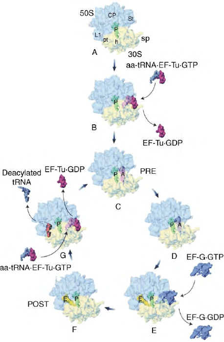
elongation cycle is shown in Figure 1, where the three
basic reactions are depicted: (1) A site occupation, (2)
peptide bond formation, and (3) the translocation
reaction.
A site occupation is separated into two subreactions:
in the first step the correct (or cognate) ternary complex
aa-tRNA·EF-Tu·GTP is selected via codon –anticodon
interaction before the aa-tRNA fully occupies the A site.
Successful decoding (decoding reaction) is sensed by the
ribosome and leads to an as yet undefined confor-
mational change within the ribosome that triggers
hydrolysis of GTP to GDP by elongation factor Tu
(EF-Tu). EF-Tu is a G protein, i.e., it can bind a GTP
molecule and is now in its “on” conformation, where
EF-Tu·GTP can bind an aa-tRNA thus forming the
ternary complex. After the ternary complex has deliv-
ered its aa-tRNA to the ribosomal A site, the ribosome
triggers the activation of the GTPase center on EF-Tu,
the resulting EF-Tu·GDP snaps into the “off” confor-
mation and falls from the ribosome.
In the second so-called accommodation step, the
release of EF-Tu·GDP from the ribosome allows the
tRNA to swing into the A site docking the aminoacyl-
residue of the aa-tRNA into the PTF center of the 50S
subunit. The aminoacyl-tRNA now occupies the A site
and is ready to accept the peptidyl-moiety from the
peptidyl-tRNA present at the adjacent P site. With A and
P sites occupied with tRNAs the ribosome is in the
so-called pretranslocational (PRE) state, although the
ribosome is not yet ready for translocation. This is
the case only after the next reaction.
In the second reaction peptidyl transfer occurs. The
peptidyl residue from the donor (P site) is linked to the
aminoacyl residue of the acceptor (A site) via a peptide
bond, forming a peptidyl-tRNA at the A site (elongated
by one amino acid) and leaving a deacylated tRNA at the
P site. Note that the ribosome is still in the PRE state
Encyclopedia of Biological Chemistry, Volume 4. q 2004, Elsevier Inc. All Rights Reserved. 214
(A and P sites are occupied), and that the tRNAs have not
changed their position after peptide-bond formation.
In the third reaction, the translocation reaction,
tRNAs are moved from the A and P site to the P and E
sites, respectively, shifting the ribosome into the post-
translocational state (POST state, P, and E sites are
occupied). The A site is vacant and ready for receiving
the next incoming ternary complex. This movement of
the tRNA
2
·mRNA complex within the ribosome by
one codon length is facilitated by elongation factor
G (EF-G), which is also a G protein with its own
GTPase center.
Functional Models of the
Elongation Cycle
Various RNA modifying techniques have been used to
probe the interactions of tRNAs within the ribosome
and to identify tRNA-related functional centers on the
ribosome. Distinct sets of rRNA bases have been
assigned to contact tRNA in each of the classical binding
site (A, P, or E site), which could be explained in the light
of high-resolution structure as resulting from either
direct contact between the tRNA and bases of rRNA or
local conformational changes of the binding regions. In
particular, the movement of tRNAs through functional
sites during a single elongation was examined using
foot-printing techniques. Two different approaches have
been applied; interestingly both led to distinct models of
the elongation cycle, although they are not mutually
exclusive. The hybrid-site model proposed by Noller and
colleagues is based on the protections of rRNA bases
from chemical modification (kethoxal, dimethylsulfate
(DMS) or hydroxyl radicals) by ribosome bound tRNA.
Nierhaus and co-workers applied the phoshorothio-
ate technique for tRNA leading to the
a
–1 model of the
elongation cycle. The latter model focuses not only on
the path of tRNA through the ribosome but also on
mechanistic features of the ribosome associated with
decoding and maintenance of the reading frame.
THE HYBRID SITE MODEL
The essence of this model (Figure 2A) is a creeping
movement of tRNAs through the ribosome. The
diagnostic feature of this model is the movement of the
tRNAs exclusively on the large subunit after peptide-
bond formation and before translocation, while the 30S
bound part of the tRNAs remain in the same site. This
results in a hybrid site: The peptidyl tRNA moves after
peptide-bond formation from an A/A site to an A/P site,
and the deacylated tRNA from P/P site to P/E (the site
before the slash indicates the site on the small subunit,
FIGURE 1 Overview of the translational elongation cycle. Multiple
cryo-EM studies determined the tRNA and elongation factor binding
positions on the 70S ribosome from E. coli during the different stages
of the elongation cycle. The schematic view of the elongation cycle
starts with an initiation complex (A) with the initiator fMet-tRNA in
the P site that represents the last stage of initiation. A ternary
complex aa-tRNA·EF-TU·GTP enters the vacant A site and after
decoding and GTP hydrolysis the binary complex of EF-Tu·GDP
leaves the ribosome (B). The A-site aa-tRNA is accommodated into
the A site and a pretranslocation complex (PRE state) is formed that
is characterized by occupied A and P sites (C). At this stage the
peptidyl residue is linked to the aminoacyl-tRNA via a peptide bond.
The result is a deacylated tRNA at the P site and a peptidyl-tRNA –
prolonged by one aminoacyl-residue – at the A site. The tRNA
positions do not change after peptide-bond formation (D). In the
next step EF G·GTP binds to the PRE complex and facilitates the
translocation of the A and P site tRNAs to the P and E sites,
respectively (E). After hydrolysis of GTP EF-G·GDP dissociates from
the ribosome, the ribosome is now in the posttranslocational state
(POST state) (F). The POST complex is ready for the newly incoming
aminoacyl-tRNA coming as ternary complex (aa-tRNA·EF-Tu·GTP).
After decoding and GTP hydrolysis the binary complex of EF-
Tu·GDP leaves the ribosome, the deacylated tRNA from the E-site is
released via a reciprocal coupling between A and E sites and the PRE
complex is formed ðG ! CÞ: (Adapted from Agrawal, R. K., Spahn,
C. M. T., Penczek, P., Grassucci, R. A., Nierhaus, K. H., and Frank,
J. (2000). Visualization of tRNA movements on the Escherichia coli
70S ribosome during the elongation cycle. J. Cell. Biol., 150,
447–459.)
TRANSLATION ELONGATION IN BACTERIA 215
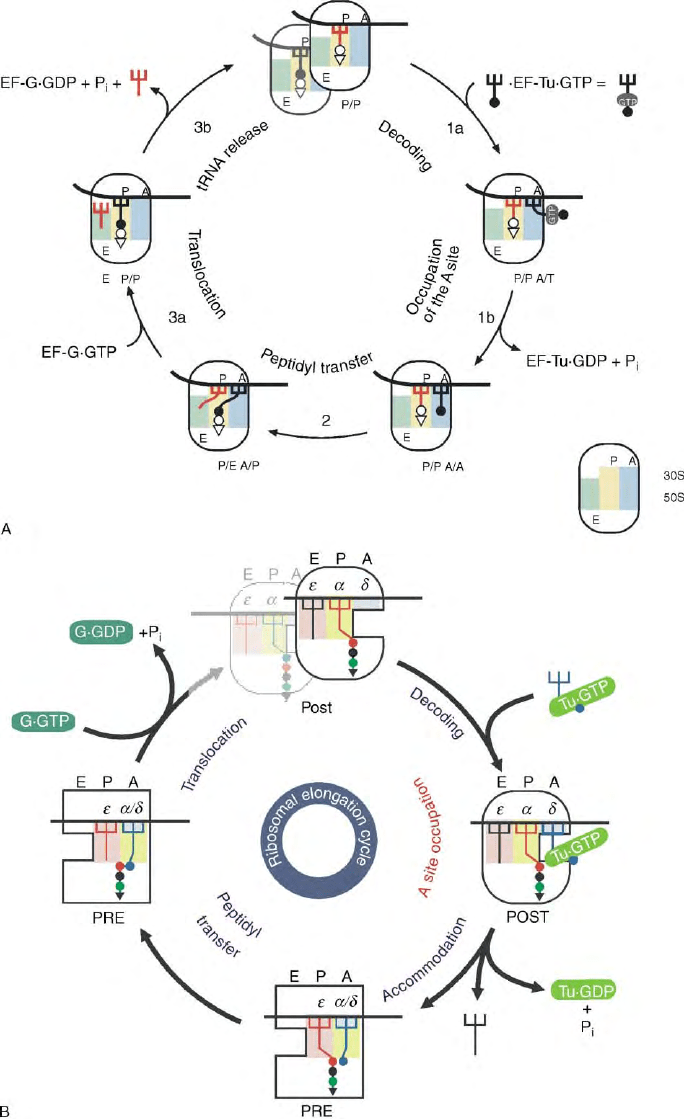
FIGURE 2 Models of the elongation cycle. (A) The hybrid-site model according to Moazed, D., and Noller, H. F. (1989). Intermediate states in
the movement of transfer RNA in the ribosome. Nature 342, 142– 148; for explanation see text. The basic feature of this model is a creeping
movement of tRNAs through the ribosome starting with a tRNA movement only on the 50S ribosome after peptide bond formation and before
translocation. This movement is uncoupled from that on the 30S subunit. (B) The
a
–1 model of the elongation cycle according to Dabrowski, M.,
Spahn, C. M., Schafer, M. A., Patzke, S., and Nierhaus, K. H. (1998). Protection patterns of tRNAs do not change during ribosomal translocation.
J. Biol. Chem. 273, 32793– 32800. The essential features are moveable ribosomal
a
- and 1-domains that connect both subunits through the
intersubunit space, bind both tRNAs and carry them in concert from A and P sites to P and E sites, respectively, during the translocation step
facilitated by EF-G. The model keeps all the features of the allosteric three side model (see text), but explains the reciprocal linkage between A and E
sites by the fact that the moveable domain moves out of the A site during translocation leaving the decoding center alone at the A site in the POST
state ribosome. The occupation of the E site generates a low-affinity A site, which is important for the selection of the newly incoming ternary
complex at the A site. Yellow and pink, the two binding regions of the
a
–1 domain, blue the decoding center (
d
) at the A site.
216 TRANSLATION ELONGATION IN BACTERIA
and after the slash that on the large subunit). In terms of
the hybrid-site model, six different protection patterns
were correlated to tRNA binding positions. The
translocation reaction brings the peptidyl-tRNA from
the A/P hybrid site to the P/P site and the deacylated
tRNA from the P/E to the E/E site.
A number of criticisms can be raised, the most serious
one is that a hybrid site was not observed when
functional states of the elongating ribosome were
systematically investigated by cryo-electron microscopy
(cryo-EM). Note that a hybrid site can be easily
detected, since 85% of a tRNA is in contact with the
large subunit with the consequence that an even partial
movement of a tRNA on the large subunit would result
in a substantial change in the overall position of the
tRNA. Even a movement of the CCA-ends of the tRNAs
at the PTF center could not be observed in crystal
structures of 50S complexes. This prompted a revision in
the hybrid-site model so as to keep the tRNAs in the
classical A/A and P/P sites after peptide-bond formation,
and only after an undefined time span the tRNAs are
then shifted to the hybrid sites A/P and P/E, respectively.
However, it seems likely that a peptidyl-tRNA at the A
site moves via a transient hybrid position A/P into the P
site and likewise a deacylated tRNA from P to E via
hybrid position P/E during the translocation reaction,
which has been resolved into a ratchet-like forth-and-
back movement between the subunits. Analyses applying
cryo-EM suggested indeed, that a tRNA moves from P to
the E site via a hybrid P/E position during translocation.
THE
a
–
1
MODEL
The essential feature of the
a
–1 model of elongation
cycle is a movable domain, called
a
–1 domain, which
binds both tRNAs of an elongating ribosome and carries
them from the A and P sites to the P and E sites,
respectively, during translocation (see Figure 2B). But
the model also includes the features of the previously
described allosteric-three-site model.
1. The ribosome contains three tRNA-binding sites:
A, P, and E site.
2. E and A site are coupled in a reciprocal manner:
An occupied E site decreases the affinity at the A site and
vice versa. The decreased affinity at the A site by an
occupied E site might be important for preventing an
interference of noncognate ternary complexes with the
selection process of the cognate ternary complex from
near cognate ternary complexes at the A site. During the
accommodation of the aa-tRNA into the A site (stable A
site occupation) the E site tRNA is released. The
reciprocal interaction between the A and the E sites
explains why statistically two tRNAs are found on
polysomes that contain a mixture of both PRE and
POST state ribosomes.
3. Both tRNAs on a ribosome are bound via codon–
anticodon interactions in both the PRE and the POST
states. Especially the codon–anticodon interaction at
the E site seems to be essential for: first, establishing the
POST state which is the proper substrate for the ternary
complex, second, reducing the error rate of protein
synthesis, and third, keeping the reading frame.
It is probable that the
a
–1 model will be modified as
soon as higher resolution structures of the ribosomal
PRE and POST states become available.
A comprehensive analysis of the translocation reac-
tion by cryo-EM to date challenges an essential feature
of the
a
–1 model: PRE states of ribosomes were
analyzed by cryo-EM carrying an fMet-Phe-tRNA at
the A site and a deacylated tRNA
Phe
at the P site. In these
complexes a tRNA is also seen at the E site, although no
tRNA was specifically bound to this site. The authors
assume that the tRNA binds from the free pool of
deacylated tRNA in solution to the “high affinity” E site.
Either way the presence of well populated A and E site is
at odds with the
a
–1 model. However, neither the origin
of the E-site tRNA nor the tRNA:70S stoichiometry is
known, therefore it would be premature to disregard
carefully controlled experiments upon which the
a
–1
model is based.
Selection of the Ternary Complex:
Decoding and A Site Occupation
In Escherichia coli, there are 45 different species of
tRNAs, where a species is defined as a tRNA with a
unique anticodon sequence. The tRNA species can be
separated into three classes with respect to the codon
displayed in the A site. The “cognate” class contains one
aminoacyl-tRNA with an anticodon complementary to
the A-site codon. The “near-cognate” class contains four
to six aminoacyl-tRNA that carry anticodons similar to
that of the cognate one (never more than one mismatch).
The “noncognate” class contains the bulk of aminoacyl-
tRNAs with a dissimilar anticodon (usually more than
one mismatch). As the tertiary structure of tRNA is
highly conserved the ribosome has to distinguish between
tRNAs that hardly differ. The problem is compounded by
the fact, that the substrate for the A site is not an isolated
aa-tRNA but rather the much larger ternary complex aa-
tRNA·EF-Tu·GTP, i.e., EF-Tu p GTP is twice as large as a
tRNA. The ribosome must therefore discriminate
between relatively large ternary complexes (72 kDa)
that are extremely similar, on the basis of a small
discriminatory region, namely the anticodon (1 kDa).
In view of the predominance of the nondiscrimina-
tory fraction of free energy of binding over the
discriminatory energy, protein synthesis must be either
slow and accurate or fast and imprecise. This is not what
TRANSLATION ELONGATION IN BACTERIA 217
we see in vivo, where protein synthesis is fast and
accurate, incorporating up to 10–20 amino acids per
second with an accuracy of one misincorporation per
3000 amino acid incorporations. How is the ribosome
solving this paradox?
A first hint gave the observation that the A site
occupation occurs in two steps as already mentioned
(see Figure 2B): a decoding step, where the selection of
the cognate ternary complex takes place, is followed by
an accommodation step. The
a
–1 model has integrated
this observation in the following way: the decoding
takes place at an A site with low affinity for tRNA,
which reduces the binding energy of the ternary complex
to mainly codon–anticodon interactions and excludes
contacts of the tRNA outside the anticodon and of EF-
Tu with the ribosome. In this state the free energy of
binding is small, and since it is restricted to codon–
anticodon interaction it is more or less identical with
the discrimination energy. This feature explains why the
majority of “noncognate” aa-tRNA (, 90% of the
aa-tRNA species) do not interfere with the decoding
process: their anticodon is different from that of
the cognate aa-tRNA, and interactions outside the
anticodon are prevented by the low-affinity A site.
This fast initial step is followed by an accommodation
step, where the aminoacyl-tRNA is tightly bound and
accommodated into the A site. This step is accompanied
by some gross conformational changes, since during this
step the E-site tRNA is released and the A site switches
into its high-affinity state. Therefore, the second step is
probably slow in comparison to the decoding the step.
The A site occupation is therefore a coupled system of
two reactions, the first of which is fast and the second
slow. An important consequence of this arrangement is
that the first runs at equilibrium even under steady-state
conditions and thus can exploit the discrimination
potential of the decoding process.
The reciprocal linkage between A and E site seems to
be a universal feature of ribosomes and has been demon-
strated not only in bacteria but also in eukarya (yeast).
After considering the competition cognate versus
noncognate aa-tRNAs, still a discussion on how
the ribosome discriminates between cognate and near-
cognate tRNAs arises. Two models have been proposed:
(1) the kinetic proofreading model, and (2) the
Potapov model.
In the late 1970s, stability measurements of anti-
codon:anticodon interactions within a complex of two
tRNAs have demonstrated that the corresponding energy
cannot explain a selection accuracy of better than 1:10.
Therefore, proofreading models have been developed
according to which the stability energy is exploited
several times in order to explain the observed accuracy
of aa-tRNA selection at the ribosomal A site of about
1:3000. One proofreading event requires one EF-Tu
dependent GTP hydrolysis, so that a measurement of
the number of GTPs hydrolyzed by EF-Tu per incorpor-
ation of a near-cognate amino acid indicates the
importance of proofreading for the selection process.
Precise measurements revealed that the importance of
proofreading is much less than originally thought, initial
binding of the ternary complex gives a precision of about
1:300 up to 1:1000, whereas the corresponding proof-
reading factor is not better than 1:10.
How initial binding is able to achieve such an
accuracy is explained by the Potapov model. This
model suggests that the decoding center on the ribosome
does not measure the stability of codon–anticodon
interaction, but rather the stereochemical correctness of
the three Watson– Crick base pairs, just as an enzyme
recognizes its substrate. With this assumption the
correct position of the sugar pucker contributes to the
accuracy, and it could be demonstrated that indeed
the 2
0
OH groups of the codon bases are of utmost
importance for the accuracy of the selection process.
The detailed molecular mechanism could be unra-
velled by the Ramakrishnan group who determined the
crystal structure of 30S subunits carrying either a cognate
or near-cognate anticodon stem-loop structures. Indeed,
the correct positions of the 2
0
OH groups of the codon–
anticodon complex is checked by forming hydrogen
bonds with universally conserved bases of the 16S rRNA
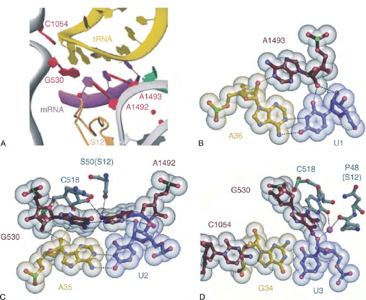
(Figure 3). The first base pair of codon–anticodon
interaction at the A site is analyzed via the so-called A-
minor motif type I and the second by an A-minor motif
type II (Figure 3B and 3C), whereas the third wobble
position has more freedom to accommodate also non-
Watson–Crick base pairs (Figure 3D). Furthermore, the
head and shoulder of the 30S subunit move relative to
each other defining an open and closed 30S configuration.
In the “open” configuration binding of cognate (but not
a near-cognate) substrate to the decoding center flips
out the bases A1492 and A1493 from the helix 44,
brings G530 from a “syn” into an “anti” conformation
(Figure 3A), and shifts the subunit into a “closed”
configuration providing a molecular basis for an under-
standing of mutations that increase or decrease accuracy.
A molecular dynamic simulation agrees with the main
conclusions and shows in addition that the kink between
the A and P site codons of about 1358 influences the
accuracy pattern.
AN ADDITIONAL ROLE OF EF-TU
It is well known that EF-Tu binds an aa-tRNA at the
amino acid acceptor stem thus shielding the labile ester
bond between the aminoacyl residue and the tRNA,
and delivers the aa-tRNA to the A site on the ribosome.
However, a second function of EF-Tu was identified by
Uhlenbeck and co-workers. Measuring the affinities of
various cognate aa-tRNA (e.g., Val-tRNA
Val
) and some
mispairs (e.g., Ala-tRNA
Val
) they recognized that either
218
TRANSLATION ELONGATION IN BACTERIA
the amino acid or the tRNA should bind to EF-Tu with
high affinity in order to form stable ternary complexes
aa-tRNA·EF-Tu·GTP. For example, EF-Tu·GTP forms
easily a ternary complex with Asp-tRNA
Asp
(weak aa
and strong tRNA) or Asn-tRNA
Asn
(strong aa and weak
tRNA), but not with Asp-tRNA
Asn
, since in the latter
case both moieties bind with low affinities. This
observation explains an important case that was an
enigma hitherto. In most organisms, there are not 20
different synthetases corresponding to the 20 natural
amino acids, but only 19 or sometimes 18. For example,
many organisms do not contain a synthetase specific for
asparagine (Asn-RS). In this case the Asp-RS is charging
also the tRNA
Asn
with aspartic acid yielding Asp-
tRNA
Asn
, which is recognized by enzymes amidating
Asp to Asn on the tRNA. The mis-charged Asp-tRNA
Asn
does not form a stable ternary complex with EF-Tu·GTP
and thus Asp is not incorporated at codons specifying
Asn. This discrimination process via EF-Tu was termed
thermodynamic compensation.
Peptide-Bond Formation
The PTF reaction, a central step in protein synthesis, is
the catalytic activity of the large subunit. Even if the
substrates are large, the reaction that occurs is quite
simple–the aminolysis of an ester bond to form a
peptide bond. The nucleophilic
a
-amino group of the
amino acid moiety of the aminoacyl tRNA at the A
site attacks the electrophilic carbonyl carbon atom of
the ester-bonded peptide moiety of the P site tRNA.
This forms an tetrahedral intermediate, which
breaks down to an uncharged (deacylated) tRNA in
the P site and a peptidyl-tRNA prolonged by one
amino acid at the A site (Figure 4A). The PTF center of
the 50S has been identified by using a transition state
analogue of the PTF reaction, which was soaked
into 50S crystals of the archaea H. marismortui. This
analogue, CCdA-phosphate-puromycin (CCdApPmn),
is a mimic of the CCA end of a tRNA in the P
site attached to puromycin in the A site, where the
FIGURE 3 Molecular details of the decoding process, principles of decoding according to Ogle et al. 2001, Science, 292, 897–902.
(A) Conformational changes at the decoding center upon binding of a cognate anticodon-stem-loop (ASL) with permission. (B) Recognition of
the Watson–Crick interaction at the first position of codon–anticodon by a type I A minor motive, A1493 is clinging into the minor groove of
the first base pair of codon-anticodon interaction. (C) Recognition of the Watson– Crick interaction at the second position of codon–
anticodon by type II A minor motive, A1492 and G530 are filling the minor groove at the second position, forming a hydrogen bond network
to 2
0
OH groups, bases of codon–anticodon, and G518 and serine50 of S12. (D) The Watson–Crick geometry at position three is less
restricted, G530, C518, and P48 are stabilizing the third codon position, but giving freedom, e.g., the G:U wobble base pair. Adapted from
Ramakrishnan, 2002.
TRANSLATION ELONGATION IN BACTERIA 219
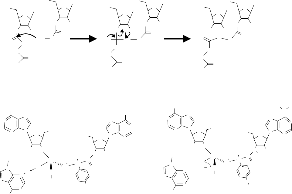
3
0
-terminal deoxy-adenosine and the phosphate residue
resemble the tetrahedral intermediate formed during
peptidyl transfer (Figure 4B). It was shown previously
by the Yarus group that this analogue is a strong
inhibitor of the PTF reaction competitively inhibiting
binding of the A-site substrate.
A long-standing discussion has been about the
composition of the PTF center and the “players”
involved in the PTF reaction, but now it is clear from
the crystal structure – the ribosome is a ribozyme, i.e.,
no protein is directly involved in catalysis. The PTF
center is tightly packed with rRNA, mainly derived from
the domain V of 23S RNA (the so-called PTF ring),
which are highly conserved over all domains of life.
Although there are 15 proteins interacting with
domain V, proteins are absent from the PTF within a
distance of at least 18A
˚
. The nearest proteins to the PTF
in H. marismortui structure are L2, L3, L4, and L10e
and interestingly, all four are also present in the vicinity
of PTF in the eubacterial ribosome, when it is taken into
account that eukaryotic L10e is evolutionary related to
L16 in prokaryotes. These proteins have been identified
previously, together with the 23S rRNA, as the major
candidates for the PTF activity by single omission tests in
a total reconstitution system of the large subunit.
Although the structure reveals no direct involvement
of protein in the catalysis, proteins might have a role in
aligning the substrates and functioning as a “glue” to
stabilize rRNA tertiary arrangement necessary for the
peptide bond formation since complete removal of
P site A site P site A site P site A site
tRNA
tRNA
tRNA
tRNA
tRNA
tRNA
O
O
O
O
OH
O
O
O
O
O
O
O
O
O
O
O
O
O
OH
HN
HN
HN
R1
R1
R1
H
2
N
CHR
3
CHR
3
CHR
2
CHR
2
CHR
3
CHR
2
A
A
O
O
–
O
A
A
A
A
OH
OH
OH
OH
H
+
NH
HO
H
N
A
}
Transition state
CCdApPmn
tRNA
tRNA
O
O
O
H
O
OH
OH
peptide
HO
O
O
O
O
N
N
N
N
N
N
N
N
N
N
N
N
N
NH
2
NH
2
NH
2
+
–
C
C
AA-Ca
C
H
H
P-site A-site
A-siteP-site
A2451A2451
R
N
H
N
N
N
NH
2
NH
2
CH
3
CH
3
H
3
C
R
3Å
1
3
3
1
O
2
O
1
O
O
O
O
HO
O
P
N
N
N
N
pCpC5′
OH
H
N
N
N
N
N
N
N
H
H
C
C
Transition state of PTF reaction Yarus inhibitor
B
FIGURE 4 Mechanism of peptide bond formation. (A) PTF reaction: The a- amino group of the A site bond aa-tRNA attacks the electrophilic
carbonyl carbon, which is attached via ester bond to the 3
0
OH of the adenosine residue of P site bond peptidyl-tRNA, forming a tetrahedral
transition state that breaks down to a prolonged peptidyl-tRNA at the A site and a deacylated tRNA at the P site. (B) Comparison of the
putative transition state of the PTF reaction with a possible transition-state analogue created by the Yarus group. From Parnell, K. M., Seila, A. C.,
and Strobel, S. A. (2002). Evidence against stabilization of the transition state oxyanion by a rka-perturbed RNA base in the peptidyl transferase
center. Proc. Natl Acad. Sci. USA 99, 11658– 11663.
220 TRANSLATION ELONGATION IN BACTERIA
proteins could only accomplished under conditions that
unfolded the rRNA and totally abolished the
PTF activity, whereas the removal of up to over
80% of proteins of the 50S subunit from Thermus
aquaticus maintained activity. Detailed analysis of the
neighborhood of the residues within close proximity to
the CCA end analogues of tRNA led to the proposal for
an acid-base catalysis mechanism for the PTF reaction
involving the universal conserved A2451 in analogy to
the back reaction of the mechanism used by serine
proteases. This proposal was immediately under attack
from a number of groups who presented biochemical
and genetic data to the contrary.
Crystal structures of the 50S of Deinococcus radio-
durans revealed some significant differences in the
arrangement of nucleotides within the PTF center and
the presence of a protein (L27), which is thought to play
a role in placement of the CCA ends of both tRNAs. The
debate on the mechanism of PTF reaction is on going. By
re-examination of the transition state analogue it turned
out that this analogue within the 50S structure can not
answer all the mechanistic questions about the PTF
reaction, since it is not a true mimic of the intermediate
of the PTF reaction: modeling the missing oxygen atom
at the 2
0
position of the desoxy-adenosine caused a
sterical clash with the phosphate group. Although the
reaction seems to be quite simple, the kind of
contribution of the ribosome in catalysis remains open.
Two main principles of enzymatic reactions can be
distinguished and could be involved in PTF reaction.
The first principle is the physical or template model,
where the enzyme, here the ribosome, arranges the
two substrates in optimal stereo-chemical positions
for the reaction to proceed. Such an arrangement of
substrates is sufficient to allow for a dramatic
acceleration of the reaction rate by six to nine orders
of magnitude. This strategy is certainly used by the
ribosome, since there are binding sides for both tRNA
substrates fixing the substrates in a defined position
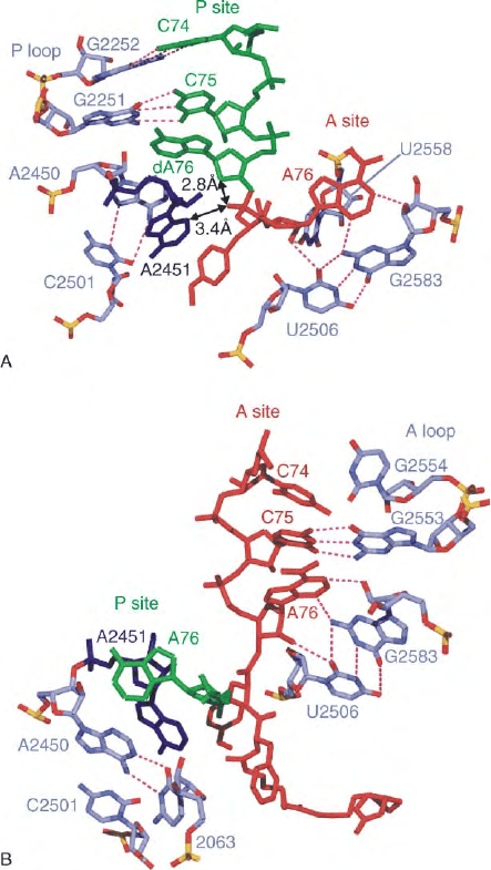
by interaction between 23S rRNA (A loop and P
loop) and tRNAs (see Figure 5), placing the corres-
ponding CCA ends into the PTF center.
In addition it is possible that a chemical concept is
utilized by the ribosome, i.e., transiently covalent bonds
are formed and broken between the enzyme and the
substrates. There are three groups in close proximity to
the reactive amino group within the PTF center in the
H. marismortui crystal structure, that could form
hydrogen bonds with it, namely (1) the 2
0
-OH of the
peptidyl-tRNA, (2) the N3 of A2451 (E. coli nomen-
clature) of 23S rRNA, and (3) the 2
0
OH of A2451.
The hydrogen bonds these groups could form with
the
a
-amino group of the aa-tRNA at the A site may
help to fix and optimally align the reactive
a
-amino
group within the PTF center. And if one of these
groups have an elevated pK
a
, its hydrogen bond would
facilitate the nucleophilic attack from the
a
-amino
group of the aminoacyl-tRNA at the A side to the
carbon of the carbonyl group of peptidyl-tRNA at the P
site. A major candidate for an enhancement of
nucleophily of the
a
-amino group is N3 of A2451
(see Figure 6).
FIGURE 5 Tight fixation of the CCA ends of the P- and A-tRNAs
observed in 50S subunit from H. marismortui in complex with (A) the
Yarus inhibitor, and (B) the products following peptide bond
formation. The CCA-ends of the tRNAs in the A and P sites are
colored red and green, respectively. The N3 of A2451 (dark blue)
is 3.4A
˚
from the O2 of the Yarus inhibitor, while the same O2 is
only 2.8A
˚
from the 2
0
-deoxy of A76 (arrowed). Selected rRNA
residues of domain V of the 23S rRNA are colored light blue, including
the A- and P-loop bases that participate in A and P site CCA end
fixation (E. coli numbering). In (B) the P site C74 and C75 have been
omitted for clarity. Dashes indicate hydrogen bonding and rRNA
nucleotides use the following color scheme: Oxygen, red; phosphorus,
yellow; nitrogen, blue; carbon, dark blue. Adapted from Nissen, P.,
Hansen, J., Ban, N., Moore, P. B., and Steitz, T. A. (2000). The
structural basis of ribosome activity in peptide bond synthesis. Science
289, 920– 930.
TRANSLATION ELONGATION IN BACTERIA 221
