Lennarz W.J., Lane M.D. (eds.) Encyclopedia of Biological Chemistry. Four-Volume Set . V. 4
Подождите немного. Документ загружается.


T-Cell Antigen Receptor
Andrea L. Szymczak and Dario A. A. Vignali
St. Jude Children’s Research Hospital, Memphis, Tennessee, USA
The T-cell antigen receptor (TCR) is a highly organized, multi-
molecular complex found exclusively on T and NK T cells. The
majority of TCRs are composed of TCR
a
and TCR
b
chains,
while a small percentage contain TCR
g
and TCR
d
chains. The
TCR chains heterodimerize and are associated with the
invariant chains of the CD3 complex, CD3
1gd
and CD247
(often referred to as the zeta chain, TCR
z
or CD3
z
). Each
chain is essential for TCR surface expression and consequently
T cell development and function. This complex mediates the
development of T cells that are able to mount a specific
immune response against a wide variety of foreign antigens and
pathogenic organisms. This unique capability is due to the
unusual genomic organization and rearrangement of TCR
genes, which generates a vast number of TCRs that recognize
almost any antigenic peptide in the context of major
histocompatibility (MHC) molecules.
TCR:CD3 Complex: Genes,
Proteins and the Receptor Complex
TCELL ANTIGEN RECEPTOR
T cells can only recognize and bind antigens when they
are presented by major histocompatibility (MHC)
molecules, a process called MHC restriction. Initially,
the T cell antigen receptor (TCR) was difficult to study
because it was membrane bound and could only
recognize antigen in the context of MHC. The first
TCR complexes isolated were composed of a disulfide-
linked heterodimer of TCR
a
and TCR
b
. Monoclonal
antibodies against this complex were either specific for a
particular T cell clone or nonspecific, indicating that the
TCR chains were similar to the immunoglobulin (Ig)
molecules, containing both variable (V) and constant (C)
regions. While the vast majority (. 95%) of T cells had
TCR
ab
heterodimers, another type of TCR containing
a TCR
g
and TCR
d
heterodimer was later identified. The
TCR chains, in particular the variable regions, are
responsible for recognizing and binding to peptide–
MHC molecules presented on the surface of antigen
presenting cells (APC).
Genes: Organization and Rearrangement
As with antibody molecules, T cells must be able to
recognize a wide variety of pathogens, therefore a
diverse repertoire of TCR must be generated that
recognize such antigens. Many similarities exist between
the Igs and the TCR. The four TCR loci (
a
,
b
,
g
, and
d
)
have a germ-line organization similar to that of Ig. The
a
- and
g
-chains are produced by rearrangement of V and
J segments, while the
b
- and
d
-chains are produced by
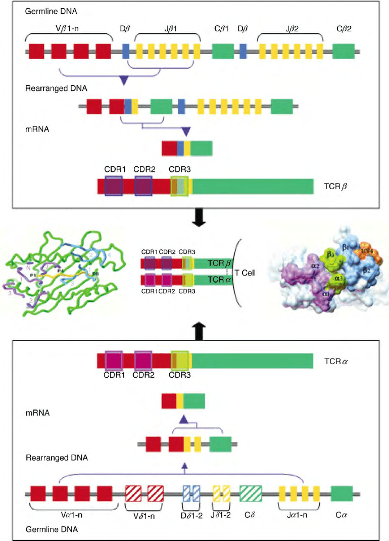
rearrangement of V, D, and J segments (Figure 1).
The segments for TCR
d
are located between the V
a
and
J
a
segments. When the TCR
a
gene is rearranged, the
TCR
d
segments are deleted, thereby ensuring the T cell
does not express TCR
gd
and TCR
ab
at the same time.
The basic domain structure is evolutionarily conserved
and is believed to have arisen through gene duplication.
Organization of the TCR genes in the mouse and human
is similar but vary in the numbers of segments (Table I).
The TCR genes encode areas of hypervariability
similar to the complementarity determining regions
(CDRs) of antibody molecules. Embedded within the
V regions are CDR1 and CDR2. There is an additional
area of hypervariability (HV4) that does not appear to
interact with antigen and is therefore not considered a
CDR (Figure 1). CDR3 is a result of VJ or VDJ joining.
This intentionally imprecise process frequently results in
the addition or deletion of nucleotides, adding signifi-
cantly to the variability in this region (N region
diversity). Occasionally, a nonproductive rearrangement
of the first gene locus occurs due to an out-of-frame
rearrangement or insertion of a stop codon. This induces
the rearrangement of the TCR locus on the other
chromosome.
Rearrangement occurs through the action of recom-
bination-activating genes (RAGs) 1 and 2 during T cell
development. Flanking each gene segment in the germ
line DNA are conserved recognition signal sequences.
The RAG enzymes recognize these heptamer and
nonamer sequences and catalyze the VJ and VDJ joining
with a mechanism analogous to that used to rearrange
the Ig locus. In addition, secondary rearrangements
termed receptor editing or revision can occur in both B
and T cells. The role of receptor editing and revision in
Encyclopedia of Biological Chemistry, Volume 4. q 2004, Elsevier Inc. All Rights Reserved. 162

FIGURE 1 Rearrangement of TCR
ab
genes. Example of TCR
a
(bottom panel) and TCRb (top panel) rearrangement. TCR
a
undergoes V to J
rearrangement while TCR
b
undergoes D to J followed by V to DJ rearrangement. Primary transcripts of rearranged DNA are processed to mRNA
that encode the mature TCR
ab
chains expressed on the T cell surface (center panel, middle). Areas encoding the CDR regions are boxed. TCR
d
segments in the TCR
a
locus are shown in stripes. The center left panel shows a view into the peptide–MHC combining site with peptide in yellow
and MHC in green superimposed with the CDR regions (CDR1, 2 and 3; HV4) of TCR
a
(purple) and TCR
b
(blue). CDR1 of V
a
and V
b
and V
b
CDR3 are clearly positioned along the central axis of the peptide. N ¼ the N-terminal residue of V
a
. (Reprinted from Garcia, K. C., Degano, M.,
Pease, L. R., Huang, M., Peterson, P. A., Teyton, L., and Wilson, I. A. (1998). Structural basis of plasticity in T cell receptor recognition of a self
peptide-MHC antigen. Science 279, 1166– 1172, with permission of AAAS.) The center right panel shows the surface of the TCR-binding site. The
surface of the loop trace of the V
a
CDRs 1 and 2 are purple; CDRs 1 and 2 of TCR
b
are blue; V
a
and V
b
CDR3s are green; and the hypervariable
region of TCR
b
(HV4) is orange. (Reprinted from Garcia, K. C., Degano, M., Stanfield, R. L., Brunmark, A., Jackson, M. R., Peterson, P. A.,
Teyton, L., and Wilson, I. A. (1996). An
ab
T cell receptor structure at 2.5A
˚
and its orientation in the TCR-MHC complex. Science 274, 209– 219,
with permission of AAAS.)
T-CELL ANTIGEN RECEPTOR 163

T cells is still unclear, but it has been suggested to
contribute to peripheral T cell tolerance.
While many similarities exist between Ig and TCR,
there is one important distinction: TCR genes do not
undergo somatic hypermutation and thus there is no
affinity maturation. While the number of TCR V region
segments is dramatically reduced compared to the Ig
genes, the number of J region gene segments is greater,
increasing the potential diversity at the V–J interface
(Table II). This is likely due to differences in the nature of
the antigen recognized by TCR and Ig.
TCR Structure and Recognition
of Peptide–MHC Complexes
The TCR is a member of the Ig super family. Structural
analysis has shown that the V
a
and V
b
domains pair via
a conserved hydrophobic core, while the C
a
and C
b
domains pair via a highly polar interface, with a skewed
distribution of acidic residues in C
a
and basic residues in
C
b
. The two chains also pair via a conserved disulfide
bond close to the membrane (Figure 2). In addition, the C
region contains a connecting peptide, a transmembrane
region and short intracellular domain. The anchoring
transmembrane domain is unusual as it contains several
highly conserved basic amino acid residues (TCR
a
–Lys/
and –Arg; TCR
b
–Lys). These positively charged
residues mediate interaction with the CD3 complex.
The cytoplasmic domains are short, consisting of only
5–12 amino acids.
TCR specificity is mediated by the V
a
and V
b
CDRs
which interact with the peptide–MHC complex. MHC
molecules are bound to the APC membrane and the
antigenic peptide is bound in a groove between two
a
-helices (Figure 2). A number of TCR:peptide –MHC
complexes have been crystallized. Although specific
contact residues vary, all have a similar mode of binding
in which the CDR2 regions contact the MHC surface
and CDR1 and CDR3 contact both the peptide and
MHC molecules (Figures 1 and 2). The V region of
TCR
a
binds closer to the N-terminal region of the
peptide while that of TCR
b
is closer to the C terminus.
The TCR binds in a diagonal orientation due to two
peaks created by the MHC
a
-helices.
The binding properties for a number of TCR:
peptide–MHC complexes have been described. In
general, the on and off rate for TCR:peptide–MHC
binding is fast and the affinity is low compared to other
receptor –ligand interactions. These biophysical proper-
ties of the TCR contrast starkly with the generally high
affinity of Ig:antigen interaction. However, in spite of
these properties, TCR interaction is sufficiently stable to
initiate signal transduction.
In most instances, only a very small fraction of
MHC molecules on a cell contain the right antigenic
peptide for an individual TCR. It has been proposed
that T cells overcome this limited ligand supply by a
process called serial ligation, where many TCRs
(perhaps as many as 200) can be ligated by a single
TABLE I
Chromosomal Location and Numbers of TCR Gene Segments
Human Mouse
TCR
a
(14)
TCR
b
(7)
TCR
g
(7)
TCR
d
(14)
TCR
a
(14)
TCR
b
(6)
TCR
g
(13)
TCR
d
(14)
V 40–50 30–57 11– 14 3 50– 100 20 –50 7 10–11
D2 3 2 2
J 60–70 13–14 5 3–4 60–100 12– 14 4 2
C1 2 2–3 1 1 2 4 1
Total number of gene segments for the human and mouse TCR.
Numbers in parenthesis indicate the chromosome on which the
gene is located. Reprinted from Allison, T. J., and Garboczi, D. N.
(2001). Structure of
gd
T cell receptors and their recognition of non-
peptide antigens. Mol. Immunol. 38, 1051– 1061, with permission
from Elsevier.
TABLE II
Table Sequence Diversity in TCR and Ig Genes
Ig TCR
ab
TCR
gd
H
ka b g d
V segments 250–1000 250 50 25 7 10
D segments 10 0 0 2 0 2
Ds read in all frames Rarely Often Often
N-region addition V–D, D–J None V–J V–D, D –J V–J V–D1, D1–D2,
D1–J
J segments 4 4 50 12 2 2
V region combinations 62 500–250 000 1250 70
Junctional combinations , 10
11
, 10
15
, 10
18
(Reprinted from Davis, M. M. (1990). T cell receptor gene diversity and selection. Annu. Rev. Biochem. 59, 475–496, with permission of Annual
Reviews.)
164 T-CELL ANTIGEN RECEPTOR

peptide–MHC complex. It is thought that the low
affinity and rapid off rate of TCR:peptide –MHC inter-
action may be instrumental in mediating this process.
INVARIANT CHAINS: CD3, CD247,
AND PRE-T
a
CD3 and CD247
The TCR is associated with the invariant chains of the
CD3 complex.: CD31, CD3
g
, and CD3
d
are members
of the Ig superfamily with one extracellular Ig domain
followed by a transmembrane domain and cytoplasmic
tail of , 40 amino acids in length. The zeta family of
molecules are unique and have a short extracellular
sequence and long cytoplasmic tail. This family consists
of CD247 (otherwise known as CD3
z
or TCR
z
), its
splice variant
h
, and the
g
-chain of the Fc receptor
(FcR
g
). The CD247
z
- and
h
-chains are alternatively
spliced gene products and are identical except for
the carboxyl-terminal region of the cytoplasmic tail
(113 and 155 amino acids long for
z
and
h
, respectively).
Analysis of CD3 and CD247 knockout mice demon-
strates their importance in TCR expression and T cell
development and function. Mice lacking CD247 (CD3
z
)
and CD3
g
exhibit a substantial block in early stages of
T cell development, whereas mice lacking CD3
d
develop
a block at a later stage. CD31 knockout mice display
a complete arrest in T cell development, likely due to its
requirement to form heterodimers with CD3
g
and CD3
d
.
The CD3 and CD247 chains contain a number of
amino acid residues and motifs that are important for
assembly, signal transduction, and regulation of cell
surface expression (Figure 2). The transmembrane
regions contain negatively charged amino acids that
interact with the positively charged residues of TCR
ab
(CD3
d
, CD31, and CD3
z
-Asp; CD3
g
-Glu). The cyto-
plasmic tails contain immuno-receptor tyrosine-based
activation motifs (ITAM) that when phosphorylated
provide docking sites for SH2 domain-containing
proteins, which are important for downstream signal
transduction. Each CD31
gd
chain contains one ITAM
while CD247 (CD3
z
) contains three. CD3
d
and CD3
g
also contain a dileucine-based motif. Both the YXXL
sequence in the ITAM and the dileucine sequence have
been shown to play a role in internalization and down-
modulation of many types of receptors from the cell
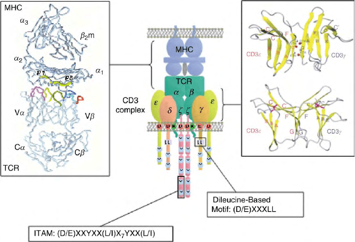
FIGURE 2 TCR:CD3 complex and its interaction with peptide–MHC. Schematic of the presumed stoichiometry of the TCR:CD3 complex.
Highlighted are charged transmembrane residues, the ITAM sequence and the dileucine-based motif. In the upper left panel is a backbone structure of
TCR:peptide–MHC complex. TCR is on the bottom and the MHC on top. The peptide (P1–8) is shown as a large tube in yellow. V
a
CDRs 1 and 2
are in light and dark purple, respectively;
a
HV4 in white; V
b
CDRs 1 and 2, in light and dark blue, respectively;
b
HV4 in orange; and V
a
and V
b
CDR3s in light and dark yellow, respectively. (Reprinted from Garcia, K. C., Degano, M., Stanfield, R. L., Brunmark, A., Jackson, M. R., Peterson, P.
A., Teyton, L., and Wilson, I. A. (1996). An
ab
T cell receptor structure at 2.5A
˚
and its orientation in the TCR-MHC complex. Science 274,
209–219, with permission of AAAS.) In the upper right panel is a ribbon diagram of CD31
g
. The beta strands are in yellow and the text colored
red for CD31 and blue for CD3
g
. Three pairs of atoms involved in the hydrogen bond formation are designated with amide protons in gray
and carbonyl oxygen atoms in red. Two disulfide-linked cysteine residues are shown. (Reproduced from Sun, Z. J., Kim, K. S., Wagner, G., and
Reinherz, E. L. (2001). Mechanisms contributing to T cell receptor signaling and assembly revealed by the solution structure of an ectodomain
fragment of the CD3
g
heterodimer. Cell 105, 913–923, with permission from Elsevier.) In the lower left and right panels are the ITAM and dileucine-
based consensus motifs, respectively.
T-CELL ANTIGEN RECEPTOR 165
surface. It has been suggested that these motifs are also
utilized for TCR transport. A number of studies using
T cell lines in vitro have shown a role for the CD3
g
dileucine-based motif in TCR down-modulation, how-
ever its role in vivo remains to be defined.
Pre-T
a
During T cell development in the thymus, TCR
ab
precursors express a pre-TCR. The pre-TCR is formed
by the functionally rearranged TCR
b
chain, the CD3
complex and a surrogate TCR
a
chain, pre-TCR
a
(pT
a
).
There are two notable differences between pT
a
and
TCR
a
. First, pT
a
only has one invariant extracellular Ig
domain. Second, the cytoplasmic tail of pT
a
is longer and
contains two potential serine and threonine phosphoryl-
ation sites and a potential SH3 domain-binding motif.
The requirements for surface expression of pT
a
are still
unclear. However, it is clear that a number of signaling
events are initiated through the pre-TCR. Pre-TCR
signaling confirms the successful rearrangement TCR
b
and induces the suppression of further TCR
b
locus
rearrangement, a process called allelic exclusion.
Rearrangement of the TCR
a
locus then occurs leading
to progression of T cell development.
TCR:CD3 COMPLEX
Expression of the TCR on the cell surface requires all six
chains of the complex. While the exact stoichiometry
and make-up of the TCR:CD3 complex is unclear, it is
generally accepted that the complex consists of four
dimers: TCR
ab
or
gd
, heterodimers of CD31
g
and
CD31
d
, and a zeta family dimer. The majority of
TCR:CD3 complexes (80–90%) contain a CD247
z
homodimer. Heterodimers of
z
-
h
or
z
-FcR
g
or homo-
dimers of FcR
g
have been observed in a small percentage
of TCR complexes. An organizing principle has been
proposed in which a single negatively charged amino acid
in the TCR dimer interacts with two positively charged
residues in the CD31
g
, CD31
d
, and CD247
z
dimers. In
this model, the TCR
a
lysine interacts with the CD31
d
dimer, the TCR
b
lysine interacts with the CD31
g
dimer
and the TCR
a
arginine interacts with the CD247
z
dimer.
Role of the TCR in T Cell Biology
and Signaling
TCELL DEVELOPMENT
T cells that exit the thymus have been through a
selection process based largely on TCR affinity. T cells
with TCR that recognize self peptide–MHC too
strongly are negatively selected and deleted. T cells
that have too low an affinity to productively interact
with self-MHC are ignored and ultimately die of neglect.
However, T cells that display a moderate affinity for the
self-MHC molecules are positively selected and allowed
to exit into the periphery.
CELL BIOLOGY
It is known that each of the six protein chains is required
for correct assembly and surface expression of
the TCR:CD3 complex. Once expressed on the cell
surface, the TCR:CD3 complex is constitutively inter-
nalized and recycled back to the cell surface via the
endosomal network. Once ligated by peptide–MHC
complexes, the TCR is down-modulated from the cell
surface and diverted to lysosomes for degradation.
Assembly and Surface Expression
of the TCR:CD3 Complex
Assembly of the complex is a highly ordered process that
takes place in the ER. Most of the evidence for assembly
order supports a model in which CD31
g
and CD31
d
heterodimerize followed by sequential addition of TCR
a
and TCR
b
chains and the
z
2
homodimer (or hetero-
dimer). Once the correct stoichiometry is achieved, the
intact complex is transported from the ER. It has been
shown that the
z
-chain can exit the ER independently of
the rest of the receptor complex and remain in the Golgi
complex. CD3
g
and CD31 have been reported to partly
exist as heterodimers in association with calnexin while
the TCR
a
and
b
-chains have been found to associate with
calreticulin. It has been suggested that these molecules
may serve as chaperones to prevent the transport of
partial complexes. A number of residues are present
within the chains that serve as ER retention signals or to
target partial complexes to lysosomes for degradation,
thereby ensuring that only complete TCR:CD3 com-
plexes are expressed on the cell surface. The charged
residues in the transmembrane regions increase their
susceptibility to ER degradation. A number of additional
residues have been described as possible ER retention or
degradation signals, however a detailed mechanism of
complex assembly, including specific interactions
between individual chains, remains to be defined.
Internalization and Down-Modulation
of the TCR:CD3 Complex
The TCR:CD3 complex is constitutively internalized
and recycled back to the cell surface. Although the exact
mechanism of this process has not yet been defined, it is
likely to involve the interaction of the CD3/CD247
molecules with adaptor protein (AP) complexes that
are associated with clathrin at the plasma membrane
and intracellular recycling vesicles of the endosomal
network. The dileucine-based motif in CD3
g
has been
166
T-CELL ANTIGEN RECEPTOR
shown to be capable of binding to a member of the AP
family of complexes, AP-2. In addition to dileucine
based motifs, AP-2 can recognize YXXL-based
sequences in a number of receptor systems. Given that
the TCR:CD3 complex contains 20 such sequences, it is
possible that one or more may be utilized for TCR
internalization.
Upon ligation with peptide –MHC complexes, the
TCR:CD3 complex is down-modulated from the cell
surface and recycling is prevented. While the exact
mechanism of this important process is unknown, it may
involve two related E3 ubiquitin ligases, c-Cbl and
Cbl-b, as T cells from mice lacking both proteins fail to
down-modulate their TCR following ligation. While
both internalization and down-modulation are hall-
marks of TCR biology, their physiological importance
and function remains to be determined.
SIGNALING THROUGH THE TCR
Initiation of T cell activation occurs when the TCR
recognizes peptide–MHC complexes. TCR
ab
consists of
the ligand-binding unit while the CD3 complex trans-
duces signals into the T cell. Clustering of TCR:peptide–
MHC complexes brings in the coreceptors CD8 or CD4
which bind to MHC class I and II molecules, respectively.
Both coreceptors are associated with Src-related protein
tyrosine kinase (PTK) p56
lck
, while another PTK, p59
fyn
,
interacts with the CD3 complex. In resting T cells,
these kinases are inactive due to the interaction of the
C-terminal phosphotyrosine residues binding to the
N-terminal SH2 domain. This intramolecular interaction
prevents substrate access to the kinase (SH1) domain. T
cell:APC interaction induces the removal of these
inhibitory phosphates by the transmembrane phospha-
tase CD45. Cross phosphorylation of active site tyrosine
residues further potentiates p56
lck
and p59
fyn
kinase
activity and results in the phosphorylation of the ITAM
tyrosine residues in the CD3/CD247 cytoplasmic tails.
Phosphorylation of both ITAM tyrosine residues is
required for docking of a specialized PTK, zeta associated
protein-70 (ZAP-70), which has two tandem SH2
domains. ZAP-70 kinase activity is further potentiated
through phosphorylation by p56
lck
and p59
fyn
.
Activated ZAP-70 initiates a number of signaling
pathways. A key target of ZAP-70 is the raft-resident
linker for activated T cells (LAT) which is heavily
phosphorylated and recruits a wide variety of down-
stream signaling molecules. Phosphorylation of phos-
pholipase C
g
1
(PLC
g
1
) leads to the production of potent
second messengers, diacylglycerol (DAG) and inositol
triphosphate (IP
3
), whose actions lead to protein kinase
C (PKC) activation and NF
k
B nuclear translocation as
well as Ca
2þ
release and nuclear factor of activated
T cells (NFAT) translocation. These events lead to the
transcription of genes required for T cell proliferation
and interleukin-2 (IL-2) production. Signaling through
ZAP-70 also initiates activation of the Ras pathway and
the MAPK signaling cascade which also results in up-
regulation of genes required for proliferation. A wide
array of additional signaling molecules and adaptor
proteins have been shown to contribute to the signaling
cascade initiated following TCR ligation and have been
reviewed extensively elsewhere.
SEE ALSO THE FOLLOWING ARTICLES
Immunoglobulin (Fc) Receptors † Mitogen-Activated
Protein Kinase Family † Protein Kinase C Family † Src
Family of Protein Tyrosine Kinases
GLOSSARY
CD3 Complex of polypetides containing three dimers: 1
g
hetero-
dimers, 1
d
heterodimers and, most frequently,
zz
homodimer
(CD247). It is associated with the TCR through charged
transmembrane residues and is involved in transducing signals
into the T cell upon TCR:peptide–MHC interaction.
complementarity-determining region (CDR) Areas in the variable
regions of antibody and TCR genes. In the TCR, the CDR regions
contact the peptide and MHC molecule on antigen presenting cells.
immunoglobulin superfamily Group of proteins that contain
immunoglobulin-fold domains of , 100 amino acids folded into
two
b
-pleated sheets and stabilized by a central disulfide bond.
Included in the family are MHC molecules, TCRs and a number of
CD antigens.
major histocompatibility complex (MHC) A complex of poly-
morphic genes that encode histocompatibility antigens termed H2
in the mouse and HLA in humans. Two main classes of MHC
antigens are found as surface glycoproteins on antigen presenting
cells that bind and present peptides to T cells.
TCR Heterodimer of TCR
ab
or TCR
gd
expressed on the surface of
T cells that is associated with the CD3 complex. The TCR binds to
peptide–MHC molecules.
FURTHER READING
Allison, T. J., and Garboczi, D. N. (2001). Structure of
gd
T cell
receptors and their recognition of non-peptide antigens. Mol.
Immunol. 38, 1051–1061.
Call, M. E., Pyrdol, J., Wiedmann, M., and Wucherpfennig, K. W.
(2002). The organizing principle in the formation of the T cell
receptor-CD3 complex. Cell 111, 967–979.
Davis, M. M. (1990). T cell receptor gene diversity and selection.
Annu. Rev. Biochem. 59, 475–496.
Germain, R. N., and Stefanova, I. (1999). The dynamics of T cell
receptor signaling: Complex orchestration and the key roles of
tempo and cooperation. Annu. Rev. Immunol. 17, 467–522.
Glusman, G., Rowen, L., Lee, I., Boysen, C., Roach, J. C., Smit, A. F. A.,
Wang, K., Koop, B. F., and Hood, L. (2001). Comparative genomic of
the human and mouse T cell receptor loci. Immunity 15, 337–349.
Goldsby, R. A., Kindt, T. J., and Osborne, B. A. (eds.) (2000). In Kuby
Immunology. W. H. Freeman, New York.
Garcia, K. C., Degano, M., Stanfield, R. L., Brunmark, A., Jackson,
M. R., Peterson, P. A., Teyton, L., and Wilson, I. A. (1996). An
ab
T cell receptor structure at 2.5A
˚
and its orientation in the TCR-
MHC complex. Science 274, 209–219.
T-CELL ANTIGEN RECEPTOR 167
Garcia, K. C., Degano, M., Pease, L. R., Huang, M., Peterson, P. A.,
Teyton, L., and Wilson, I. A. (1998). Structural basis of plasticity in
T cell receptor recognition of a self peptide-MHC antigen. Science
279, 1166–1172.
Hennecke, J., and Wiley, D. C. (2001). T cell receptor-MHC
interactions up close. Cell 104,1–4.
Kruisbeek, A. M., Haks, M. C., Carleton, M., Michie, A. M., Zuniga-
Pflucker, J. C., and Wiest, D. L. (2000). Branching out to gain
control: How the pre-TCR is linked to multiple functions.
Immunol. Today 21, 637– 644.
Samelson, L. E., Harford, J. B., and Klausner, R. D. (1985).
Identification of the components of the murine T cell antigen
receptor complex. Cell 43, 223–231.
Sun, Z. J., Kim, K. S., Wagner, G., and Reinherz, E. L. (2001).
Mechanisms contributing to T cell receptor signaling and assembly
revealed by the solution structure of an ectodomain fragment of the
CD3
g
heterodimer. Cell 105, 913– 923.
Valitutti, S., Muller, S., Cella, M., Padovan, E., and Lanzavecchia, A.
(1995). Serial triggering of many T-cell receptors by a few peptide-
MHC complexes. Nature 375, 148–151.
BIOGRAPHY
Andrea Szymczak is a graduate student in the Department
of Pathology at the University of Tennessee in Memphis, under
the guidance of Dr. Dario Vignali at St. Jude Children’s Research
Hospital. She received her B.Sc. degree in biology at the University
of Tennessee at Chattanooga. At present, her research interest is in
T cell biology, specifically TCR:CD3 complex internalization and
down-modulation.
Dario Vignali is an Associate Member in the Department
of Immunology at St. Jude Children’s Research Hospital. His
research interests are the TCR:CD3 complex, regulation of T cell
function and type 1 diabetes. He holds a Ph.D. from the London
School of Hygiene and Tropical Medicine in England, and was a
postdoctoral fellow at the German Cancer Research Center,
Heidelberg, Germany, and Harvard University. His laboratory has
made important contributions to the understanding of how the TCR
recognizes MHC class II:peptide complexes and how T cells traffic
TCR:CD3 complexes.
168 T-CELL ANTIGEN RECEPTOR

Tec/Btk Family Tyrosine Kinases
Shuling Guo and Owen N. Witte
Howard Hughes Medical Institute, University of California, Los Angeles, California, USA
Nonreceptor tyrosine kinases (NRTKs) are cytoplasmic
enzymes that phosphorylate tyrosine residues when activated
and thereby play critical roles in many signal transduction
pathways in multicellular organisms. These kinases are
grouped into families including Src, Syk, Abl, and Fak families,
according to their protein sequence and structure similarities.
In 1993, mutations in Bruton’s tyrosine kinase (Btk) were
demonstrated to cause human X-linked agammaglobulinemia
(XLA) and murine X-linked immunodeficiency (xid). Since
then, more proteins similar to Btk were discovered and
Tec/Btk family tyrosine kinases became a new NRTK
subfamily. This family is now the second largest nonreceptor
tyrosine kinase family after the Src family.
Tec Family Kinase Members
and Expression Pattern
The Tec family is composed of five mammalian
members: Btk, Tec, Itk, Txk, and Bmx. These kinases
are differentially expressed and most of them are found
primarily in hematopoietic cells. This family is
also expressed in other species, including Drosophila
melanogaster, skate, and zebrafish. In addition, a Btk
orthologue designated NRTK3 has been identified in the
sea urchin Anthocidaris crassispina.
1. Btk is also known as Atk, Bpk, or Emb. Btk is
expressed in all stages of B cell development except
plasma cells. Btk is also expressed in myeloid and mast
cells as well as early erythroid and megakaryocytic
precursors, but Btk is not expressed in T cells. In tissues,
Btk is found in bone marrow, spleen, lymph node, and
fetal liver.
2. Tec (tyrosine kinase expressed in hepatocellular
carcinoma) is expressed in bone marrow, spleen,
thymus, and liver. In cell lines, Tec is primarily found
in T cells, myeloid cells, and hepatocarcinoma cells.
3. Itk (interleukin-2 inducible T-cell specific kinase,
also known as Tsk or Emt) is primarily expressed in
T cells, natural killer (NK) cells, and mast cells. The
expression of Itk in T cells is developmentally regu-
lated. Its expression begins at early fetal thymus and
the expression level is higher in murine thymus than
peripheral T cells.
4. Txk (T and X cell expressed kinase, also known as
Rlk) is found in T cells, NK cells, as well as myeloid and
mast cell lines.
5. Bmx (bone marrow kinasegeneontheX
chromosome, also known as Etk) was originally
identified from a bone marrow library and subsequently
in prostate cancer cells. This kinase is the only member
of the Tec family that is not primarily expressed in
hematopoietic cells. Bmx is mainly found in endothelial,
epithelial, fibroblast, neutrophil, and carcinoma cells.
Tec Family Kinase
Domain Structure
The general domain structure for Tec family kinases is
formed of the amino-terminal pleckstrin homology (PH)
domain, a Tec homology (TH) domain, Src homology 3
(SH3) and Src homology 2 (SH2) domains, and the
kinase domain which is adjacent to the SH2 domain
through an SH2-kinase linker region. This differs from
the Src family kinases that have a lipid modification
motif in the amino terminus instead of a PH domain.
Another difference between Src and Tec family kinases is
that Src kinases contain a carboxyl-terminal tyrosine
phosphorylation site as a negative regulation mechanism
that the Tec kinases lack (Figure 1).
PH DOMAIN
The core structure of PH domain is a 7-
b
-sheet structure
that is mainly involved in protein-lipid interactions. For
the PH domain of Btk, the high-affinity ligands are
phosphatidylinositol 3,4,5-trisphosphate (PI(3,4,5)P
3
)
and inositol 1,3,4,5-tetrakisphosphate (I(1,3,4,5)P
4
).
PI(3,4,5)P
3
is the product of PI 3-kinase and acts as
the second messenger to recruit cytoplasmic Btk to the
plasma membrane. This recruitment is a critical step for
the activation of Btk, so it is not surprising that many
mutations are found in the PH domain in XLA patients.
Encyclopedia of Biological Chemistry, Volume 4. q 2004, Elsevier Inc. All Rights Reserved. 169

The xid mutation (R28C) is also in the PH domain
and the mutant PH domain has a greatly reduced ability
to bind its ligand. On the other hand, a constitutively
active mutant of Btk (E41K) showed improved mem-
brane association capability. All Tec kinases contain the
N-terminal PH domain except Txk. Txk has a unique
region with a palmitoylated cysteine string, which serves
a similar membrane-translocating function as the PH
domain. As a result, Txk is not activated by PI 3-kinase.
TH DOMAIN
The TH domains in Tec family kinases consist of a Btk
motif (BM) and proline-rich region. The Btk motif is a
27-amino-acid stretch containing a zinc-binding fold
formed by conserved cysteine and histidine residues
homologous to Ras GTPase activating protein (GAP).
This Btk motif is absent in Txk. Following Btk motif are
two consecutive proline-rich motifs in Btk and Tec,
while only one proline-rich motif is found in Itk, Txk,
and Bmx. These proline-rich motifs are able to bind SH3
domain, so they may participate in intermolecular or
intramolecular interactions.
SH3 AND SH2 DOMAINS
SH3 domains in Tec family kinases are adjacent to the
proline-rich motif(s). The Itk SH3 domain has been
crystallized with the N-terminal proline-rich motif and
the intramolecular proline-rich motif binds to the SH3
domain, suggesting that this interaction may serve as a
mechanism for the regulation of enzyme activity. In
contrast, Bmx has a truncated SH3 domain. SH2
domain binds phosphotyrosine, providing a docking
site for regulatory proteins or effector proteins. In the Itk
SH2 domain, there is a proline residue that is not
conserved in other Tec family kinases. This proline
residue may undergo cis –trans conformational switch,
possibly catalyzed by a peptidyl-prolyl isomerase cyclo-
philin A. This conformational change controls the
orientation of the protein-binding surface of the SH2
domain and may affect the catalytic activity of Itk.
KINASE DOMAIN
The kinase domains of Tec kinases are highly conserved.
The Btk kinase domain contains a small lobe and a large
lobe, with the active site in between. This structure is
very similar to Src family kinases and other tyrosine
kinases. However, the 30-amino acid linker region
between SH2 domain and the kinase domain is less
conserved in the Tec family kinases and very different
between the Tec and Src family kinases. This linker
region has been shown to regulate the intracellular
interaction of Src family kinases, yet the function of this
linker region in Tec family kinases is not yet known.
Although these kinases are primarily cytoplasmic
kinases, there has been evidence that Btk, Itk, and Txk
can also be shuttled into the nucleus. Itk is translocated
into the nucleus through the interaction with a nuclear
import chaperone karyopherin
a
. A shorter form of Txk
originated from internal initiation of translation gets
into the nucleus via nuclear localization sequence (NLS)-
dependent mechanism, while Btk is found in the nucleus
through an NLS-independent way.
Tec Family Kinase Functions
To date, Btk is the only Tec family kinase that is
involved in human disease when mutated. However, the
physiological importance of all these kinases has been
investigated through the murine knockout models.
PH SH3 SH2
Kinase
Btk, Tec
PH
SH3 SH2 Kinase
PH
SH3
SH2 Kinase
Itk
Bmx
L
SH3 SH2 Kinase
Txk
SH3 SH2 Kinase
Myristylation
pY Src
L
SH2-kinase linker
unique
Palmitoylation
Btk motif
Proline-rich motif
L
L
L
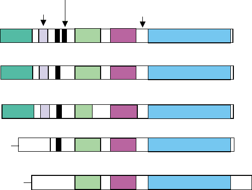
FIGURE 1 Domain structures of Tec family kinases. Src kinase is shown at the bottom for comparison.
170 Tec/Btk FAMILY TYROSINE KINASES
Btk is essential for B-cell development and function,
as demonstrated by XLA. XLA patients lack mature B
cells in the periphery and do not have immunoglobulins
as a result. However, inactivating Btk in the mouse
causes only a mild defect in B cell development and
function. This mimics the phenotype of xid mice, caused
by a spontaneous mutation (R28C) in the Btk
PH domain. In these mouse models, mature B-cell
number is reduced and these B cells have a defect in
response to B-cell receptor (BCR) stimulation. A minor
B-cell population, B-1 cells, is absent in these mice.
Moreover, the serum immunoglobulins IgG3 and IgM
levels are greatly reduced in these mice and they are
not able to respond to T-independent type-II antigens.
Interestingly, even though Tec-/- mice have no
major phenotypic alterations of the immune system,
Btk-/-Tec-/- mice showed a severe defect in B-cell
development, similar to human XLA. This suggests
that Tec may compensate partially for the lack of Btk in
murine B-cell development.
In T cells, three Tec family members coexist: Itk,
Txk, and Tec. Inactivating Itk results in slightly
decreased number of mature thymocytes, especially
CD4
þ
cells, while inactivating Txk does not affect
either T-cell development or activation. Double
mutants, on the other hand, showed improved mature
T-cell number, while still maintaining a decreased
CD4/CD8 ratio, compared to Itk-/- mice. However,
T cells in the double knockout mice have a severe
defect in T-cell receptor (TCR) induced proliferation
and cytokine production.
When the Bmx gene was replaced by the LacZ
reporter gene, the homozygous mice lacking Bmx
activity showed no obvious phenotype. But the
expression of the reporter gene is strong in endothelial
cells of large arteries and in the endocardium from
embryogenesis to adult mice. Moreover, Bmx is acti-
vated through endothelial receptor tyrosine kinases
Tie-2, vascular endothelial growth factor receptor 1
(VEGFR-1) and tumor necrosis factor (TNF) receptor.
These data suggest a redundant role of Bmx in
endothelial signal transduction.
Tec Family Kinases in
Signal Transduction
Tec family kinases not only play a critical part in T-cell
receptor or B-cell receptor signaling, but are also
involved in cytokine receptor signaling as well as mast
cell Fcepisilon receptor I (Fc1RI) signaling. Here we will
use Btk as an example to discuss the signaling functions
of these kinases.
Each domain of Btk is essential for the function
of the kinase, as suggested by mutation analysis in
XLA patients. To date, over 400 Btk mutations
(missense mutation, frameshift, truncation) from 556
XLA families have been reported and these mutations
cover all the domains. However, missense mutations
are found in each domain except the SH3 domain,
suggesting that the SH3 domain may tolerate such
alterations. Each domain has been shown to bind
regulatory and/or effector proteins. In addition to
PI(3,4,5)P
3
, Btk PH domain can also bind protein
kinase C
b
(PKC
b
), Fas, F-actin and a transcription
factor TFII-I. TH domain binds G protein subunits
and Src family kinases as Lyn or Hck. SH3 domain
interacts with proteins as Cbl, WASP, and Vav, while
SH2 domain binds B-cell linker protein (BLNK)
through interaction with phosphotyrosine. The cataly-
tic domain has also been shown to bind G protein
bg
-subunit, and the kinase activity is activated by this
interaction. Although some of the interactions have
been confirmed in cellular context, the physiological
importance of many still needs to be carefully
evaluated.
B-cell development and activation is a tightly
regulated process and Btk plays an important role. It is
involved in a number of signaling pathways that are
activated when B cells are stimulated through the BCR,
accessory molecules such as CD19 and CD38, or
cytokine receptors such as IL-5R and IL-10R. In normal
B cells, cross-linking of the BCR activates PI 3-kinase
leading to the translocation of cytoplasmic Btk to the
lipid raft on the plasma membrane. Src family tyrosine
kinase Lyn, which is also activated upon BCR stimu-
lation, phosphorylates Y551 in the Btk kinase domain
and activates Btk kinase activity. These serial activations
lead to the assembly of a B-cell signalosome and proteins
like Btk, Lyn, PI 3-kinase, BLNK, PLC
g
2, BCAP (B-cell
adapter for PI 3-kinase), and PKC are among the players
in the B-cell signalosome. Once activated, Btk trans-
duces signals to a number of effectors, including
phospholipase C gamma (PLC
g
), calcium response,
transcription factors such as TFII-I, genes involved in
apoptosis (bcl-2 and bcl-xl), and cell cycle control
(cyclins). Interestingly, the Btk-deficient and xid pheno-
type is phenocopied by the deficiency of PI 3-kinase
regulatory subunit p85 or the catalytic subunit p110
d
,
BCAP, PKC
b
, BLNK, or PLC
g
2. These data indicate
that these proteins are involved in the same signaling
pathway and likely in the same B-cell signalosome
(Figure 2).
There are several mechanisms to down-regulate Btk.
Autophosphorylation at Y223 in the SH3 domain
has been implicated to negatively regulate Btk function,
and phosphorylation of S180 in the TH domain by PKC
b
also serves to down-regulate the membrane association
of Btk. Additionally, a recent study identified an inhibitor
for Btk (IBtk). IBtk directly binds the PH domain of
Tec/Btk FAMILY TYROSINE KINASES 171
