Lennarz W.J., Lane M.D. (eds.) Encyclopedia of Biological Chemistry. Four-Volume Set . V. 4
Подождите немного. Документ загружается.


Btk and down-regulates the kinase activity, calcium
response and NF-
k
B-induced transcription.
In addition, Btk is involved in myeloid lineage cell
signaling, including macrophages and mast cells.
Although mast-cell development is normal in xid
mice, Btk is phosphorylated and translocated to the
plasma membrane after cross-linking of Fc
1
RI in
mast cells.
Future Directions
Extensive studies have been carried out on Tec family
kinases since the 1990s, yet the detailed function of these
kinases is far from resolved. In the coming years, further
efforts will be put on the participation of these kinases in
signalosome formation, the components of signalosome,
and the regulation of the signal transduction. In
addition, the complete structure of the proteins in this
family will help the understanding of intramolecular
domain–domain interaction as well as protein–protein
interaction, and shed light on the regulation of kinase
activity and function.
SEE ALSO THE FOLLOWING ARTICLES
c-fes Proto-Oncogene † Epidermal Growth Factor
Receptor Family † Phosphatidylinositol Bisphosphate
and Trisphosphate † Phosphatidylinositol-3-Phosphate
GLOSSARY
Bruton’s tyrosine kinase (Btk) A member of the Tec/Btk tyrosine
kinase family. Mutations of Btk cause X-linked agammaglobuline-
mia (XLA) in human and x-linked immunodeficiency (xid) in
mouse. This protein was named after Dr. Ogden Bruton, who
described XLA in 1950s.
signalosome Multiprotein signaling complex formed in the cell that is
regulated both spatially and temporally.
Tec/Btk tyrosine kinase family A family of the non-
receptor tyrosine kinases including Btk, Tec, Itk, Txk, and Bmx,
which play important signaling roles primarily in hematopoietic
cells.
X-linked agammaglobulinemia (XLA) A human genetic disease
caused by the mutations in a proteins tyrosine kinase Btk. Patients
lack mature B cells in the periphery and suffer from recurrent
infections.
X-linked immunodeficiency (xid) An immunodeficiency found in
CBA/N mice that is caused by a spontaneous point mutation in Btk
(R28C). Xid mice have reduced mature B cells and defective B-cell
response.
FURTHER READING
Fruman, D. A., Satterthwaite, A. B., and Witte, O. N. (2000). Xid-like
phenotypes: a B cell signalosome takes shape. Immunity 13,1–3.
Lewis, C. M., Broussard, C., Czar, M. J., and Schwartzberg, P. L.
(2001). Tec kinases: Modulators of lymphocyte signaling and
development. Curr. Opin. Immunol. 13, 317–325.
Miller, A. T., and Berg, L. J. (2002). New insights into the regulation
and functions of Tec family tyrosine kinases in the immune system.
Curr. Opin. Immunol. 14, 331–340.
Pillai, S., and Moran, S. T. (2002). Tec kinase pathways in lymphocyte
development and transformation. Biochim. Biophys. Acta 1602,
162–167.
BLNK
BCR
Ig /
Syk
Lyn
*
*
*
*
Btk
*
Y551
PLC 2
*
*
*
*
PI(4,5)P
2
I(1,4,5)P
3
DAG
PKC
PI(3,4,5)P
3
*BCAP
p85
p110
Calcium flux
Proliferation Apoptosis Transcription
regulation
Cytoskeletal
rearrangement
*: phosphotyrosine
PI(4,5)P
2
PI(3,4,5)P
3
α
β
γ
β
δ
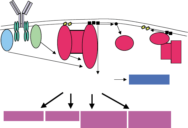
FIGURE 2 Signal transduction pathway after B cell receptor stimulation. Btk is activated through translocation to the plasma membrane through
binding of PI(3,4,5)P
3
and phosphorylation at Y551 by Lyn. The B cell signalosome is assembled and downstream effectors will be sequentially
activated. Proteins that have a xid-like phenotype when knocked out are shown in red. Adapted from Fruman, D. A., Satterthwaite, A. B., and
Witte, O. N. (2000), Xid-like phenotypes: a B cell signalosome takes shape. Immunity 13,1–3.
172 Tec/Btk FAMILY TYROSINE KINASES
Qiu, Y., and Kung, H. J. (2000). Signaling network of the Btk family
kinases. Oncogene 19, 5651–5661.
Satterthwaite, A. B., and Witte, O. N. (2000). The role of Bruton’s
tyrosine kinase in B-cell development and function: A genetic
perspective. Immunol. Rev. 175, 120–127.
Smith, C. I., Islam, T. C., Mattsson, P. T., Mohamed, A. J., Nore, B. F.,
and Vihinen, M. (2001). The Tec family of cytoplasmic tyrosine
kinases: Mammalian Btk, Bmx, Itk, Tec, Txk, and homologs in
other species. Bioessays 23, 436–446.
Vihinen, M., Kwan, S. P., Lester, T., Ochs, H. D., Resnick, I., Valiaho, J.,
Conley, M. E., and Smith, C. I. (1999). Mutations of the human
BTK gene coding for bruton tyrosine kinase in X-linked agamma-
globulinemia. Hum. Mutat. 13, 280–285.
BIOGRAPHY
Owen N. Witte is an Investigator of Howard Hughes Medical Institute
and a Professor at the University of California Los Angeles. His
primary research interest is signal transduction in hematopoietic
cells and related diseases. Dr. Witte is a pioneer in the study of tyrosine
kinases. His group and others were the first to report Btk mutations
cause Xid and XLA (Rawlings DJ et al., Science 1993; Tsukada S et al.,
Cell, 1993; Thomas JD et al., Science 1993; Vetrie D et al.,
Nature 1993).
Shuling Guo received her Ph.D. from Duke University and is currently
a postdoctoral fellow in Dr. Witte’s laboratory.
Tec/Btk FAMILY TYROSINE KINASES 173

Telomeres: Maintenance
and Replication
Alessandro Bianchi and David Shore
University of Geneva, Geneva, Switzerland
All linear eukaryotic chromosomes terminate in a specialized
nucleoprotein structure, the telomere. Telomeres perform at
least two essential functions: they provide a protective “cap”
on chromosome ends that prevents their degradation or
deleterious fusion, and they provide a special mechanism for
replicating the DNA at chromosome ends. In most organisms,
telomeres are composed of a tandem array of simple DNA
repeats to which a large set of protein factors is bound. The
telomeric DNA repeats are generated by a specialized reverse
transcriptase, called telomerase that uses an endogenous RNA
template. Defects in the maintenance of telomeric DNA, for
example through inactivation of the telomerase enzyme, lead
to the progressive loss of telomeric repeats and their bound
factors, which eventually causes a catastrophic “uncapping” of
telomeres that results in fusion of chromosome ends. The
telomere complex is thus essential to ensure genome stability.
The Telomeric Complex,
a Specialized Nucleoprotein
Structure at Chromosome Ends
Pioneering studies in the fruit fly and in maize, carried
out respectively by H. Muller and B. McClintock in the
early 20th century, first identified the telomere as a
special genetic entity that protects chromosome ends
from degradation and fusion, a property absent in DNA
ends that result from random chromosomal breakage. A
wealth of subsequent genetic and biochemical studies
have led to the understanding that telomeres are
specialized nucleoprotein complexes composed of a
large number of protein factors assembled onto telo-
meric DNA.
TELOMERIC DNA
In most eukaryotes, the terminal DNA sequences at each
chromosome are composed of arrays of variable length
of simple tandem repeats, as first recognized by Black-
burn and Gall in the ciliate Tetrahymena termophila.
These telomeric repeats are typically 5– 8 nucleotides
long, but can be up to 25 bp (Figure 1) and usually have
a higher G content in the DNA strand that runs with a 5
0
to 3
0
polarity towards the end of the chromosome. In all
species, the majority of the telomeric repeats appear to
be in double-stranded form and their total length varies
from a few nucleotides in some species of ciliates (28 bp
of duplex telomeric repeats in Euplotes, for example) to
tens of thousands of bp in some strains of laboratory
mice. In both budding and fission yeasts, telomeres are
, 300 bp in length, whereas in human cells telomeres are
generally 6–12 kbp long. With the exception of several
ciliate species in which the length of telomeric DNA is
fixed, in most species the length of each telomere is
variable from cell to cell, individual to individual, and
during the life of the organism. In many species, it has
been shown that telomeres terminate in a single-
stranded overhang of the G-rich strand. This overhang
can range from only a few nucleotides long (in some
ciliates) to about 100–200 bases in mammals, and is
believed to represent a general feature of telomeres. G-
rich telomeric overhangs in vitro can adopt a variety of
intra- and intermolecular paired structures involving
most commonly four strands interacting through
Hoogsteen-type G–G base-pairing (G-quartets), but
the in vivo occurrence and significance of such structures
remains unclear. In mammals, some ciliates, and
Trypanosomes, telomeric DNA appears to fold back in
a structure, the t-loop, where the single-stranded over-
hang is tucked into the duplex portion of the telomeric
tract, by base pairing with the C-rich strand and thus
displacing a short portion of the G-strand.
In most organisms, the short telomeric repeats
described above are preceded by less conserved DNA
elements, called subtelomeric repeats or telomere-
associated sequences (TAS). These elements are of
variable lengths but are generally at least a few hundred
base pairs long and have been described in most studied
organisms. In a particular species several classes of these
repeats can be present, and elements of each class are
associated with a particular chromosome with great
variation in number and organization. The assembly of
these subtelomeric repeats is highly dynamic and subject
Encyclopedia of Biological Chemistry, Volume 4. q 2004, Elsevier Inc. All Rights Reserved. 174

to active recombination, resulting in a large variation of
subtelomeric regions between strains of yeast or between
human individuals. TAS do not appear to carry out an
essential telomeric function, as both human and yeast
chromosomes that are devoid of them are replicated and
segregated properly both through mitosis and meiosis.
TELOMERASE
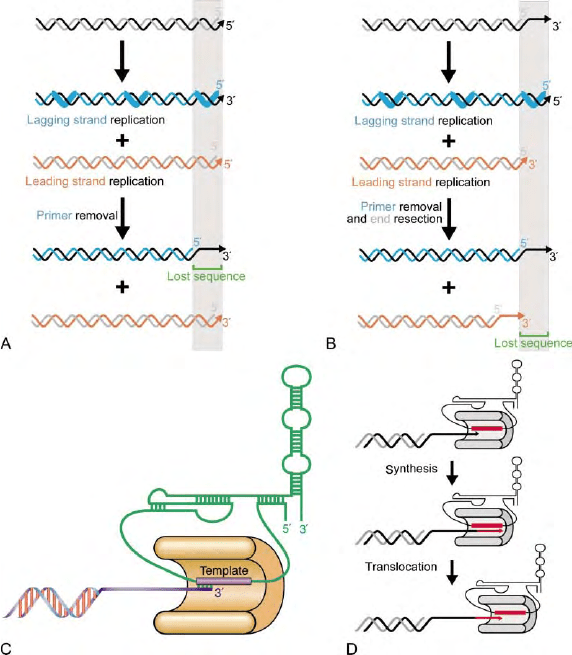
DNA polymerases replicate DNA uniquely in a 5
0
to 3
0
direction. As a consequence, as the DNA replication
“bubble” moves along the DNA, the two strands are
replicated differently: one strand (the “leading” strand) is
replicated in the same direction as the movement of the
polymerase, while the other strand (the “lagging” strand)
is replicated backwards in small installments, each one
initiating from an RNA primer laid down by the primase
enzyme (Figure 2A). Due to the requirement for an
initiating RNA primer, which is later removed, replica-
tion of linear DNA molecules by the conventional DNA
replication machinery would result in a terminal gap in
the lagging strand end at the telomere (Figure 2A). The
presence of overhangs at telomeres could in principle
mask this problem at the lagging strand, but a terminal
gap would then occur at the strand replicated by leading
strand synthesis after processing of the end to generate
the single-stranded overhang (Figure 2B). Thus, in the
absence of a specialized mechanism to replicate the ends,
loss of genetic material is expected to occur at each cell
division. The most general solution to this problem in
eukaryotes is represented by the specialized replication of
telomeric repeats by the telomerase enzyme.
Telomerase was first identified by Greider and Black-
burn in the ciliate Tetrahymena termophila based on its
ability to add telomeric repeats in a terminal transferase-
like fashion to a telomeric DNA primer in an in vitro
reaction. The enzyme is in fact a ribonucleoprotein,
composed of a protein and an RNA moiety, both of
which are required for catalytic function (Figure 2C).
Isolation of the protein component from the ciliate
Euplotes by Lingner and Cech in 1997 has revealed that
telomerase shares extensive homology with reverse
transcriptases. Thus telomerase is a specialized reverse
transcriptase that utilizes an endogenous RNA template
for the synthesis of telomeric repeats. The enzyme
appears to be capable of synthesizing several repeats on
Telomeric repeats
Vertebrates
Arthropodes
Homo, Mus, Rattus, Gallus TTAGGG
Insects
Bombix mori
TTAGG
TTAGGC
TTAGGC
TTTAGGG
TTTTAGGG
ACGGATTTGATTAGGTATGTGGTGT
T(G)
1–3
Nematodes
Ascaris
Caenorhabditis elegans
Arabidopsis thaliana
Chlamydomonas
Plants
Green algae
Fungi
Ascomycota
Hemiascomycetes
Saccharomyces cerevisiae
Kluyveromyces lactis
Archeascomycetes
Schizosaccharomyces pombe
Euascomycetes
Neurospora
Protists
Alveolata
Plasmodium
Ciliates
Tetrahymena
Oxytricha
Euplotes
Diplomonadida
Giardia
Euglenozoa
Trypanosoma
TTACAGG(G)0–4
TTAGGG
TT[T/C]AGGG
TTGGGG
TTTTGGGG
TTTTGGGG
TAGGG
TTAGGG
FIGURE 1 Representative list of telomeric repeats in several eukaryotic organisms, including some of the most extensively studied ones with
regard to telomere biology.
TELOMERES: MAINTENANCE AND REPLICATION 175

a particular substrate through repeated steps of
elongation and translocation (Figure 2D) and, like
reverse transcriptases, appears to be a dimer.
TELOMERE PROTEINS
Telomeric repeats serve as binding sites for telomeric
DNA-binding proteins, which act as a scaffold for the
recruitment of additional protein factors to telomeres,
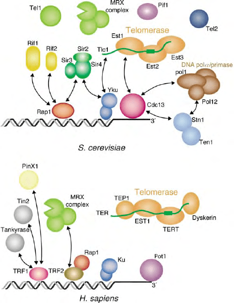
including telomerase (Figure 3). The analysis of telo-
meric proteins in a wide range of organisms has revealed
important similarities, though significant differences
have also emerged.
The major double-stranded DNA-binding activity in
budding yeast is Rap1, a protein also involved in
transcriptional regulation at many other nontelomeric
chromosomal sites. S. cerevisiae Rap1 binds directly
to yeast telomeric repeats through two Myb-like
DNA-binding domains. Rap1 is instrumental in recruit-
ing to yeast telomeres a set of proteins involved in the
regulation of telomere length (the Rap1 interacting
factors 1 and 2, Rif1 and Rif2) and in the assembly of a
complex (which includes the proteins Sir2, 3, and 4) that
results in the transcriptional repression of genes posi-
tioned near telomeres (telomere position effect, or TPE).
In contrast, mammalian and fission yeast Rap1 appears
to be recruited to telomeres indirectly, through the
binding to a different class of double-stranded DNA-
binding factors, TRF1 and TRF2 in vertebrates and Taz1
in fission yeast. These proteins bind as dimers and
contain a single Myb repeat that is essential for DNA
binding. They share among themselves a large domain
(TRFH, for TRF homology) that is responsible for
homodimerization. Rap1 is recruited to mammalian and
fission yeast telomeres through binding with TRF2 and
Taz1, respectively. Human TRP2 also recruits the MRX
FIGURE 2 (A) Schematic view of the end replication problem of a linear DNA molecule. The RNA primers laid down by the primase enzyme for
the synthesis of the so-called ‘lagging’ strand are indicated by thicker blue lines. After removal of the primers, replication of a double-stranded DNA
molecule would result in a single-stranded terminal gap in the DNA strand replicated by lagging strand synthesis. (B) Schematic view of the end
replication problem at a telomere terminating in a single-stranded overhang. Exonucleolytic resection of the ends is hypothesized to generate the
overhangs after replication. In this scenario, a terminal gap would be created in the strand replicated by leading strand synthesis. (C) Representation
of the structure of the core telomerase enzyme. The protein, based on its homology to known viral reverse transcriptases, is expected to be folded in
a structure with a central cleft (palm) bearing the active site and flanked by two domains (thumb and fingers). The template region of the RNA is
placed in this cleft, and the secondary structure of the RNA represented here is derived from ciliated Protozoa species and appears to be largely
conserved in higher Eukaryotes. (D) Representation of the processive mechanism of action of telomerase, where a round of synthesis of telomeric
repeats is followed by translocation of the enzyme on the DNA and a subsequent new round of synthesis.
176 TELOMERES: MAINTENANCE AND REPLICATION

complex to telomeres. TRF1 in mammals also is
responsible for the recruitment of protein factors,
including the telomere length regulators Tin2 and
tankyrase, the telomerase inhibitor PinX1, and the
single-stranded DNA-binding protein Pot1.
In addition to double-stranded DNA-binding activi-
ties, single-stranded DNA-binding factors also appear to
be an essential and general feature of telomeres. Over-
hang-binding proteins were initially characterized in
ciliates, where they form tenacious salt-resistant com-
plexes with the single-stranded DNA termini. In bud-
ding yeast, the Cdc13 protein is bound to the single
stranded overhangs created in S-phase in this organism
and it carries out essential functions in telomere
replication, through interaction with the telomerase
and DNA replication complex, and in telomere protec-
tion, through interactions with the Stn1 and Ten1
proteins. In fission yeast and mammals, Pot1, an
orthologue of the ciliate end factors, is involved in
telomere protection in fission yeast and in telomere
length regulation in mammals. Although Cdc13 does
not share sequence homology with the ciliate, fission
yeast and mammalian end factors, all these proteins bind
DNA through an oligonucleotide/oligosaccharide-
binding (OB) domain.
An additional factor, present at telomeres from yeast
to humans, is the DNA repair protein Ku, which might
bind to telomeres through its nonsequence-specific DNA
end-binding activity. However protein –protein inter-
actions between Ku and Sir4, and between Ku and TRF2
have been described in yeast and humans, respectively.
Finally, the highly conserved telomere length regulator
Tel2, has been shown (for the yeast protein) to be able to
bind both single- and double-stranded telomeric repeats
in vitro.
Telomere Replication
Telomeres are normally maintained through the action
of the telomerase enzyme. Although telomerase activity
in an in vitro assay requires only the reverse trans-
criptase-like protein motif and the RNA component,
it is now clear that the action of the enzyme at
telomeres is subject to complex regulation in cis at each
individual telomere.
POSITIVE REGULATION OF
THE
TELOMERASE ENZYME
In budding yeast, a set of genes is required for telomerase
activity in vivo in addition to the catalytic core
components Est2 and Tlc1. These include Est1, which
appears to bridge an interaction between telomerase and
Cdc13 required to either recruit or activate the enzyme
at the telomere, and telomerase-associated Est3, whose
mode of action is unclear. The combined action of the
DNA damage checkpoint kinases Tel1 and Mec1 is also
necessary for telomerase activity in budding yeast,
possibly by promoting a structural transition in the
telomere complex from a “closed” to an “open” state
that is accessible to telomerase. A similar requirement
exists in fission yeast for the Tel1 and Mec1 homologues
Tel1 and Rad3. In mammalian cells dyskerin binds the
telomerase RNA and appears to affect in vivo telomerase
activity by influencing accumulation of the RNA.
Significantly, budding yeast telomerase action is strictly
limited to the S-phase of the cell cycle and requires active
DNA replication.
ANEGATIVE REGULATORY LOOP
STABILIZES TELOMERE LENGTH
In cells expressing telomerase, telomere length is main-
tained around a constant average value that is species
specific, as mentioned above. Evidence from a variety of
organisms, including the budding and fission yeasts, and
human cells, has led to the proposal that telomere length
is controlled by a protein-counting mechanism. In this
FIGURE 3 Representation of some of the characterized components
of the budding yeast and human telomeric complexes. All proteins
depicted have either been shown to affect telomere behavior and/or to
interact with other telomere components.
TELOMERES: MAINTENANCE AND REPLICATION 177

scenario, the number of double-stranded DNA-binding
factors that are associated with the telomere at any
given time is somehow sensed by the telomere length
regulation machinery. An increase in the number of
these negative regulators results in the inhibition of
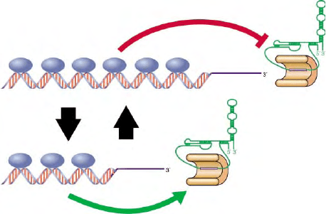
telomerase activity in cis at the telomere (Figure 4). The
inhibitory function of the DNA-binding factors (Rap1
in budding yeast, Taz1 in fission yeast, and TRF1 and
2 in mammals) is apparently mediated by a set of
interacting factors (Rif1 and 2 in budding yeast, Pot1 in
humans). The precise mechanism of the proposed cis
inhibition of telomerase is unknown, but might, at least
in some organisms, involve formation of the t-loop
structure described previously.
TELOMERASE-INDEPENDENT
MAINTENANCE OF TELOMERES
In the absence of telomerase, alternative pathways
for the maintenance of telomeric repeats exists that
are based on homologous recombination. In budding
yeast, telomere-associated sequences can contribute
to this recombinational process. In mammals, several
tumor cell lines have been shown to maintain their
telomeres without detectable telomerase activity
through a mechanism named ALT, for alternative
lengthening of telomeres. Finally, the fruit fly
Drosophila melanogaster dispenses altogether with
short telomeric repeats and forms telomeres through
the repeated insertion of telomere-specific trans-
posable elements (HeT-A and TART) onto the ends
of its chromosomes.
Telomerase Repression in Somatic
Cells: Implications for Replicative
Senescence and Maintenance of
Genome Integrity
Mice engineered to lack the gene encoding telomerase
RNA (mTRT) exhibit a gradual (, sixth generation) loss
of fertility, defects in cell proliferation, and an increase in
the frequency of end-to-end chromosome fusions. These
phenotypes result from telomere repeat erosion and
consequent loss of telomere function. Since active
telomerase enzyme is primarily detected in mouse
germline cells, these data indicate that telomerase-
dependent telomere length “resetting” in the germline
is essential for long-term survival in the mouse, and by
inference in all mammals. In humans, the gene encoding
the catalytic subunit of telomerase is also repressed in
most somatic cells, causing telomeres to undergo
progressive shortening with each cell division. This
process leads to cellular senescence in culture and can be
reversed, at least in some primary cell lines, by the
ectopic expression of an active telomerase gene in the
senescing cells. These observations in cultured primary
cells have led some researchers to propose that
telomerase repression in humans, and consequent
telomere erosion, acts as a kind of “mitotic clock” that
determines aging of the whole organism.
An attractive alternative view is that the telomere
“mitotic clock” in humans might instead represent a
critical barrier against malignant transformation.
Recent experiments suggest that either normal telo-
mere erosion, or uncapping caused by mutational
alteration of key telomere proteins, will induce a DNA
damage checkpoint and apoptosis (programmed cell
death). This mechanism may place a limit on
continued cell proliferation, which is a prerequisite
for tumor formation. However, telomere uncapping
also leads to telomere-telomere fusions and rampant
genomic instability, which is thought to be a driving
force in oncogenesis. Telomere dysfunction might
therefore represent an important step in the early
stages of tumorigenesis, particularly in cells with
compromised checkpoint (DNA damage surveillance)
function. The proper regulation of telomerase and
telomere capping thus appear to be remarkably critical
cellular functions.
SEE ALSO THE FOLLOWING ARTICLES
Chromosome Organization and Structure, Overview †
DNA Replication: Eukaryotic Origins and the Origin
Recognition Complex † Homologous Recombination in
Meiosis † Meiosis † Mitosis
FIGURE 4 Depiction of the protein-counting mechanism for
telomere length regulation. In this model, longer telomeres have higher
numbers of inhibitory factors bound, resulting in repression of
telomerase activity in cis. Diminished action by telomerase results in
telomere shortening, loss of the bound inhibitor, and telomerase
activation. In this manner telomeres are maintained in a state of
dynamic equilibrium. The organism in which our understanding of
the telomeric complex is the most complete, both with regard to the
identification of protein components and to their function, is the
budding yeast Saccharomyces cerevisiae (Figure 1).
178 TELOMERES: MAINTENANCE AND REPLICATION
GLOSSARY
telomerase A nuclear-encoded ribonucleoprotein complex homolo-
gous to viral reverse transcriptases that adds single-stranded
telomeric repeats to the 3
0
ends of duplex DNA using its own
RNA template; responsible for telomere maintenance in most
eukaryotic organisms.
telomere The DNA–protein complex at the extreme ends of linear
eukaryotic chromosomes that orchestrates telomere replication and
capping.
telomere associated sequence (TAS) The highly variable, often
repeated DNA sequences found immediately internal to the
terminal simple telomere repeat sequences.
telomere capping Refers to the ability of the telomere DNA–protein
complex to prevent chromosome end degradation or fusion (DNA
joining) reactions.
telomeric repeats Simple, short (usually 5–8, but as long as 25
nucleotides) DNA repeat sequences (T
2
AG
3
in vertebrates and minor
variants in many other eukaryotes) that constitute the DNA
component of telomeres. Synthesized by the telomerase enzyme,
with the TG-rich sequence extending towards the 3
0
DNA end.
FURTHER READING
Blackburn, E. H., and Greider, C. W. (eds.) (1995). Telomeres. Vol 29,
Cold Spring Harbor Monographs, Cold Spring Harbor Press,
Plainview, NY.
Chakhparonian, M., and Wellinger, R. J. (2003). Telomere mainten-
ance and DNA replication: How closely are these two connected?
Trends Genet. 19(8), 439– 446.
de Lange, T. (2002). Protection of mammalian telomeres. Oncogene
21(4), 532–540.
Evans, S. K., and Lundblad, V. (2000). Positive and negative regulation
of telomerase access to the telomere. J. Cell Sci. 113(Pt),
3357–3364.
Kass-Eisler, A., and Greider, C. W. (2000). Recombination in telomere-
length maintenance. Trends Biochem. Sci. 25(4), 200–204.
Lingner, J., and Cech, T. R. (1998). Telomerase and chromosome end
maintenance. Curr. Opin. Genet. Dev. 8(2), 226– 232.
Lundblad, V. (2002). Telomere maintenance without telomerase.
Oncogene 21(4), 522–531.
Maser, R. S., and DePinho, R. A. (2002). Connecting chromosomes,
crisis, and cancer. Science 297(5581), 565–569.
McEachern, M. J., Krauskopf, A., and Blackburn, E. H. (2000).
Telomeres and their control. Annu. Rev. Genet. 34, 331–358.
Wright, W. E., and Shay, J. W. (2001). Cellular senescence as a tumor-
protection mechanism: the essential role of counting. Curr. Opin.
Genet. Dev. 11(1), 98–103.
Zakian, V. A. (1996). Structure, function, and replication of Sacchar-
omyces cerevisiae telomeres. Annu. Rev. Genet. 30, 141–172.
BIOGRAPHY
David Shore is a Professor in the Department of Molecular Biology at
the University of Geneva (Switzerland) and member of the NCCR
program “Frontiers in Genetics.” His principal research interests are
in gene regulation, chromatin structure, and telomere biology. He
holds a Ph.D. from the Department of Biochemistry at Stanford
University Medical School and did postdoctoral work at the MRC
Laboratory of Molecular Biology in Cambridge (UK).
Alessandro Bianchi received his Ph.D. at the Rockefeller University
(New York) and is currently a postdoctoral fellow at the University
of Geneva.
TELOMERES: MAINTENANCE AND REPLICATION 179

Thyroid-Stimulating
Hormone/Luteinizing
Hormone/Follicle-Stimulating
Hormone Receptors
Deborah L. Segaloff, Dario Mizrachi and Mario Ascoli
The University of Iowa, Iowa City, Iowa, USA
The thyroid-stimulating hormone receptor (TSHR), luteiniz-
ing hormone receptor (LHR), and follicle-stimulating hormone
receptor (FSHR) are collectively referred to as the glycoprotein
hormone receptors because they bind the structurally similar
glycoprotein hormones. The glycoprotein hormones consist of
the pituitary hormones thyroid-stimulating hormone (thyro-
tropin, TSH), luteinizing hormone (lutropin, LH), and follicle-
stimulating hormone (follitropin, FSH) as well as the placental
hormone chorionic gonadotropin (choriogonadotropin, CG).
They are each composed of two dissimilar subunits (
a
and
b
)
that are noncovalently associated. Within a given species the
a
-subunit is identical, and the
b
-subunits are distinct but
homologous. Due to the nearly identical nature of LH and CG,
the LHR binds either LH or CG. However, the FSHR binds
only FSH and the TSHR binds only TSH. Because the LHR
and FSHR are localized primarily to the gonads, these two
receptors are also referred to as the gonadotropin receptors.
The glycoprotein hormone receptors are members of the
superfamily of G protein-coupled receptors (GPCRs) and,
despite their different physiological roles, share a similar
structural organization and mechanism of action. In addition,
the glycoprotein hormone receptors are also members of a
subfamily of GPCRs known as the leucine-rich repeat-contain-
ing GPCRs (LGR). This subfamily of GPCRs is characterized
by the presence of a large extracellular domain composed of
several leucine-rich repeats.
Expression and Physiological
Roles of the Glycoprotein
Hormone Receptors
THE THYROID-STIMULATING
HORMONE RECEPTOR
TSHR is expressed primarily in the follicular cells of the
thyroid. In more recent years, it has also been detected in
lymphocytes, thymus, pituitary, testis, kidney, brain,
adipose tissue and fibroblasts, heart, and bone.
In response to thyroid-stimulating hormone (TSH),
the TSH receptor (TSHR) stimulates the synthesis and
secretion of thyroid hormone by the follicular cells of the
thyroid. Autoantibodies to the TSHR that are stimu-
latory are present in individuals with Grave’s disease and
stimulate the TSHR causing excessive and unregulated
secretion of thyroid hormone. Conversely, inhibitory
autoantibodies to the TSHR are found in individuals
with Hashimoto’s thyroiditis. These antibodies bind to
the TSHR and inhibit the binding of TSH, thus blocking
the synthesis and secretion of thyroid hormones.
The TSHR present in the adipose and connective
tissue of the orbit of the eye is thought to play a role in
the development of exopthalmos found in individuals
with Grave’s disease. The physiological roles of the
TSHR in other nonthyroid tissues is not yet known.
THE LUTEINIZING HORMONE RECEPTOR
The luteinizing hormone receptor (LHR) is expressed
primarily in the ovaries and the testes. Within the ovary,
the LHR is present on theca and interstitial cells and on
mature granulosa cells. After ovulation, the granulosa
and theca cells of the ruptured follicle differentiate into
the luteal cells, and express higher levels of LHR. In
males, the LHR is expressed on the Leydig cells. In both
males and females, nongonadal expression of the LHR
has also been reported in the reproductive tract and
many other tissues.
In the nonpregnant postpubertal female, ovarian
theca cells respond to luteinizing hormone (LH)
with increased synthesis of androgens, which are used
as substrates for estrogen production by follicle-
stimulating hormone (FSH)-stimulated granulosa cells.
Mature granulosa cells respond to LH with increased
Encyclopedia of Biological Chemistry, Volume 4. q 2004, Elsevier Inc. All Rights Reserved. 180
progesterone production. In response to the ovulatory
surge of LH, the ovarian LHR receptors mediate
ovulation. If pregnancy ensues, then the LHR of the
corpus luteum responds to placental chorionic gonado-
tropin (CG) with increased progesterone synthesis. As
such, the LHR is essential for the maintenance of
pregnancy, particularly during the first trimester.
In males, the testicular LHR plays an important
physiological role during fetal development. Maternal
hCG stimulates the fetal Leydig cells to synthesize
testosterone, which is required for the differentiation of
the external male genitalia and for the descent of the
testes into the scrotum. Testosterone levels in boys
decrease after birth (due to the absence of maternal LH)
and the LHR remains unstimulated until the time of
puberty. After puberty the testicular LHR responds to
pituitary LH with increased testosterone synthesis.
The physiological roles of nongonadal LHR are not
known. It should be pointed out, though, that the only
functional consequences of loss-of-function or gain-
of-function mutations of the LHR described in males
or females have been restricted to abnormalities in
reproductive physiology.
THE FOLLICLE-STIMULATING
HORMONE RECEPTOR
The follicle-stimulating hormone receptor (FSHR) is
expressed in ovarian granulosa cells and in the Sertoli
cells within the seminiferous tubules of the testes. In
post-pubertal females, the FSHR mediates follicular
growth and controls estrogen synthesis. In post-pubertal
males, pituitary FSH facilitates spermatogenesis by
stimulating the Sertoli cells that are adjacent to the
developing sperm to synthesize and secrete components
needed for spermatogenesis. Although it is accepted that
optimal spermatogenesis requires the actions of FSH, it
is controversial as to whether FSH is essential for this
process.
Structural Organization of the
Glycoprotein Hormone Receptors
SERPENTINE REGIONS
The glycoprotein hormone receptors are members of the
rhodopsin-like family of GPCRs. As such, they all
contain the seven membrane-spanning regions proto-
typical of the superfamily of GPCRs. Residues that are
conserved within the transmembrane (TM) domains of
the rhodopsin-like GPCRs are also generally conserved
in the glycoprotein hormone receptors. The recent
solving of a high resolution crystal structure of
rhodopsin has provided a template for creating models
of the TM regions of the glycoprotein hormone
receptors and permitting investigators to envision the
interhelical interactions maintaining the receptors in
their inactive states. The crystal structure of rhodopsin
also revealed the presence of an eighth
a
-helix that
extends from TM7 and lies parallel to the inner face of
the plasma membrane. In rhodopsin, helix 8 extends
until a cysteine residue that is palimitoylated and serves
to anchor the helix to the plasma membrane. This
cysteine is conserved as a single residue or as a pair in the
glycoprotein hormone receptors. An alignment of the
human TSHR, LHR, and FSHR is shown in Figure 1.
The amino acid identity is greatest between the
glycoprotein hormone receptors in the transmembrane
regions and helix VIII.
EXTRACELLULAR DOMAINS
A unique feature to the glycoprotein hormone receptors
is their relatively large (i.e., 300–400 amino acids)
extracellular domains. This is the receptor domain that
is responsible for the selective recognition and high-
affinity binding of each of the glycoprotein hormones.
The extracellular domains are N-glycosylated and a fully
conserved tyrosine residue has been shown to be sulfated
in the TSHR. The TSHR has the largest extracellular
domain which is clipped once the receptor is inserted at
the plasma membrane. This proteolytic cleavage results
in the formation of an
a
-subunit containing a portion of
the N-terminal extracellular domain and a
b
-subunit
containing the remaining of the N-terminal extracellular
domain and the transmembrane and C-terminal
domains. Although the
a
- and
b
-subunits are initially
bound by disulfide bonds, these are reduced and the
a
-
subunit is released from the membrane bound
b
-subunit.
The extracellular domains of the glycoprotein hor-
mone receptors can be subdivided into a short,
N-terminal cysteine-rich region which is followed by
nine leucine rich repeats (LRR) and a C-terminal
cysteine-rich region. LRR motifs are found in a variety
of proteins and are composed of 20 –30 amino acids.
Based on the known three-dimensional structure of LRR
motifs present in the ribonuclease inhibitor, each LRR is
proposed to be formed by a
b
-strand and an
a
-helix
joined by short loops and positioned in a nearly
antiparallel orientation. Tandem arrays of these units
are believed to form a horseshoe-like structure with
consecutive
b
-strands forming a parallel
b
-sheet at the
concave surface of the horseshoe. By analogy with the
known structure of the ribonuclease–ribonuclease
inhibitor complex, it is assumed that hormone binding
occurs mostly through contact points with the
b
-sheets
present at the concave surface.
Homology cloning and data mining have now
uncovered additional GPCRs with large extracellular
domain containing a variable number of LRRs. Four of
these (designated LGR4–8) are found in mammals.
TSH, LH AND FSH RECEPTORS 181
