Jan Lindhe. Clinical Periodontology
Подождите немного. Документ загружается.

TREATMENT
OF
FURCATION-INVOLVED
TEETH • 715
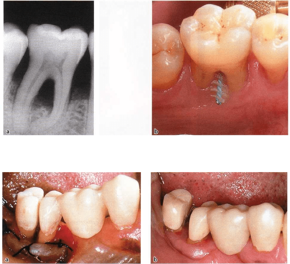
Fig. 29-20. Tunnel preparation of a degree III-involved mandibular molar. Radiograph (a) and photograph (b)
showing a wide interradicular space where self-performed plaque control can be obtained by the use of an inter
-
proximal brush.
Fig. 29-21. Effect of orthodontic treatment of a separated mandibular molar with a small root divergence. (a) After
root separation. (b) 3 months after completion of orthodontic therapy
ino 1980). A tooth with a short root trunk is a good
candidate for RSR; the amount of remaining periodon-
tal tissue support following separation and resection
is
often sufficient to ensure the stability of the remaining
root cone. If the root trunk
is long,
the furcation
involvement occurs later in the disease process, but
once established the amount of periodontal tissue
support left apical of the furcation may be insufficient
to allow RSR.
The divergence between the root cones
The distance between the root cones must be consid-
ered. Roots with a short divergence are technically
more difficult to separate than roots which are wide
apart. In addition, the smaller the divergence is, the
smaller also is the interradicular (furcation) space. In
cases where the divergence between two roots is
small,
the possibility of increasing the interradicular
distance
with an orthodontic root movement may be
considered
(Fig. 29-21a,b).
The furcation space may also be increased by odon-
toplasty performed during surgery. Fig. 29-22a-c illus-
trates that
odontoplasty
was performed on (1) the distal
part of the mesial root and (2) the mesial part of the
distal root and deep finishing lines prepared for the
subsequent restoration (Di Febo et al. 1985).
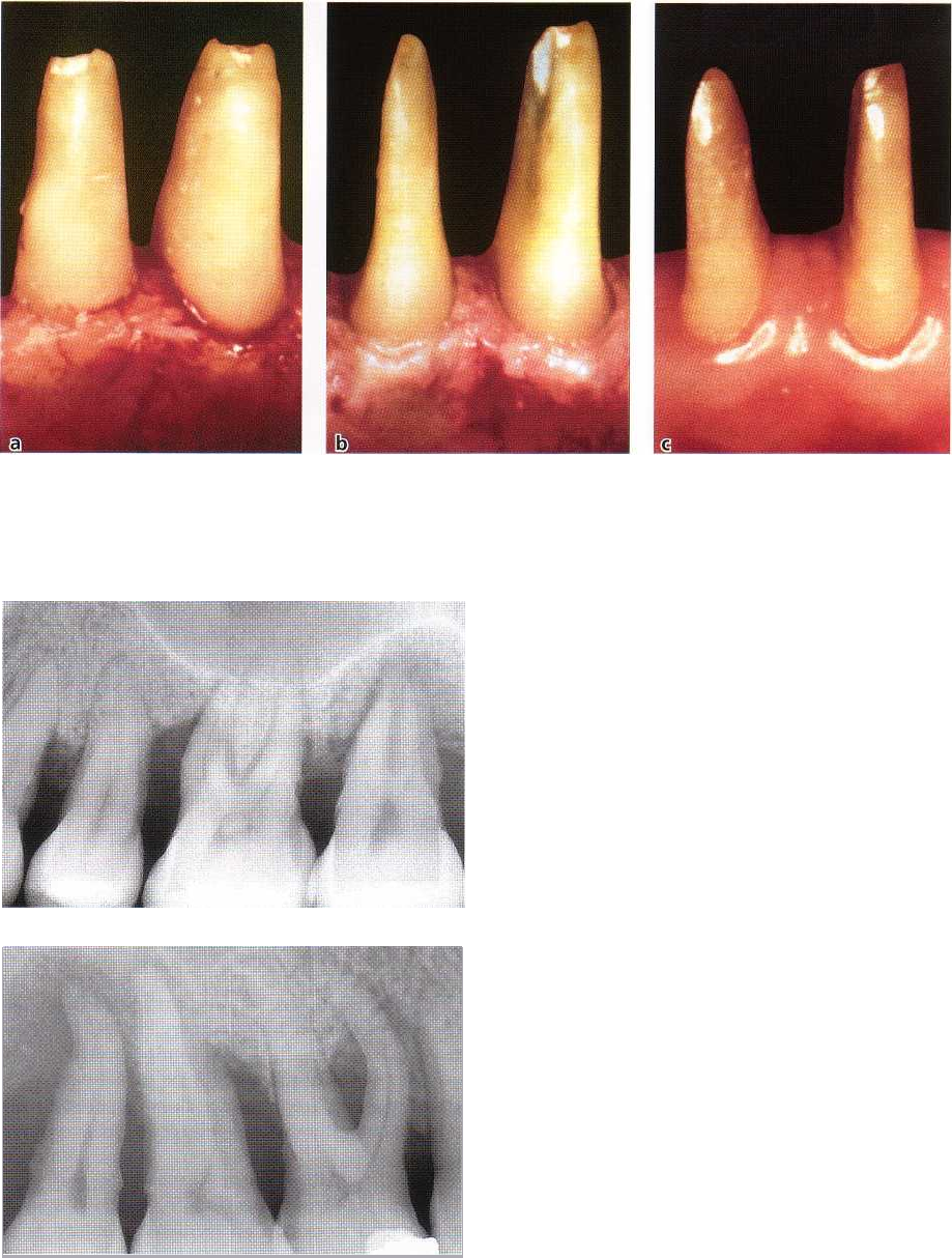
The length and the shape of the root cones
Short and small root cones (Fig. 29-23) following sepa-
ration tend to exhibit an increased mobility. Such
roots, in addition, have narrow root canals which are
difficult to ream. Short and small roots consequently
should be regarded as poor abutments for prosthetic
restorations.
Fusion between root cones
When a decision has been made to perform RSR, it is
important that the clinician first determines that the
cones within the root complex are not fused. This is
generally an uncomplicated diagnostic problem for
716 • CHAPTER 29
Fig. 29-22. Odontoplasty of a separated mandibular molar performed during surgery to increase the furcation
space. After flap elevation and exposure of the alveolar bone, it is evident that the distance between the two roots is
small (a). By preparing the interradicular surfaces during surgery (b) the furcation space is increased and is suffi
cient for self-performed plaque control measures (c).
Fig. 29-23. Radiograph showing maxillary molars with
thin, short and conical roots.
Fig. 29-24. Radiograph indicating the presence of a de
-
gree III involvement of the buccal furcation of the max
-
illary first molar. This tooth is a candidate for root re-
section.
mandibular molars or for the buccal furcation of max- raised to allow the operator to get proper access to the
illary molars (Fig.
29-24).
At such teeth the separation approximal tooth surfaces. The mesial (or distal) en-area
between the roots can easily be identified both trance of the furcation must be probed to a depth of with the
probe and in a radiograph. It is more difficult 3-5 mm to ascertain that a fusion does NOT exist
to identify a
separation line between mesiobuccal (or between the roots scheduled for RSR.
distobuccal) and palatal roots of a maxillary molar or
maxillary first premolar with a narrow root complex.
In such situations, a soft tissue flap must often be
TREATMENT
OF
FURCATION-INVOLVED TEETH •
7
1
7
Table 29-1. Root resective treatment possibilities in molars with furcation involvement
Furcation involvement
Root resection
Root resection plus separation
of the remaining roots
1
Buccal
Mesial
Distal
Mesiobuccal, Distobuccal
Mesiobuccal, Palatal
Distobuccal, Palatal
2
Buccal & Distal
Buccal & Mesial
Mesial & Distal
Distobuccal, Mesiobuccal & Palatal
Mesiobuccal, Distobuccal & Palatal
Palatal, Mesial & Distobuccal
Palatal
Palatal, Distobuccal
Distobuccal
3
Buccal, Distal & Mesial
Distobuccal & Palatal, Mesiobuccal
& Palatal, Mesio & Distobuccal
Palatal, Distobuccal
Fig. 29-25. Occlusal view of a restoration using the
mesial root of a maxillary first molar as abutment.
Note the alignment of the mesial root and the adjacent
premolars.
Amount of remaining
support around individual
roots
This should be determined by probing the entire cir-
cumference of the separated roots. It should be ob-
served that a localized deep attachment loss at one
surface of one particular root (e.g. on the buccal sur-
face of the palatal root, or the distal surface of the
mesiobuccal root of a maxillary molar) may compro-
mise the long-term prognosis for an otherwise ideal
root.
Stability
of individual roots
Must be examined following root separation. Rule of
thumb: the more mobile the root cone is, the less
periodontal tissue support remains.
Access for oral hygiene devices
The site must after completion of therapy have an
anatomy which facilitates proper self-performed
toothcleaning.
Maxillary molars
General example
Several decisions must be made when RSR is planned
for a furcation-involved maxillary molar. Since such
teeth have three root cones, one or two cones may be
retained after separation. Different treatment alterna-
tives exist. They are listed in Table 29-1.
Prior to RSR, the morphology of the individual
roots as well as the surface area of each root must be
carefully analyzed.
The
distobuccal root
of a maxillary molar (1) is the
shortest of the three roots; (2) the root trunk is com-
paratively long. Thus, the distal root has a small quan
tity of bone support and once separated, the cone may
exhibit increased mobility. The distobuccal root is,
therefore, often removed as part of RSR (Rosenberg
1978, Ross & Thompson 1980).
The
mesiobuccal root
has (1) a wide buccopalatal
dimension, (2) an hour-glass cross section, and there
-
fore a large root surface area. In fact, the mesiobuccal
cone often has a total root surface area that is equal to
or greater than that of the palatal root cone. The
mesiobuccal root (1) is located centrally in the alveolar
process, (2) is properly aligned with the maxillary
premolars and is in an ideal position to function as a
separate unit (Fig. 29-25). For these reasons, the mes
-
iobuccal root may be preferred for retention when the
clinician is selecting between the mesiobuccal or pala
-
tal root. It should be remembered, however, that the
root canals of the mesiobuccal root are narrow and
more difficult to treat than the single and wide canal
of the palatal root.
The tissue destruction in the furcation area often
causes deep attachment and bone loss at the distal-
718 • CHAPTER 29
Fig. 29-26. Palatal root of a root-resected maxillary molar serving as a single abutment for a crown restoration (a). A
mesiobuccal root was included in the restoration for esthetic reasons (b).
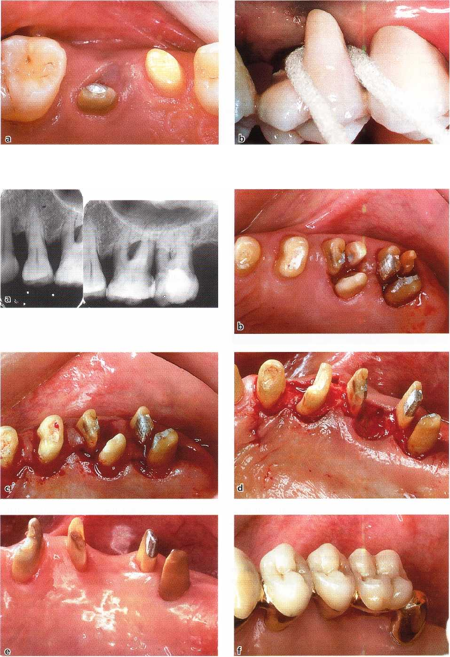
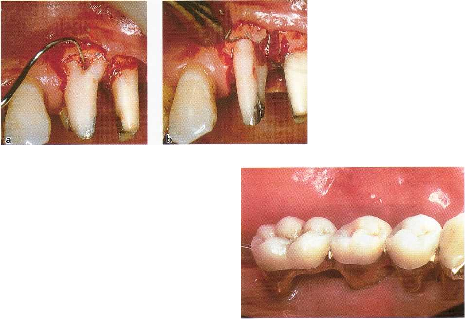
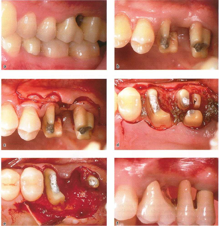
Fig. 29-27. The sequential stages of root resection of two maxillary molars with degree III involvement. Radiograph
showing the pre RSR situation (a). The roots were separated before flap elevation (b). The distal roots of both mo
lars
and the palatal root of the first molar were extracted and the teeth prepared (c,d). After 3 months of healing (e).
The
final prosthetic restoration of the site (f).
TREATMENT OF FURCATION-INVOLVED TEETH •
719
Fig. 29-28. Resection of the dis
tobuccal root of a three-rooted
maxillary first premolar.
palatal surface of the mesiobuccal root. In such situ-
ations the palatal root remains as the only candidate
for retention (Fig. 29-26a,b).
The series of illustrations presented in Fig. 29-27
demonstrates two left maxillary molars (teeth 26 and
27) with degree III involvement of all six furcation
entrances. Both teeth were, following a detailed ex-
amination and diagnosis, scheduled for treatment
with RSR. Note that in this case the second premolar
was missing. In cases of advanced periodontal disease
at maxillary molars, it is often necessary to separate
all three roots of the individual tooth to obtain access
to the interradicular area for assessment of the height
of the remaining bone at (1) the buccal surface of the
palatal root and (2) the palatal surfaces of the buccal
roots. Fig. 29-27b illustrates the two maxillary molars
with all six roots separated. Because of anatomic con
-
siderations and increased mobility, the distobuccal
roots of 26 and 27 were extracted (Fig. 29-27c). The
palatal root of the first molar had a deep area of
localized attachment loss on its buccal surface, was
considered to be a poor candidate for a bridge abut-
ment and was extracted. The mesiobuccal root of the
first molar as well as the mesiobuccal and palatal roots
of the second molar (27) were stable and exhibited
moderate probing depth. It was anticipated that at all
three roots the anatomy following healing after treat-
ment would allow proper plaque control. The three
roots were maintained (Fig. 29-27d). Fig. 29-27e shows
the area after 3 months' healing and Fig. 29-27f illus-
trates the segment properly restored. Since in this
segment one premolar was missing, the mesiobuccal
root of the first molar was used as second premolar in
the prosthetic reconstruction and the two roots of the
second molar served as abutments for a crown resto-
ration in the position of a molar.
Maxillary premolars
Root resection of maxillary first premolars is possible
only in rare instances due to the anatomy of the root
complex (Joseph et al. 1996) (Fig. 29-28a,b). The furca
-
tion of this premolar is often located at such an apical
level that the maintenance of one root serves no mean
-
ingful purpose. In most cases, therefore, the presence
Fig. 29-29. Results of the root resection of a mandibular
first molar of which the distal root was retained.
of a deep furcation involvement of degree II or degree
III in a maxillary first premolar calls for tooth extrac-
tion.
Mandibular molars
If RSR must be applied in a furcation-involved
mandibular molar, three treatment alternatives exist:
1.
separate the two roots, but maintain both roots
(
premolarization)
2.
separate and extract the mesial root
3.
separate and extract the distal root
In some situations, both roots may be maintained
following separation.
If one root is to be removed, the following facts
must be considered:
The
mesial
root has a significantly greater root sur-
face area than the distal root. The mesial root, how-
ever, has an hour-glass-shaped cross section which
may be difficult to manage (1) in the self-performed
plaque control and (2) in the restorative procedure. In
addition, the mesial root frequently has two narrow
root canals. The root canals are often close to the
external root surface. This may complicate root prepa
-
ration during the subsequent restorative treatment.
The
distal
root has an oval cross section and, as a
rule, only one, wide root canal. The distal root (1) is
720 • CHAPTER 29
Fig. 29-30. Combined photograph and radiograph showing the "conservativ
e
"
approach both regarding the access
to the pulp chamber (a) and the shaping and filling of the root canal system (b). Schematic illustration showing the
temporary restoration of the endodontically treated tooth (c).
Fig. 29-31. Radiograph illustrating the damage which
occurred to the interradicular septum during root sepa
-
ration.
comparatively large, providing a greater mass of
dentin to resist root fracture (Langer et al. 1981); (2) is
a good candidate for pin or post placement. Further,
when the resected mandibular molar is a terminal
abutment for a bridge, the retention of the distal root
will result in a longer dental arch than would be the
case had the mesial root been retained (Fig. 29-29).
Sequence of treatment at RSR
Once anatomic and pathologic characteristics of the
root complex (es) of multirooted teeth have been docu
-
mented, treatment should follow a logical plan (see
also Chapter 19).
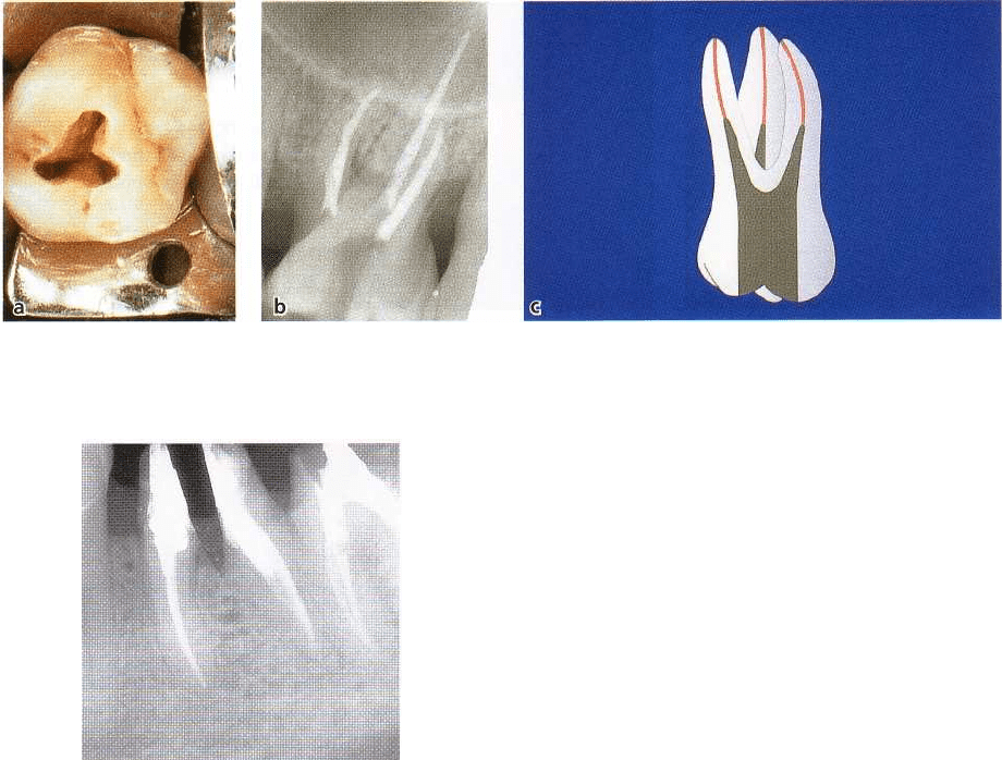
Endodontic
treatment
If the tooth to be resected is vital or if an improper root
canal filling was placed in a non-vital tooth, RSR starts
with endodontic therapy. Rubber dam can be placed,
and optimal conditions thus be established for the
important management (cleaning and shaping) of the
root canal. The structural integrity of the root must be
maintained and minimal amounts of root dentin
should be removed (Fig. 29-30a,b). Direct filling with
amalgam or chemically cured composite of the endo-
dontically treated tooth should be performed before
RSR (Fig. 29-30c). Each root should have individual
retention for a restoration which should not break or
detach during RSR, removal and relining of the pro-
visional restorations, impressions and prosthetic trys
-
in. Endocanal posts or endodontic screws are used
only if natural retention needs improvement.
Occasionally, a furcation involvement may first be
identified during periodontal surgery. In this emer-
gency situation RSR may be completed but the root
canal entrance(s) of the remaining root(s) must be
properly sealed. Definitive root canal therapy must be
completed within two weeks (Smukler & Tagger
1976)
.
Provisional restoration
Alginate impressions of the area to be treated are taken
and sent to the laboratory together with a wax record
of the intercuspal position. A provisional restoration
is prepared.
RSR
Root separation and root resection may be performed
as part of the preparation of the segment for prosthetic
rehabilitation ("prosthetic preparation"), i.e. prior to
periodontal surgery (Carnevale et al. 1981). During
the prosthetic preparation it is important to
avoid
•
exposing the interradicular bone to undue
mechani
cal trauma (Fig. 29-31)
•
leaving behind parts of the furcation fornix (Fig. 29-
32a-d)
•
perforating the root canals
•
preparing the vertical surfaces of the remaining
roots with sharp angles (Fig. 29-33).
Situation 1:
mandibular molar
Following separation, both roots are maintained. The
distal surface of the distal root and the mesial surface
of the mesial root must be prepared parallel with each
other to increase the retention for a subsequent resto
-
ration. The mesial surface of the distal root and the
distal surface of the mesial root should be prepared
TREATMENT OF FURCATION-INVOLVED TEETH • 721
Fig. 29-32. Radiographs of a mandibular first molar to be extracted and of a second molar to be root resected (a).
During hemisection an overhang is left behind as a result of an oblique sectioning of the tooth distal to the furcation (
b). In a radiograph obtained 2 years later, the presence of an angular bony defect can be seen adjacent to the "over-
hang" (c). The lesion was resolved and the angular defect disappeared following removal of the "overhang". Radio-
graph
after 2 years (d).
Fig. 29-33. Maintenance of the two fused buccal roots
of a maxillary first molar. The buccal roots were sepa-
rated from the palatal root. Note the rounded line an-
gles and the wide space created between the separated
roots.
Fig. 29-34. Mandibular molar after root separation.
Note the diverging angle of preparation performed to
increase the interradicular space between the mesial
and distal roots and the parallel approximal surfaces.
722 • CHAPTER 29
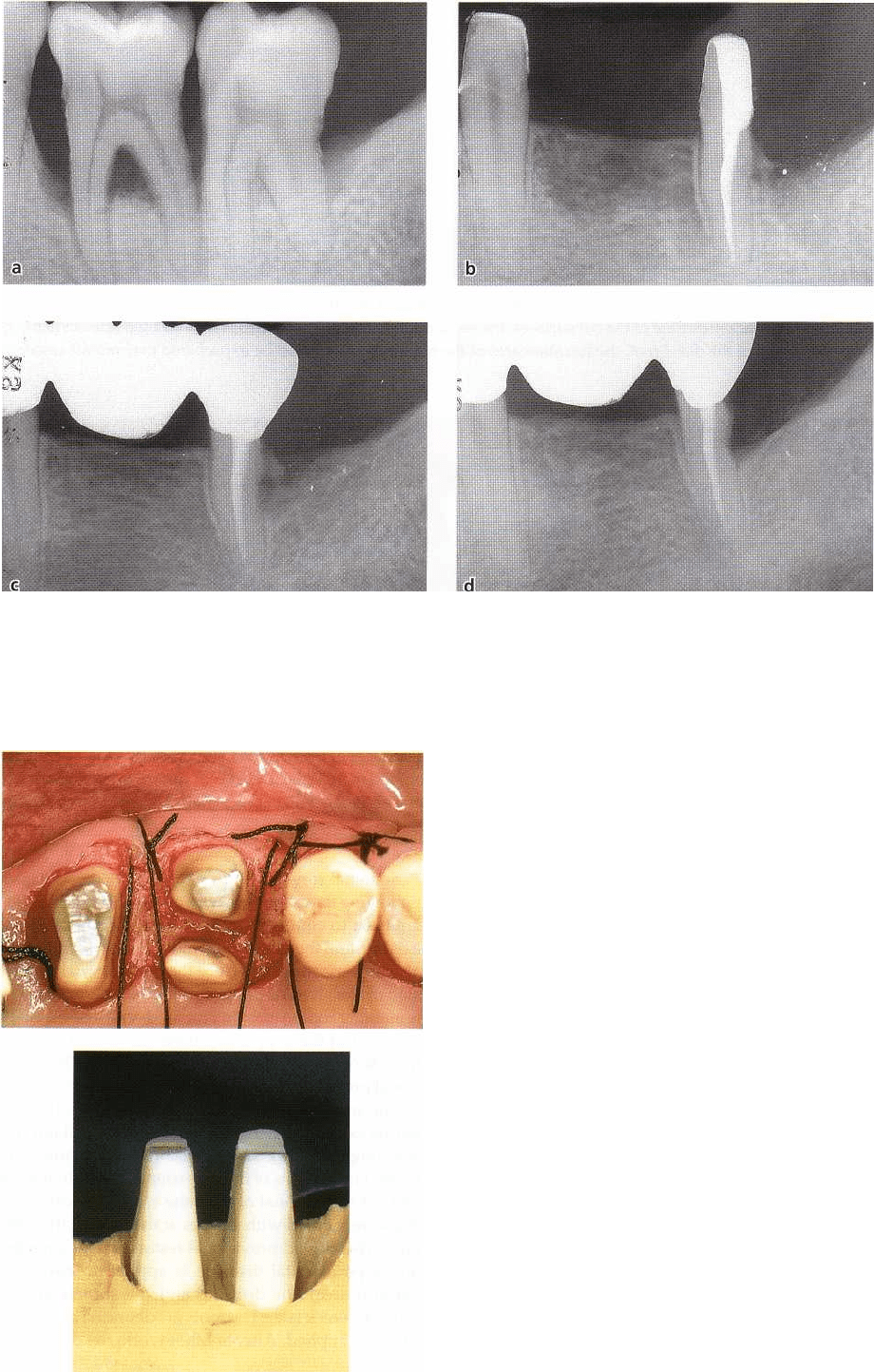
Fig. 29-35. The sequential stages of root resection and extraction of the distal root of a maxillary molar. In order to
minimize the concave outline of the cut surfaces, the sectioning should be performed with a straight line cut (a,b).
After extraction of the distal root, the furcation area of the remaining roots must be re-prepared to eliminate under
-
cuts (c, d).
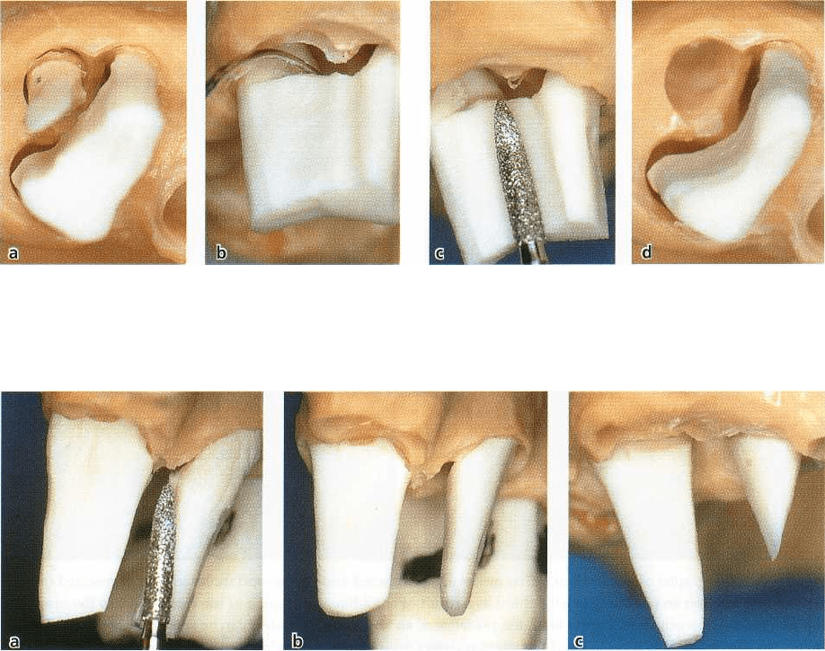
Fig. 29-36. Preparation, during separation, of the mesiobuccal and palatal roots after the distobuccal root of a maxil
-
lary molar had been extracted. The internal (furcation) surfaces of the two roots should be prepared with diverging
angles to increase the interradicular space, while the external surfaces of the two roots should be prepared parallel
to
each other to increase the subsequent retention of the restoration (a,b). When the palatal surface of the palatal
root
is not prepared parallel to the buccal surface (c), the palatal abutment will become shortened and not self-reten
tive.
with diverging angles to increase the space available
between the separated roots (Fig. 29-34).
Situation
2: maxillary molar
Following separation, the distobuccal root was ex-
tracted. The distal surface of the crown is prepared
with
a bevel cut and in such a way that the concave
curvature (in apicocoronal direction) is eliminated
(Fig.
29-35a-d).
If the mesiobuccal and the palatal roots of this
molar
must be separated but maintained, it is important that
the buccal surface of the mesiobuccal root and
the
palatal surface of the palatal root are prepared
parallel
with each other. This will enhance the reten
tion of the
subsequent restoration. The palatal surface
of the
mesiobuccal root and the buccal surface of the
palatal
root must be prepared at diverging angles to increase
the space available between the separated
roots (Fig. 29-
36a-c).
The provisional restoration is at this stage relined
with cold cured acrylic and cemented after RSR.
Periodontal surgery
Following flap elevation, osseous resective techniques
are used to eliminate angular bone defects that may
exist around the maintained roots. Bone resection may
also be performed to reduce the buccolingual dimen-
sion of the alveolar process of the extraction site. The
remaining root(s) may be prepared with a bevel cut to
the level of the supporting bone (Levine 1972, Ram-
fjord
& Nissle 1974, Carnevale et al. 1983). This addi
tional
preparation may serve the purpose of (1) elimi
nating
residual soft and hard deposits and (2) eliminating
existing undercuts to facilitate the final impres
sion (Fig.
29-37a-f). The provisional restoration is re-
lined. The
margins of the provisional restoration must
end >_ 3
mm coronal of the bone crest. The soft tissue
flaps are
secured with sutures at the level of the bone
crest. The
relined provisional restoration is cemented
and a
periodontal dressing is applied to cover the
surgical
area. The dressing and the sutures are re-
moved 1 week
later. The roots are debrided and a new
dressing applied.
After another week, the dressing is
TREATMENT
OF
FURCATION-INVOLVED TEETH • 723
Fig. 29-37. Sequential stages of root resection at maxillary first and second molars. The extraction of the distal root
of the first molar was performed during tooth preparation and prior to the insertion of the provisional restoration
(a,b). During the surgical procedure, after flap elevation, the furcation-involved second molar was separated,
the
mesial and palatal roots were extracted and the osseous defects were eliminated (c, d, e). Healing with the
defini
tive prosthetic restoration in place (f).
finally removed and the patient instructed in proper
plaque control techniques.
Final prosthetic restoration
Since the prosthetic preparation of the roots was com-
pleted during surgery, the clinician concerns him/her-
self with only minor adjustments. The preparation
margins are located supragingivally, which improves
the precision of the definitive crown restoration. The
framework of the restoration must be rigid to compen-
sate for the compromised abutments (roots) with a
compromised periodontal tissue support. The occlu-
sion should be designed to minimize the infliction of
lateral deflective forces (see Chapter 30) (Fig. 29-
38a,b).
Regeneration of furcation defects
The possibility of regenerating and closing a furcation
defect has been investigated (see Chapter 28).
Following an early case report publication
(Gottlow
et al. 1986), where histologic documentation
of new
attachment formation in human furcation de-
724 •
CHAPTER
29
Fig. 29-38. Soft tissue healing at a separated maxillary first molar and at a root-resected second molar (a). The final
prosthetic restoration in place with the occlusion designed to minimize the lateral stresses on the roots left as abut
-
ments (b).
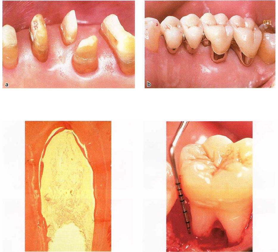
Fig. 29-39. Histologic mesiodistal section of a previous
degree II furcation involvement of a human mandibu
-
lar molar, treated with GTR. The section demonstrates
that the newly formed cementum covers the entire cir
-
cumference of the furcation defect.
fects (Fig. 29-39) treated by "guided tissue regenera-
tion" (GTR) therapy was provided, the results of sev-
eral investigations on this form of treatment in furca-
tion-involved teeth have been presented. In these re-
ports, a reasonably predictable outcome of GTR ther-
apy was demonstrated only in degree II furcation-in-
volved mandibular molars, where a clinical soft tissue
closure or a decreased probing depth of the furcation
defect was recorded (Pontoriero et al. 1988, Lekovic et
al. 1989, Caffesse et al. 1990).
Less favorable results have been reported when
GTR therapy was used in other types of furcation
defects such as degree III furcation-involved mandib-
ular and maxillary molars (Pontoriero et al. 1989, Pon-
toriero & Lindhe 1995a) and degree II furcations in
maxillary molars (Metzeler et al. 1991, Pontoriero &
Lindhe 1995b).
The reason for the limited predictability of GTR
Fig. 29-40. Position of the furcation fornix in relation to
the level of the supporting bone and attachment appa
ratus in a lingual degree II furcation-involved
mandibular molar.
therapy in furcation-involved teeth may be related to
several factors:
•
The morphology of the periodontal defect, which in
the root complex often has the character of a "hori-
zontal lesion". New attachment formation is hence
dependent on coronal upgrowth of periodontal
ligament tissue (Fig. 29-40).
•
The anatomy of the furcation, with its complex
internal morphology, may prevent proper instru-
mentation and debridement of the exposed root
surface (Fig. 29-41).
•
The varying and changing location of the soft tissue
margins during the early phase of healing with a
possible recession of the flap margin and early ex-
posure of both the membrane material and the for-
nix of the furcation (Fig. 29-42).