Jan Lindhe. Clinical Periodontology
Подождите немного. Документ загружается.


ANATOMY OF THE PERIODONTIUM • 43
Fig. 1-92.
a.a.i.
Fig. 1-93.
a.s.
a.m.
rr.p.
a.i.
a.d.
a.ap.
a.p.
Fig. 1-94. Fig. 1-95.
BLOOD SUPPLY OF THE
PERIODONTIUM
Fig. 1-92. The schematic drawing depicts the blood
supply to the teeth and the periodontal tissues. The
dental artery (a.d.), which is a branch of the superior or
inferior alveolar artery (a.a.i.), dismisses the intraseptal
artery (a.i.) before it enters the tooth socket. The termi-
nal branches of the intraseptal artery (rami perforantes, rr.
p.) penetrate the alveolar bone proper in canals at all
levels of the socket (see Fig. 1-76). They anastomose in
the periodontal ligament space, together with blood
vessels originating from the apical portion of the peri-
odontal ligament and with other terminal branches,
from the intraseptal artery (a.i.). Before the dental
artery (a.d.) enters the root canal it puts out branches
which supply the apical portion of the periodontal
ligament.
Fig. 1-93. The gingiva receives its blood supply mainly
through supraperiosteal blood vessels which are termi-
nal branches of the sublingual artery (a.s.), the mental
artery (a.m.), the buccal artery (a.b.), the facial artery (a.f.),
the greater palatine artery (a.p.), the infra orbital artery (a.i.)
and the posterior superior dental artery (a.ap.).
Fig. 1-94 depicts the course of the greater palatine
artery (a.p.) in a specimen of a monkey which at
sacrifice was perfused with plastic. Subsequently, the
soft tissue was dissolved. The greater palatine artery (
a.p.), which is a terminal branch of the ascending
palatine artery (from the maxillary, "internal maxillary",
artery), runs through the greater palatine canal (arrow)
to the palate. As this artery runs in a frontal direction
it puts out branches which supply the gingiva and the
masticatory mucosa of the palate.
Fig. 1-95. The various arteries are often considered to
supply certain well-defined regions of the dentition.
In reality, however, there are numerous anastomoses
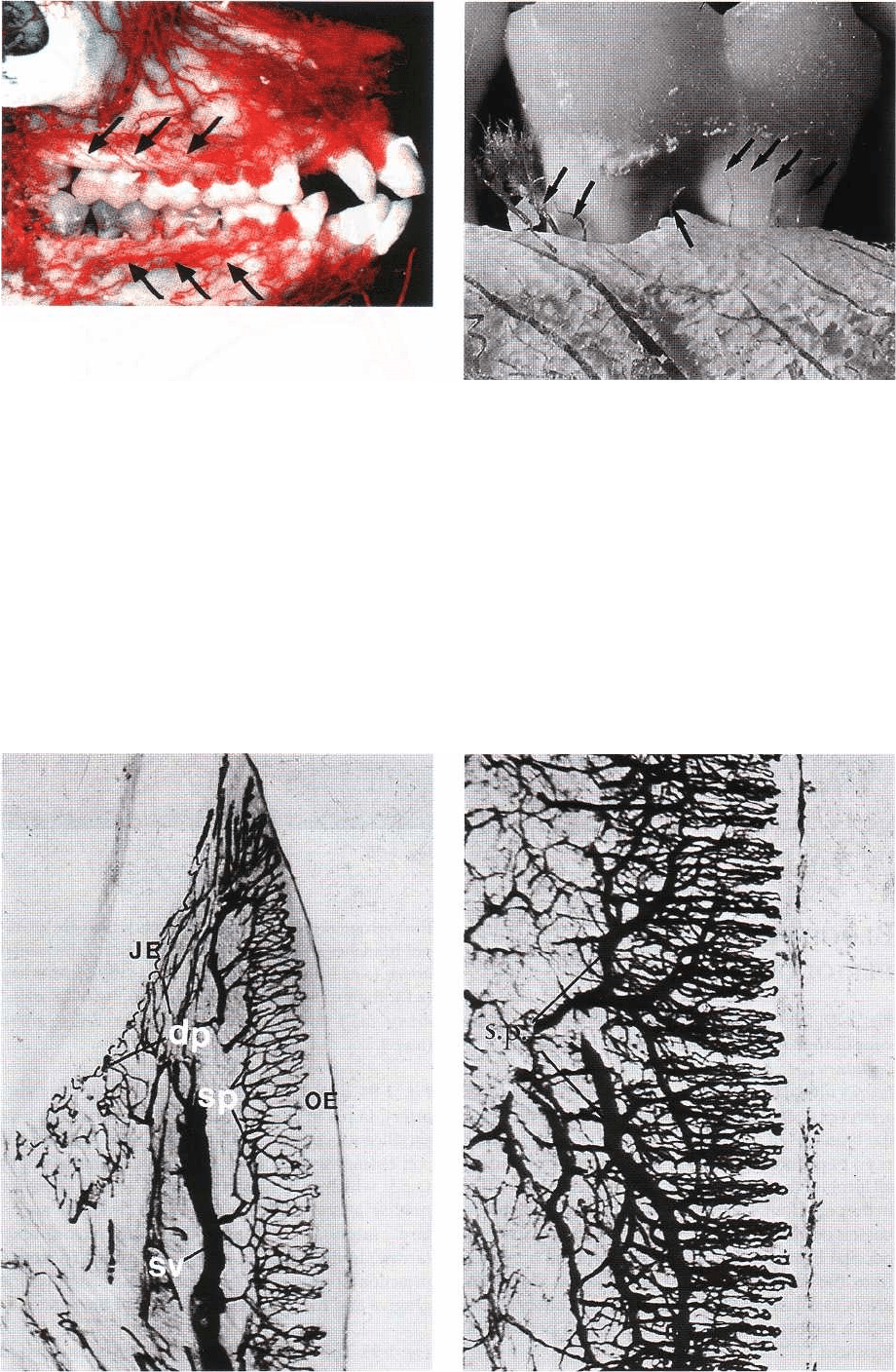
Fig. 1-98. Fig. 1-99.
44 • CHAPTER 1
Fig. 1-96.
present between the different arteries. Thus, the entire
system of blood vessels, rather than individual groups of
vessels, should be regarded as the unit supplying the
soft and hard tissue of the maxilla and the mandible,
e.g. in this figure there is an anastomosis (arrow)
between the facial artery (a.f.) and the blood vessels of
the mandible.
Fig. 1-96 illustrates a vestibular segment of the maxilla
and mandible from a monkey which at sacrifice was
perfused with plastic. Notice that the vestibular
gingiva is supplied with blood mainly through su-
praperiosteal blood vessels (arrows).
Fig. 1-97. As can be seen, blood vessels (arrows) origi-
Fig. 1-97.
nating from vessels in the periodontal ligament are
passing the alveolar bone crest and contribute to the
blood supply of the free gingiva.
Fig. 1-98 shows a specimen from a monkey which at
the time of sacrifice was perfused with ink. Sub-
sequently, the specimen was treated to make the tissue
transparent (cleared specimen). To the right, the su-
praperiosteal blood vessels (sv) can be seen. During
their course towards the free gingiva they put forth
numerous branches to the subepithelial plexus (sp) lo-
cated immediately beneath the oral epithelium of the
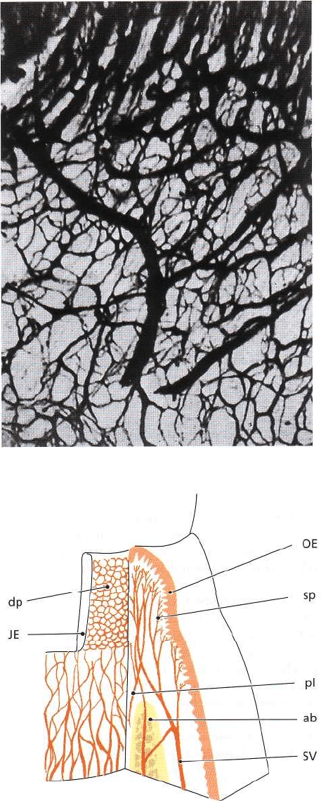
ANATOMY OF THE PERIODONTIUM • 45
Fig. 1-100.
free and attached gingiva. This subepithelial plexus in
turn yields thin capillary loops to each of the connective
tissue papillae projecting into the oral epithelium (
OE). The number of such capillary loops is constant
over a very long time and is not altered by application
of epinephrine or histamine to the gingival margin.
This implies that the blood vessels of the lateral por-
tions of the gingiva, even under normal circum-
stances, are fully utilized and that the blood flow to
the free gingiva is regulated entirely by velocity altera-
tions. In the free gingiva, the supraperiosteal blood
vessels (sv) anastomose with blood vessels from the
periodontal ligament and the bone. Beneath the junc-
tional epithelium (JE) seen to the left, is a plexus of
blood vessels termed the dentogingival plexus (dp). The
blood vessels in this plexus have a thickness of ap-
proximately 40 urn, which means that they are mainly
venules. In healthy gingiva, no capillary loops occur
in the dentogingival plexus.
Fig. 1-99. This specimen illustrates how the subepi-
thelial plexus (sp), beneath the oral epithelium of the
free and attached gingiva, yields thin capillary loops
to each connective tissue papilla. These capillary loops
have a diameter of approximately 7 µm, which means
they are the size of true capillaries.
Fig. 1-100 illustrates the dentogingival plexus in a
section cut parallel to the subsurface of the junctional
epithelium. As can be seen, the dentogingival plexus
consists of a fine-meshed network of blood vessels. In
the upper portion of the picture, capillary loops can be
detected belonging to the subepithelial plexus be-
neath the oral sulcular epithelium.
Fig. 1-101 is a schematic drawing of the blood supply
to the free gingiva. As stated above, the main blood
supply of the free gingiva derives from the suprape-
riosteal blood vessels (SV) which, in the gingiva, anas-
tomose with blood vessels from the alveolar bone (ab)
and periodontal ligament (pl). To the right in the draw-
ing, the oral epithelium (OE) is depicted with its un-
derlying subepithelial plexus of vessels (sp). To the left
beneath the junctional epithelium (JE), the dentogingi-
val plexus (dp) can be seen, which, under normal
conditions, comprises a fine-meshed network without
capillary loops.
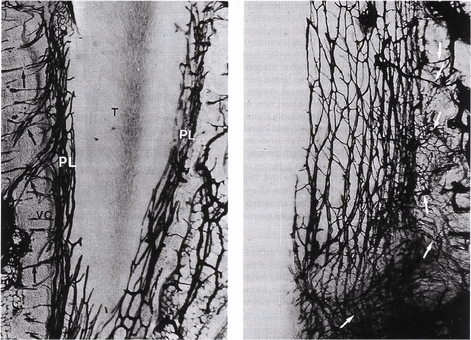
Fig. 1-102 shows a section prepared through a tooth Fig. 1-101.
(T) with its periodontium. Blood vessels (perforating
46 • CHAPTER 1
Fig. 1-102.
rami; arrows) arising from the intraseptal artery in the
alveolar bone run through canals (Volkmann's canals)
in the socket wall (VC) into the periodontal ligament (
PL), where they anastomose.
Fig. 1-103 shows blood vessels in the periodontal liga-
ment in a section cut parallel to the root surface. After
entering the periodontal ligament, the blood vessels (
perforating rami; arrows) anastomose and form a
polyhedral network which surrounds the root like a
stocking. The majority of the blood vessels in the
periodontal ligament are found close to the alveolar
bone. In the coronal portion of the periodontal liga-
ment, blood vessels run in coronal direction, passing
the alveolar bone crest, into the free gingiva (see Fig.
1-97).
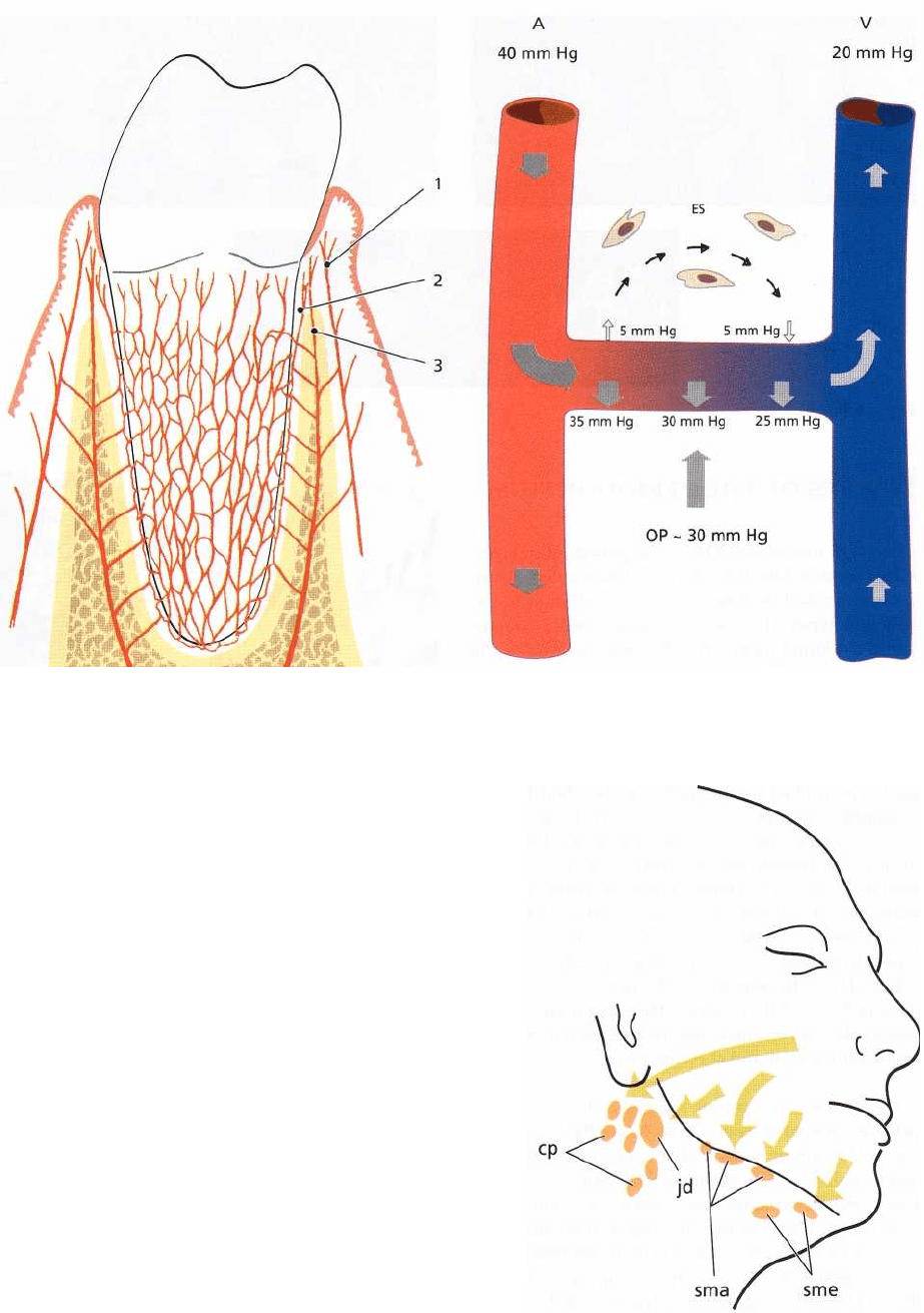
Fig. 1-104 is a schematic drawing of the blood supply
of the periodontium. The blood vessels in the peri-
odontal ligament form a polyhedral network sur-
rounding the root. Note that the free gingiva receives
its blood supply from (1) supraperiosteal blood ves-
sels, (2) the blood vessels of the periodontal ligament
and (3) the blood vessels of the alveolar bone.
Fig. 1-103.
Fig. 1-105 illustrates schematically the so-called ex-
travascular circulation through which nutrients and
other substances are carried to the individual cells and
metabolic waste products are removed from the tis-
sue. In the arterial (A) end of the capillary, to the left
in the drawing, a hydraulic pressure of approximately
35 mm Hg is maintained as a result of the pumping
function of the heart. Since the hydraulic pressure is
higher than the osmotic pressure (OP) in the tissue (
which is approximately 30 mm Hg), a transportation
of substances will occur from the blood vessels to the
extravascular space (ES). In the venous (V) end of the
capillary system, to the right in the drawing, the hy-
draulic pressure has decreased to approximately 25
mm Hg (i.e. 5 mm lower than the osmotic pressure in
the tissue). This allows a transportation of substances
from the extravascular space to the blood vessels.
Thus, the difference between the hydraulic pressure
and the osmotic pressure (OP) results in a transporta-
tion of substances from the blood vessels to the ex-
travascular space in the arterial part of the capillary
while, in the venous part, a transportation of sub-
stances occurs from the extravascular space to the
blood vessels. Hereby an extravascular circulation is
established (small arrows).
ANATOMY OF THE PERIODONTIUM • 47
LYMPHATIC SYSTEM OF THE
PERIODONTIUM
Fig. 1-106. The smallest lymph vessels, the lymph cap-
illaries, form an extensive network in the connective
tissue. The wall of the lymph capillary consists of a
single layer of endothelial cells. For this reason such
capillaries are difficult to identify in an ordinary his-
tologic section. The lymph is absorbed from the tissue
fluid through the thin walls into the lymph capillaries.
From the capillaries, the lymph passes into larger
lymph vessels which are often in the vicinity of corre-
sponding blood vessels. Before the lymph enters the
blood stream it passes through one or more lymph
nodes in which the lymph becomes filtered and sup-
plied with lymphocytes. The lymph vessels are like
veins provided with valves. The lymph from the peri-
odontal tissues is drained to the lymph nodes of the
head and the neck. The labial and lingual gingiva of
the mandibular incisor region is drained to the sub-
mental lymph nodes (sme). The palatal gingiva of the
maxilla is drained to the deep cervical lymph nodes (cp).
The buccal gingiva of the maxilla and the buccal and
lingual gingiva in the mandibular premolar-molar
region are drained to submandibular lymph nodes (sma).
Except for the third molars and mandibular incisors,
all teeth with their adjacent periodontal tissues are
drained to the submandibular lymph nodes (sma).
The third molars are drained to the jugulodigastric
lymph node (jd) and the mandibular incisors to the
submental lymph nodes (sme).
Fig. 1-105.
Fig. 1-106,
Fig. 1-104.
48 • CHAPTER 1
Fig. 1-107.
NERVES OF THE PERIODONTIUM
Like other tissues in the body, the periodontium con-
tains receptors which record pain, touch and pressure (
nociceptors and mechanoreceptors). In addition to the
different types of sensory receptors, nerve compo-
nents are found innervating the blood vessels of the
periodontium. Nerves recording pain, touch, and
pressure have their trophic center in the semilunar
ganglion and are brought to the periodontium via the
trigeminal nerve and its end branches. Owing to the
presence of receptors in the periodontal ligament,
small forces applied on the teeth may be identified.
For example, the presence of a very thin (10-30 µm)
metal foil (strip) placed between the teeth during
occlusion can readily be identified. It is also well
known that a movement which brings the teeth of the
mandible in contact with the occlusal surfaces of the
maxillary teeth is arrested reflexively and altered into
an opening movement if a hard object is detected in
the chew. Thus, the receptors in the periodontal liga-
ment, together with the proprioceptors in muscles and
tendons, play an essential role in the regulation of
chewing movements and chewing forces.
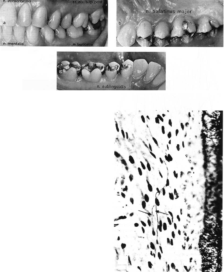
Fig. 1-107 shows the various regions of the gingiva
which are innervated by end branches of the trigemi-
nal nerve. The gingiva on the labial aspect of maxillary
incisors, canines and premolars is innervated by supe-
rior labial branches from the infraorbital nerve, n. infraor-
bitalis (Fig. 1-107a). The buccal gingiva in the maxil-
lary molar region is innervated by branches from the
posterior superior dental nerve, rr. alv. sup. post (Fig. 1-
107a). The palatal gingiva is innervated by the greater
palatal nerve, n. palatinus major (Fig. 1-107b), except
for the area of the incisors, which is innervated by the
long sphenopalatine nerve, n. pterygopalatini. The lingual
gingiva in the mandible is innervated by the sublingual
nerve, n. sublingualis (Fig. 1-107c), which is an end
branch of the lingual nerve. The gingiva at the
Fig. 1-108.
labial aspect of mandibular incisors and canines is
innervated by the mental nerve, n. mentalis, and the
gingiva at the buccal aspect of the molars by the buccal
nerve, n. buccalis (Fig. 1-107a). The innervation areas
of these two nerves frequently overlap in the premolar
region. The teeth in the mandible including their peri-
odontal ligament are innervated by the inferior alveolar
nerve, n. alveolaris inf., while the teeth in the maxilla
are innervated by the superior alveolar plexus, n. alveo-
lares sup.
I
ANATOMY OF THE PERIODONTIUM • 49
Fig. 1-108. The small nerves of the periodontium fol-
low almost the same course as the blood vessels. The
nerves to the gingiva run in the tissue superficial to
the periosteurn and put out several branches to the
oral epithelium on their way towards the free gingiva.
The nerves enter the periodontal ligament through the
perforations (Volkmann
'
s canals) in the socket wall
(see Fig. 1-102). In the periodontal ligament, the nerves
join larger bundles which take a course parallel to the
long axis of the tooth. The photomicrograph illustrates
small nerves (arrows) which have emerged from
larger bundles of ascending nerves in order to supply
certain parts of the periodontal ligament tissue. Vari-
ous types of neural terminations such as free nerve
endings and Ruffini's corpuscles have been identified
in the periodontal ligament.
REFERENCES
Ainamo, J. & Talari, A. (1976). The increase with age of the width
of attached gingiva. Journal of Periodontal Research 11, 182-188.
Anderson, D.T., Hannam, A.G. & Matthews, G. (1970). Sensory
mechanisms in mammalian teeth and their supporting struc-
tures. Physiological Review 50, 171-195.
Bartold, P.M. (1995). Turnover in periodontal connective tissue:
dynamic homeostasis of cells, collagen and ground sub-
stances. Oral Diseases 1, 238-253.
Beertsen, W., McCulloch, C.A.G. & Sodek, J. (1997). The peri-
odontal ligament: a unique, multifunctional connective tis-
sue. Periodontology 2000 13, 20-40.
Bosshardt, D.D. & Schroeder, H.E. (1991). Establishment of acel
lular extrinsic fiber cementum on human teeth. A light- and
electron-microscopic study. Cell Tissue Research 263, 325-336.
Bosshardt, D.D. & Selvig, K.A. (1997). Dental cementum: the
dynamic tissue covering of the root. Periodontology 2000 13,
41-75.
Carranza, E.A., Itoiz, M.E., Cabrini, R.L. & Dotto, C.A. (1966). A
study of periodontal vascularization in different laboratory
animals. Journal of Periodontal Research 1, 120-128.
Egelberg, J. (1966). The blood vessels of the dentogingival junc
-
tion. Journal of Periodontal Research 1, 163-179.
Fullmer, H.M., Sheetz, J.H. & Narkates, A.J. (1974). Oxytalan
connective tissue fibers. A review. Journal of Oral Pathology 3,
291-316.
Hammarstrom, L. (1997). Enamel matrix, cementum develop-
ment and regeneration. Journal of Clinical Periodontology 24,
658-677.
Karring, T. (1973). Mitotic activity in the oral epithelium. Journal
of Periodontal Research, Suppl. 13, 1-47.
Karring, T., Lang, N.R. & Loe, H. (1974). The role of gingival
connective tissue in determining epithelial differentiation.
Jou
r
nal of Periodontal Research 10, 1-11.
Karring, T. & Loe, H. (1970). The three-dimensional concept of
the epithelium-connective tissue boundary of gingiva. Acta
Odontologica Scandinavia 28, 917-933.
Karring, T., Ostergaard, E. & Loe, H. (1971). Conservation of
tissue specificity after heterotopic transplantation of gingiva
and alveolar mucosa. Journal of Periodontal Research 6, 282-293.
Kvam, E. (1973). Topography of principal fibers. Scandinavian
Journal of Dental Research 81, 553-557.
Lambrichts, I., Creemers, J. & van Steenberghe, D. (1992). Mor
-
phology of neural endings in the human periodontal liga-
ment: an electron microscopic study. Journal of Periodontal
Research 27, 191-196.
Listgarten, M.A. (1966). Electron microscopic study of the
gingivo-dental junction of man. American Journal of Anatomy
119, 147-178.
Listgarten, M.A. (1972). Normal development, structure, physi-
ology and repair of gingival epithelium. Oral Science Review 1,
3-67.
Lozdan, J. & Squier, C.A. (1969). The histology of the mucogingi
val junction. Journal of Periodontal Research 4, 83-93.
Melcher, A.H. (1976). Biological processes in resorption, deposi
-
tion and regeneration of bone. In: Stahl, S.S., ed. Periodontal
Si rgen, Biologic Basis and Technique. Springfield: C.C. Thomas,
pp. 99-120.
Page, R.C., Ammons, W.F., Schectman, L.R. & Dillingham, L.A.
(1974). Collagen fiber bundles of the normal marginal gingiva
in the marmoset. Archives of Oral Biology 19, 1039-1043.
Palmer, R.M. & Lubbock, M.J. (1995). The soft connective tissue
of the gingiva and periodontal ligament: are they unique?
Oral Diseases 1, 230-237.
Saffar, J.L., Lasfargues, J.J. & Cherruah, M. (1997). Alveolar bone
and the alveolar process: the socket that is never stable.
Periodontology 2000 13, 76-90.
Schenk, R.K. (1994). Bone regeneration: Biologic basis. In: Buser,
D., Dahlin, C. & Schenk, R. K., eds. Guided Bone Regeneration in
Implant Dentistry. Berlin: Quintessence Publishing Co.
Schroeder, H.E. (1986). The periodontium. In: Schroeder, H. E.,
ed. Handbook of Microscopic Anatomy. Berlin: Springer, pp.
47-64.
Schroeder, H.E. & Listgarten, M.A. (1971). Fine Structure of the
Developing Epithelial Attachment of Human Teeth, 2nd edn.
Basel: Karger, p. 146.
Schroeder, H.E. & Listgarten, M.A. (1997). The gingival tissues:
the architecture of periodontal protection. Periodontology 2000
13, 91-120.
Schroeder, H.E. & Miinzel-Pedrazzoli, S. (1973). Correlated mor
phometric and biochemical analysis of gingival tissue. Mor-
phometric model, tissue sampling and test of stereologic
procedure. Journal of Microscopy 99, 301-329.
Schroeder, H.E. & Theilade, J. (1966). Electron microscopy of
normal human gingival epithelium. Journal of Periodontal Re-
search 1, 95-119.
Selvig, K.A. (1965). The fine structure of human cementum. Ada
Odoutologica Scandinavia 23, 423-441.
Valderhaug, J.R. & Nylen, M.U. (1966). Function of epithelial
rests as suggested by their ultrastructure. Journal of Periodontal
Research 1, 67-78.
Acknowledgement
We thank the following for contributing to the illustrations in
Chapter 1:
M. Listgarten, R.K. Schenk, H.E. Schroeder, K.A. Selvig and K.
Josephsen.

CHAPTER 2
Epidemiology of Periodontal
Diseases
PANGS N. PAPAPANOU AND JAN LINDHE
Methodological issues
Examination methods — index systems
Critical evaluation
Prevalence of periodontal diseases
Introduction
Periodontitis in adults
Periodontitis in children and adolescents
Periodontitis and tooth loss
Risk factors for periodontitis
Introduction and definitions
Studies of putative risk factors for periodontitis
Longitudinal studies and conclusions
Periodontal infections and risk for systemic
disease
Atherosclerosis — cardiovascular/cerebrovascular
disease
Preterm birth
Diabetes mellitus
Concluding remarks
The term epidemiology is of Hellenic origin; it consists
of the preposition "epi", which means "among" or "
against" and the noun "demos" which means "peo-
ple". As denoted by its etymology, epidemiology is
defined as "the study of the distribution of disease or
a physiological condition in human populations and
of the factors that influence this distribution" (Lilien-
feld 1978). Amore inclusive description by Frost (1941)
emphasizes that "epidemiology is essentially an in-
ductive science, concerned not merely with describing
the distribution of disease, but equally or more with
fitting it into a consistent philosophy". Thus, the in-
formation obtained from an epidemiologic investiga-
tion should extend beyond a mere description of the
distribution of the disease in different populations (
descriptive epidemiology): it should be further util-
ized to (1) elucidate the etiology of a specific disease
by combining epidemiologic data with information
from other disciplines such as genetics, biochemistry,
microbiology, sociology, etc. (etiologic epidemiology);
(2) evaluate the consistency of epidemiologic data
with hypotheses developed clinically or experimen-
tally (analytical epidemiology); and (3) provide the
basis for developing and evaluating preventive pro-
cedures and public health practices (experimental/in-
terventive epidemiology).
Based on the above, epidemiological research in
periodontics must (1) fulfill the task of providing data
on the prevalence of periodontal diseases in different
populations, i.e. the frequency of their occurrence, as
well as on the severity of such conditions, i.e. the level
of occurring pathologic changes; (2) elucidate aspects
related to the etiology and the determinants of develop-
ment of these diseases (causative and risk factors); and (
3) provide documentation concerning the effective-
ness of preventive and therapeutic measures aimed
against these diseases on a population basis.
METHODOLOGICAL ISSUES
Examination methods - index systems
Examination of the periodontal status of a given indi-
vidual includes clinical assessments of inflammation
in the periodontal tissues, registration of probing
depths and clinical attachment levels and radio-
graphic assessments of remaining alveolar bone. A
variety of index systems for the scoring of these pa-
rameters has been developed over the years. A num-
ber of such systems was designed exclusively for ex-
amination of patients in a dental practice set-up, while
others were to be utilized in epidemiological research.
EPIDEMIOLOGY OF PERIODONTAL DISEASES • 51
The design of the index systems and the definition of
the various scores inevitably reflected the knowledge
on the etiology and pathogenesis of periodontal dis-
ease of the time they were introduced, as well as
concepts related to the current therapeutic approaches
and strategies. This section will not provide a com-
plete list of all available scoring systems, but rather
give a brief description of a limited number of indices
that are either currently used or are likely to be en-
countered in the recent literature. For description of
earlier scoring systems and a historical perspective of
their development, the reader is referred to Ainamo (
1989).
Assessment of inflammation of the periodontal
tissues
Presence of inflammation in the marginal portion of
the gingiva is usually recorded by means of probing
assessments, according to the principles of the Gingi-
val Index outlined in the publication by Loe (1967).
According to this system, entire absence of visual
signs of inflammation in the gingival unit is scored as
0, while a slight change in color and texture is scored
as 1. Visual inflammation and bleeding tendency from
the gingival margin right after a periodontal probe is
briefly run along the gingival margin is scored as 2,
while overt inflammation with tendency for sponta-
neous bleeding is scored as 3. A parallel index for
scoring plaque deposits (Plaque Index) in a scale from
0 to 3 (Silness & Loe 1964) was introduced, according
to which absence of plaque deposits is scored as 0,
plaque disclosed after running the periodontal probe
along the gingival margin as 1, visible plaque as 2 and
abundant plaque as 3. Simplified variants of both the
Gingival and the Plaque Index (Ainamo & Bay 1975)
have been extensively used, assessing presence/ab-
sence of inflammation or plaque respectively in a
binomial fashion (dichotomous scoring). In such sys-
tems, bleeding from the gingival margin and visible
plaque score "1", while absence of bleeding and no
visible plaque score "0".
Bleeding after probing to the base of the probeable
pocket (Gingival Sulcus Bleeding Index) has been a
common way of assessing presence of subgingival
inflammation (Mi hlemaim & Son 1971). In this di-
chotomous registration, "1" is scored in case bleeding
emerges within 15 seconds after probing. Pres-
ence/absence of bleeding on probing to the base of the
pocket tends to increasingly substitute the use of the
Gingival Index in epidemiological studies.
Assessment of loss of periodontal tissue support
One of the early indices providing indirect informa-
tion on the loss of periodontal tissue support was the
Periodontal Index (PI) developed in the 1950s by
Russell (1956), and until the 1980s it was the most
widely used index in epidemiological studies of peri-
odontal disease. Its criteria are applied to each tooth
and the scoring is as follows: a tooth with healthy
periodontium scores (0), a tooth with gingivitis
around only part of the tooth circumference (1), a tooth
with gingivitis encircling the tooth (2), pocket forma-
tion (6) and loss of function due to excessive tooth
mobility (8). Due to the nature of the criteria used, the
PI is a reversible scoring system, i.e. a tooth or an indi
vidual can, after treatment, have the score lowered or
reduced to 0.
In contrast to the PI system, the Periodontal Disease
Index (PDI), developed by Ramfjord (1959), is a sys-
tem designed to assess destructive disease, measures
loss of attachment instead of pocket depth and is, there-
fore, an irreversible index. The scores, ranging from
0-6, denote periodontal health or gingivitis (scores 0-3)
and various levels of attachment loss (scores 4-6).
In contemporary epidemiological studies, loss of
periodontal tissue support is assessed by measure-
ments of pocket depth and attachment level. Probing
pocket depth (PPD) is defined as the distance from the
gingival margin to the location of the tip of a periodon
tal probe inserted in the pocket with moderate prob-
ing force. Likewise, probing attachment level (PAL) or
clinical attachment level (CAL) is defined as the dis-
tance from the cemento-enamel junction (CEJ) to the
location of the inserted probe tip. Probing assessments
may be carried out at different locations of the tooth
circumference (buccal, lingual, mesial or distal sites).
The number of probing assessments per tooth has
varied in epidemiological studies from two to six,
while the examination may either include all present
teeth (full-mouth) or a subset of index teeth (partial-
mouth examination).
Carlos et al. (1986) proposed an index system which
records loss of periodontal tissue support. The index
was denoted the Extent and Severity Index (ESI) and
consists of two components (bivariate index): (1) the
Extent, describing the proportion of tooth sites of a
subject examined showing signs of destructive perio-
dontitis, and (2) the Severity, describing the amount of
attachment loss at the diseased sites, expressed as a
mean value. An attachment loss threshold of 1 mm
was set as the criterion for a tooth site to qualify as
affected by the disease. Although arbitrary, the intro-
duction of a threshold value serves a dual purpose: (1)
it readily distinguishes the fraction of the dentition
affected by disease at levels exceeding the error inher
ent in the clinical measurement of attachment loss,
and (2) it prevents unaffected tooth sites from contrib
uting to the individual subject's mean attachment loss
value. In order to limit the assessments to be per-
formed, a partial examination comprising the mid-
buccal and mesio-buccal aspects of the upper right
and lower left quadrants was recommended. It has to
be emphasized that the system was designed to assess
the cumulative effect of destructive periodontal dis-
ease rather than the presence of the disease itself. The
bivariate nature of the index facilitates a rather de-
tailed description of attachment loss patterns: for ex-
ample an ESI of (90, 2.5) suggests a generalized but
rather mild form of destructive disease, in which 90%,
of the tooth sites are affected by an average
attachment
52 • CHAPTER 2
loss of 2.5 mm. In contrast, an ESI of (20, 7.0) describes
a severe, localized form of disease. Validation of vari-
ous partial extent and severity scoring systems against
the full-mouth estimates has been also performed (Pa-
papanou et al. 1993).
Radiographic assessment of alveolar bone loss
The potential and limitations of intraoral radiography
to describe loss of supporting periodontal tissues were
reviewed by Lang & Hill (1977) and Benn (1990).
Radiographs have been commonly employed in cross-
sectional epidemiologic studies to evaluate the result
of periodontal disease on the supporting tissues rather
than the presence of the disease itself. Radiographic
assessments have been particularly common as
screening methods for detecting subjects suffering
from juvenile periodontitis as well as a means for
monitoring periodontal disease progression in longi-
tudinal studies. Assessments of bone loss in intraoral
radiographs are usually performed by evaluating a
multitude of qualitative and quantitative features of
the visualized interproximal bone, e.g. (1) presence of
an intact lamina dura, (2) the width of the periodontal
ligament space, (3) the morphology of the bone crest
("even" or "angular" appearance), and (4) the dis-
tance between the cemento-enamel junction (CEJ) and
the most coronal level at which the periodontal liga-
ment space is considered to retain a normal width. The
threshold for bone loss, i.e. the CEJ-bone crest distance
considered to indicate that bone loss has occurred,
varies between 1 and 3 mm in different studies. Radio-
graphic data are usually presented as (1) mean bone
loss scores per subject (or group of subjects), and (2)
number or percentage of tooth surfaces per subject (or
group of subjects) exhibiting bone loss exceeding cer-
tain thresholds. In early studies, bone loss was fre-
quently recorded using "ruler" devices, describing the
amount of lost or remaining bone as a percentage of
the length of the root or the tooth (Laystedt et al.
1975; Schei et al. 1959).
Assessment of periodontal treatment needs
An index system aimed at assessing the need for
periodontal treatment in large population groups was
developed, at the initiative of the World Health Organ-
isation (WHO), by Ainamo et al. (1982). The principles
of the Community Periodontal Index for Treatment
Needs (CPITN) can be summarized as follows:
1. The dentition is divided into six sextants (one ante-
rior and two posterior tooth regions in each dental
arch). The treatment need in a sextant is recorded
when two or more teeth – not intended for extrac-
tion – are present. If only one tooth remains in the
sextant, the tooth is included in the adjoining sex-
tant.
2. Probing assessments are performed either around
all teeth in a sextant or around certain index teeth (
the latter approach has been recommended for
epidemiologic surveys). However, only the most
severe measure in the sextant is chosen to represent
the sextant.
3. The periodontal conditions are scored as follows:
• Code 1 is given to a sextant with no pockets,
calculus or overhangs of fillings but in which
bleeding occurs after gentle probing in one or
several gingival units.
• Code 2 is assigned to a sextant if there are no
pockets exceeding 3 mm, but in which dental
calculus and plaque retaining factors are seen or
recognized subgingivally.
• Code 3 is given to a sextant that harbors 4-5 mm
deep pockets.
• Code 4 is given to a sextant that harbors pockets
6 mm deep or deeper.
4. The treatment needs are scores based on the most
severe code in the dentition as TN 0, in case of
gingival health, TN 1 indicating need for improved
oral hygiene if Code 1 has been recorded, TN 2
indicating need for scaling, removal of overhangs
and improved oral hygiene (Codes 2+3) and TN 3
indicating complex treatment (Code 4).
Although not designed for epidemiological purposes,
this index system has been extensively used world-
wide and particularly in developing countries. A sub-
stantial amount of data generated by its use has been
accumulated in the WHO Global Oral Data Bank (Mi-
yazaki et al. 1992, Pilot & Miyazaki 1994).
Critical evaluation
A fundamental prerequisite for any epidemiological
study is an accurate definition of the disease under
investigation. Unfortunately, no uniform criteria have
been established in periodontal research for this pur-
pose. Epidemiological studies have employed a wide
array of symptoms including gingivitis, probing
depth, clinical attachment level and radiographically
assessed alveolar bone loss in an inconsistent manner.
Considerable variation characterizes the threshold
values employed for defining periodontal pockets as "
deep" or "pathological", or the clinical attachment
level and alveolar bone scores required for assuming
that "true" loss of periodontal tissue support has, in
fact, occurred. In addition, the number of "affected"
tooth surfaces required for assigning an individual
subject as a "case", i.e. as suffering from periodontal
disease, has varied. These inconsistencies in the defi-
nitions inevitably affect the figures describing the dis-
tribution of the disease (Papapanou 1996). A review of
the literature charged with the task to compare disease
prevalence or incidence in different populations or at
different time periods must first be confronted with
the interpretation of the figures reported and literally
"decode" the published data in order to identify the
state of periodontal health or disease that these figures
reflect. These problems have been addressed in the
literature and two specific aspects have attracted spe-
