Jan Lindhe. Clinical Periodontology
Подождите немного. Документ загружается.

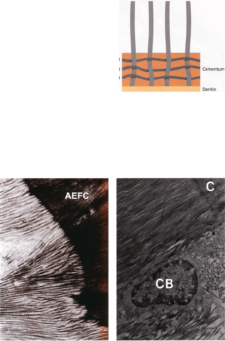
Fig. 1-69. Fig. 1-70.
ANATOMY OF THE PERIODONTIUM • 33
the surface. The presence of cementocytes allows
transportation of nutrients through the cementum,
and contributes to the maintenance of the vitality of
this mineralized tissue.
Fig. 1-68a is a photomicrograph of a section through
the periodontal ligament (PDL) in an area where the
root is covered with acellular, extrinsic fiber cemen-
tum (AEFC). The portions of the principal fibers of the
periodontal ligament which are embedded in the root
cementum (arrows) and in the alveolar bone proper (
ABP) are called Sharpey's fibers. The arrows to the
right indicate the border between ABP and the alveo-
lar bone (AB). In AEFC the Sharpey's fibers have a
smaller diameter and are more densely packed than
their counterparts in the alveolar bone. During the
continuous formation of AEFC, portions of the peri-
odontal ligament fibers (principal fibers) adjacent to
the root become embedded in the mineralized tissue.
Thus, the Sharpey's fibers in the cementum are a direct
continuation of the principal fibers in the periodontal
ligament and the supra-alveolar connective tissue.
Fig. 1-68b. The Sharpey's fibers constitute the extrinsic
fiber system (E) of the cementum and are produced by
fibroblasts in the periodontal ligament. The intrinsic
fiber system (I) is produced by cementoblasts and is
composed of fibers oriented more or less parallel to
the long axis of the root.
Fig. 1-69 shows extrinsic fibers penetrating acellular,
extrinsic fiber cementum (AEFC). The characteristic
crossbanding of the collagen fibers is masked in the
cementum because apatite crystals have become de-
posited in the fiber bundles during the process of
mineralization.
Fig. 1-70. In contrast to the bone, the cementum (C)
does not exhibit alternating periods of resorption and
apposition, but increases in thickness throughout life
by deposition of successive new layers. During this
E E
E E
Fig. 1-68b.
34 • CHAPTER 1
process of gradual apposition, the particular portion
of the principal fibers which resides immediately ad-
jacent to the root surface becomes mineralized. Min-
eralization occurs by the deposition of hydroxyapatite
crystals, first within the collagen fibers, later upon the
fiber surface and finally in the interfibrillar matrix.
The electronphotomicrograph shows a cementoblast (
CB) located near the surface of the cementum (C) and
between two inserting principal fiber bundles. Gener-
ally, the AEFC is more mineralized than CMSC and
CIFC. Sometimes only the periphery of the Sharpey's
fibers of the CMSC is mineralized, leaving an unmin-
eralized core within the fiber.
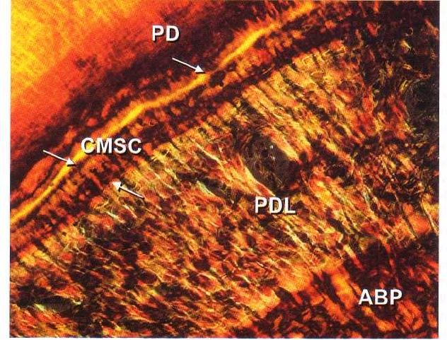
Fig. 1-71 is a photomicrograph of the periodontal liga-
ment (PDL) which resides between the cementum (
CMSC) and the alveolar bone proper (ABP). The
CMSC is densely packed with collagen fibers oriented
parallel to the root surface (intrinsic fibers) and Shar-
pey's fibers (extrinsic fibers), oriented more or less
perpendicularly to the cementum-dentine junction (
predentin (PD)). The various types of cementum in-
crease in thickness by gradual apposition throughout
life. The cementum becomes considerably wider in the
apical portion of the root than in the cervical portion,
where the thickness is only 20-50µm. In the apical root
portion the cementum is often 150-250 µm wide. The
cementum often contains incremental lines indicating
alternating periods of formation. The CMSC is formed
after the termination of tooth eruption, and after a
response to functional demands.
ALVEOLAR BONE
The alveolar process is defined as the parts of the
maxilla and the mandible that form and support the
sockets of the teeth. The alveolar process develops in
conjunction with the development and eruption of the
teeth. The alveolar process consists of bone which is
formed both by cells from the dental follicle (alveolar
bone proper) and cells which are independent of tooth
development. Together with the root cementum and
the periodontal membrane, the alveolar bone consti-
tutes the attachment apparatus of the teeth, the main
function of which is to distribute and resorb forces
generated by, for example, mastication and other tooth
contacts.
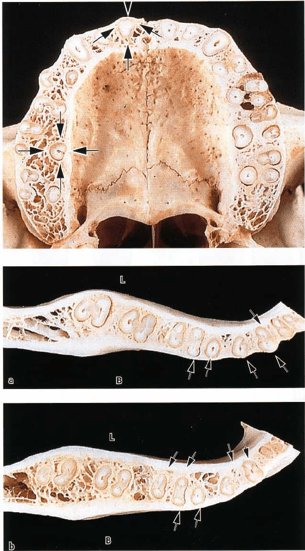
Fig. 1-72 illustrates a cross-section through the alveo-
lar process (pars alveolaris) of the maxilla at the mid-
root level of the teeth. Note that the bone which covers
the root surfaces is considerably thicker at the palatal
than at the buccal aspect of the jaw. The walls of the
sockets are lined by cortical bone (arrows), and the area
between the sockets and between the compact jaw
bone walls is occupied by cancellous bone. The cancel-
lous bone occupies most of the interdental septa but
only a relatively small portion of the buccal and pala-
tal bone plates. The cancellous bone contains bone
trabeculae, the architecture and size of which are partly
genetically determined and partly the result of the
forces to which the teeth are exposed during function.
Note how the bone on the buccal and palatal aspects
of the alveolar process varies in thickness from one
region to another. The bone plate is thick at the palatal
aspect and on the buccal aspect of the molars but thin
in the buccal anterior region.
Fig. 1-73 shows cross-sections through the mandibular
alveolar process at levels corresponding to the coronal
(Fig. 1-73a) and apical (Fig. 1-73b) thirds of the roots.
The bone lining the wall of the sockets (alveolar bone
proper) is often continuous with the compact or corti-
cal bone at the lingual (L) and buccal (B) aspects of
the
Fig. 1-71
ANATOMY OF THE PERIODONTIUM • 35
Fig. 1-72.
Fig. 1-73.
alveolar process (arrows). Note how the bone on the incisor and premolar regions, the bone plate at the buccal
and lingual aspects of the alveolar process buccal aspects of the teeth is considerably thinner than varies in
thickness from one region to another. In the
36 • CHAPTER 1
B
L
Incisors
Premolars
Molars
Fig. 1-75.
Fig. 1-76.
at the lingual aspect. In the molar region, the bone is
thicker at the buccal than at the lingual surfaces.
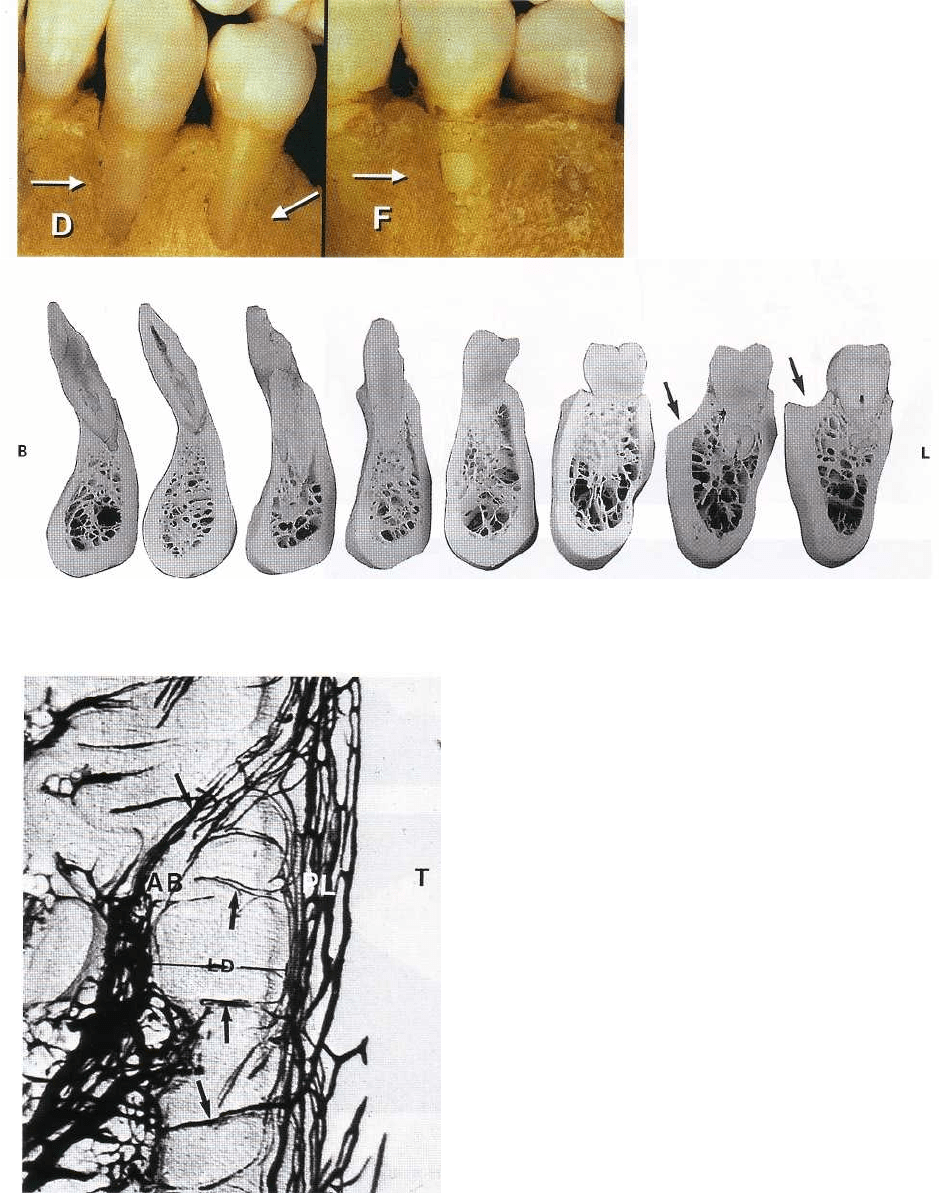
Fig. 1-74. At the buccal aspect of the jaws, the bone
coverage is sometimes missing at the coronal portion
of the roots, forming a so-called dehiscence (D). If some
bone is present in the most coronal portion of such an
area the defect is called a fenestration (F). These defects
often occur where a tooth is displaced out of the arch
and are more frequent over anterior than posterior
teeth. The root in such defects is covered only by
periodontal ligament and the overlying gingiva.
Fig. 1-75 presents vertical sections through various
regions of the mandibular dentition. The bone wall at
the buccal (B) and lingual (L) aspects of the teeth varies
considerably in thickness, e.g. from the premolar to
the molar region. Note, for instance, how the presence
of the oblique line (linea obliqua) results in a shelf-like
bone process (arrows) at the buccal aspect of the sec-
ond and third molars.
Fig. 1-76 shows a section through the periodontal
ligament (PL), tooth (T), and the alveolar bone (AB).
The blood vessels in the periodontal ligament and the
alveolar bone appear black because the blood system
was perfused with ink. The compact bone (alveolar
bone proper) which lines the tooth socket, and in a
radiograph (Fig. 1-57) appears as "lamina dura" (LD),
is perforated by numerous Volkmann's canals (arrows)
through which blood vessels, lymphatics, and nerve
fibers pass from the alveolar bone (AB) to the peri-
odontal ligament (PL). This layer of bone into which
the principal fibers are inserted (Sharpey's fibers) is
sometimes called "bundle bone". From a functional
and structural point of view, this "bundle bone" has
many features in common with the cementum layer
on the root surfaces.
ANATOMY OF THE PERIODONTIUM • 37
Fig. 1-77.
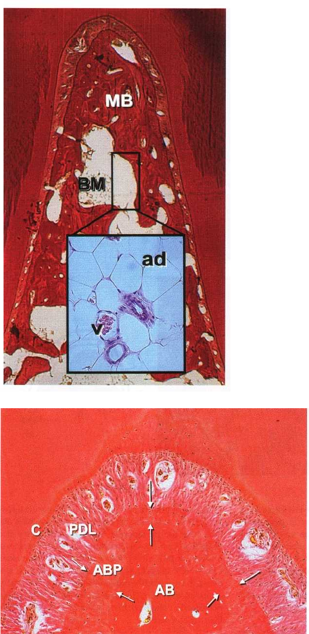
Fig. 1-77. The alveolar process starts to form early in
fetal life, with mineral deposition at small foci in the
mesenchymal matrix surrounding the tooth buds.
These small mineralized areas increase in size, fuse,
and become resorbed and remodeled until a continu-
ous mass of bone has formed around the fully erupted
teeth. The mineral content of bone, which is mainly
hydroxyapatite, is about 60% on a weight basis. The
photomicrograph illustrates the bone tissue within the
furcation area of a mandibular molar. The bone tissue
can be divided into two compartments: mineralized
bone (MB) and bone marrow (BM). The mineralized
bone is made up of lamellae — lamellar bone — while
the bone marrow contains adipocytes (ad), vascular
structures (v), and undifferentiated mesenchymal
cells (see insertion).
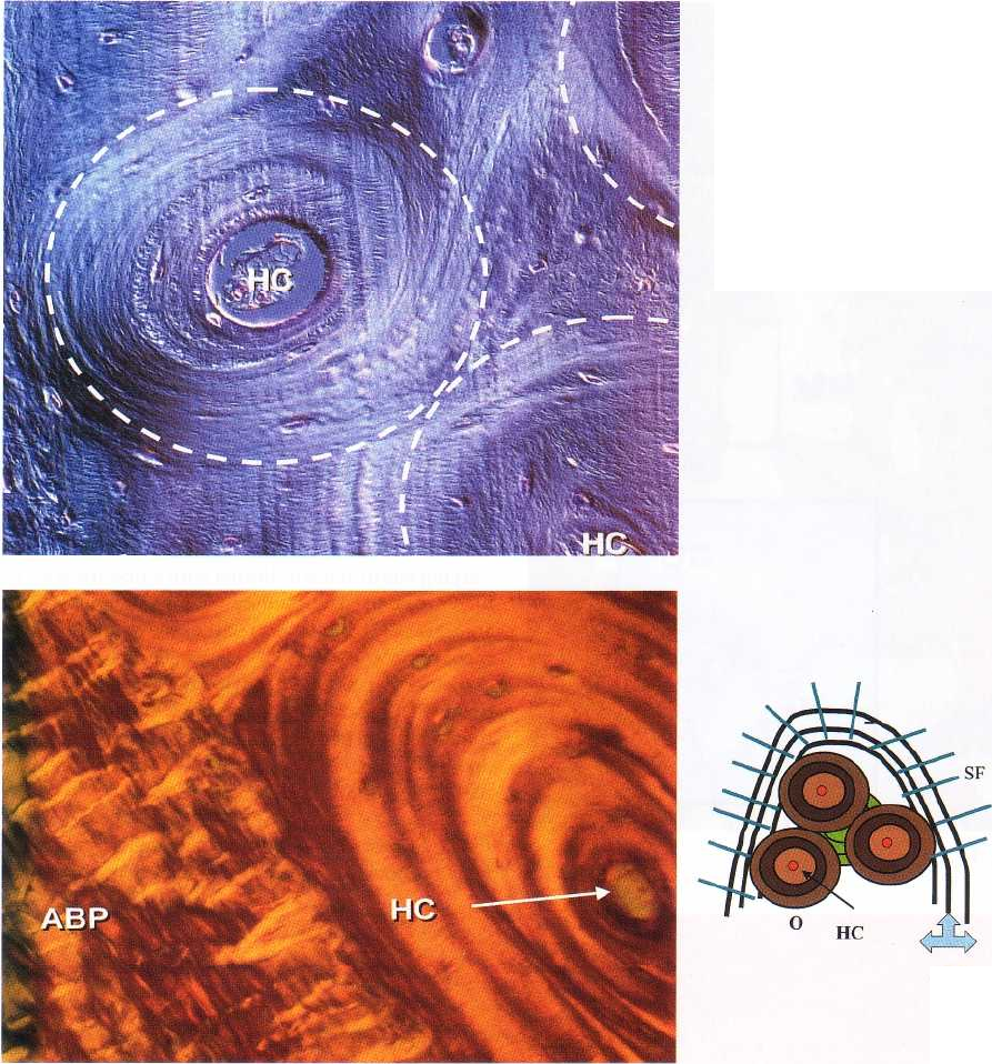
Fig. 1-78. The mineralized, lamellar bone includes two
types of bone tissue: the bone of the alveolar process
(AB) and the alveolar bone proper (ABP), which cov-
ers the alveolus. The ABP or the bundle bone has a
varying width and is indicated with white arrows. The
alveolar bone (AB) is a tissue of mesenchymal origin
and it is not considered as part of the genuine attach-
ment apparatus. The alveolar bone proper (ABP), on
the other hand, together with the periodontal liga-
ment (PDL) and the cementum (C) is responsible for
the attachment between the tooth and the skeleton. AB
and ABP may, as a result of altered functional de-
mands, undergo adaptive changes.
Fig. 1-79 describes a portion of lamellar bone. The
lamellar bone at this site contains osteons (white cir-
Fig. 1-78.
38 • CHAPTER I
Fig. 1-79.
ABP
Fig. 1-80a.
Iles) each of which harbors a blood vessel located in a
Haversian canal (HC). The blood vessel is surrounded
by concentric, mineralized lamellae to form the
osteon. The space between the different osteons is
filled with so-called interstitial lamellae. The osteons
in the lamellar bone are not only structural units but
also metabolic units. Thus, the nutrition of the bone is
secured by the blood vessels in the Haversian canals
and connecting vessels in the Volkmann canals.
Fig. 1-80. The histologic section (Fig. 1-80a) shows the
borderline between the alveolar bone proper (ABP)
and lamellar bone with an osteon. Note the presence
of the Haversian canal (HC) in the center of the
osteon.
Fig. 1-80b.
The alveolar bone proper (ABP) includes circumferen-
tial lamella and contains Sharpey's fibers which ex-
tend into the periodontal ligament. The schematic
drawing (Fig. 1-80b) is illustrating three active osteons
(brown) with a blood vessel (red) in the Haversian
canal (HC). Interstitial lamella (green) is located be-
tween the osteons (0) and represents an old and partly
remodelled osteon. The alveolar bone proper (ABP) is
presented by the dark lines into which the Sharpey's
fibers (SF) insert.
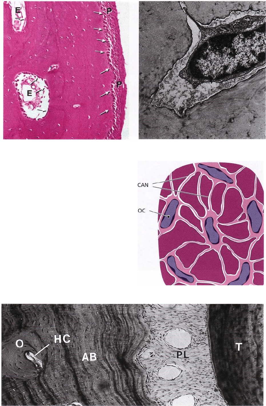
Fig. 1-81 illustrates an osteon with osteocytes (OC)
residing in osteocyte lacunae in the lamellar bone. The
osteocytes connect via canaliculi (can) which contain
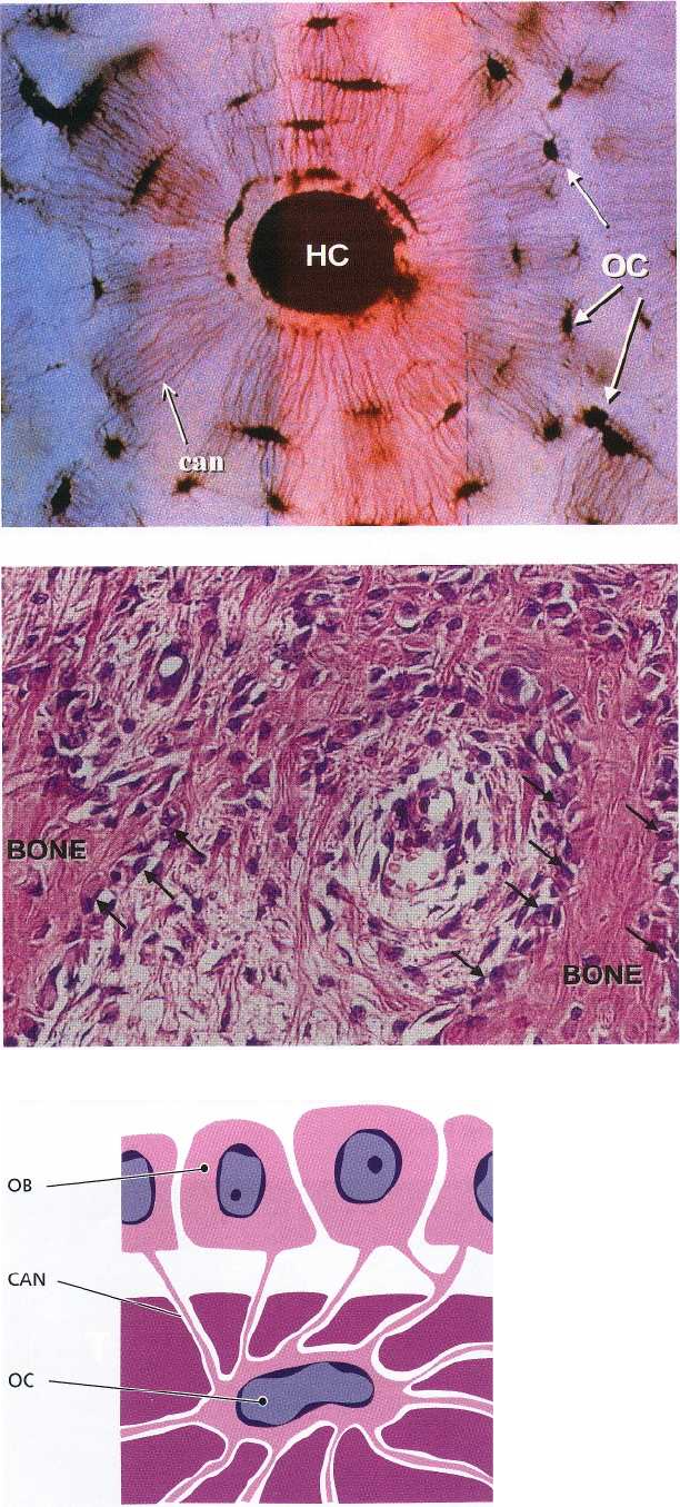
Fig. 1-83.
ANATOMY OF THE PERIODONTIUM • 39
Fig. 1-81.
Fig. 1-82.
OB
CAN
OC
cytoplasmatic projections of the osteocytes. A Haver-
sian canal (HC) is seen in the middle of the osteon.
Fig. 1-82 illustrates an area of the alveolar bone in
which bone formation occurs. The osteoblasts (ar-
rows), the bone-forming cells, are producing bone
matrix (osteoid) consisting of collagen fibers, glyco-
proteins and proteoglycans. The bone matrix or the
osteoid undergoes mineralization by the deposition of
minerals such as calcium and phosphate, which are
subsequently transformed into hydroxyapatite.
Fig. 1-83. The drawing illustrates how osteocytes, pre-
sent in the mineralized bone, communicate with
osteoblasts on the bone surface through canaliculi.
40 • CHAPTER 1
Fig. 1-84. All active bone forming sites harbor
osteoblasts. The outer surface of the bone is lined by a
layer of such osteoblasts which, in turn, are organized
in a periosteum (P) that contains densely packed col-
lagen fibers. On the "inner surface" of the bone, i.e. in
the bone marrow space, there is an endosteum (E),
which presents similar features as the periosteum.
Fig. 1-85 illustrates an osteocyte residing in a lacuna
in the bone. It can be seen that cytoplasmic processes
radiate in different directions.
Fig. 1-86 illustrates osteocytes (OC) and how their long
and delicate cytoplasmic processes communicate
through the canaliculi (CAN) in the bone. The result-
ing canalicular-lacunar system is essential for cell me
tabolism by allowing diffusion of nutrients and waste
products. The surface between the osteocytes with
their cytoplasmic processes on the one side, and the
mineralized matrix on the other, is very large. It has
Fig. 1-86.
Fig. 1-87.
Fig. 1-84.
Fig. 1-85

ANATOMY OF THE PERIODONTIUM • 41
Fig. 1-88.
been calculated that the interface between cells and
matrix in a cube of bone, 10 x 10 x 10 cm, amounts to
approximately 250 m
2
. This enormous surface of ex-
change serves as a regulator, e.g. for the serum calcium
and the serum phosphate levels via hormonal control
mechanisms.
Fig. 1-87. The alveolar bone is constantly renewed in
response to functional demands. The teeth erupt and
migrate in a mesial direction throughout life to com-
pensate for attrition. Such movement of the teeth im-
plies remodelling of the alveolar bone. During the
process of remodelling, the bone trabeculae are con-
tinuously resorbed and reformed and the cortical bone
mass is dissolved and replaced by new bone. During
breakdown of the cortical bone, resorption canals are
formed by proliferating blood vessels. Such canals,
which in their center contain a blood vessel, are sub-
sequently refilled with new bone by the formation of
lamellae arranged in concentric layers around the
blood vessel. A new Haversian system (0) is seen in
the photomicrograph of a horizontal section through
the alveolar bone (AB), periodontal ligament (PL) and
tooth (T).
Fig. 1-89.
Fig. 1-88. The resorption of bone is always associated
with osteoclasts (Ocl). These cells are giant cells special
ized in the breakdown of mineralized matrix (bone,
dentin, cementum) and are probably developed from
blood monocytes. The resorption occurs by the release
of acid substances (lactic acid, etc.) which forms an
acidic environment in which the mineral salts of the
bone tissue become dissolved. Remaining organic
substances are eliminated by enzymes and osteoclas-
tic phagocytosis. Actively resorbing osteoclasts ad-
here to the bone surface and produce lacunar pits
called Howship's lacunae (dotted line). They are mobile
and capable of migrating over the bone surface. The
photomicrograph demonstrates osteoclastic activity at
the surface of alveolar bone (AB).
Fig. 1-89 illustrates a so-called bone multicellular unit
(BMU), which is present in bone tissue undergoing
active remodeling. The reversal line, indicated by red
arrows, demonstrates to which level bone resorption
has occurred. From the reversal line new bone has
started to form and has the character of osteoid. Note
the presence of osteoblasts (ob) and vascular struc-
tures (v). The osteoclasts resorb organic as well as
inorganic substances.
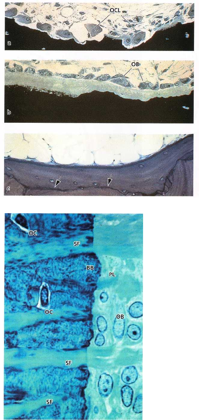
42 • CHAPTER 1
Fig. 1-90.
sorption followed by formation) in response to tooth
drifting and changes in functional forces acting on the
teeth. Remodeling of the trabecular bone starts with
resorption of the bone surface by osteoclasts (OCL) as
seen in Fig. 1-90a. After a short period, osteoblasts (
OB) start depositing new bone (Fig. 1-90b) and finally a
new bone multicellular unit is formed, clearly deli-
neated by a reversal line (arrows) as seen in Fig. 1-90c.
Fig. 1-91. Collagen fibers of the periodontal ligament (
PL) are inserting in the mineralized bone which lines
the wall of the tooth socket. This bone, which as
previously described is called alveolar bone proper or
bundle bone (BB), has a high turnover rate. The por-
tions of the collagen fibers which are inserted inside
the bundle bone are called Sharpey's fibers (SF). These
fibers are mineralized at their periphery, but often have
a non-mineralized central core. The collagen fiber
bundles inserting in the bundle bone generally have a
larger diameter and are less numerous than the
corresponding fiber bundles in the cement-um on the
opposite side of the periodontal ligament. Individual
bundles of fibers can be followed all the way from the
alveolar bone to the cementum. However, despite
being in the same bundle of fibers, the collagen adja-
cent to the bone is always less mature than that adja-
cent to the cementum. The collagen on the tooth side
has a low turnover rate. Thus, while the collagen
adjacent to the bone is renewed relatively rapidly, the
collagen adjacent to the root surface is renewed slowly
Fig. 1-90. Both the cortical and cancellous alveolar or not at all. Note the occurrence of osteoblasts (OB)
bone are constantly undergoing remodeling (i.e. re- and osteocytes (OC).
Fig. 1-91.
