Jan Lindhe. Clinical Periodontology
Подождите немного. Документ загружается.

ANATOMY OF THE PERIODONTIUM • 23
TF
Fig. 1-45.
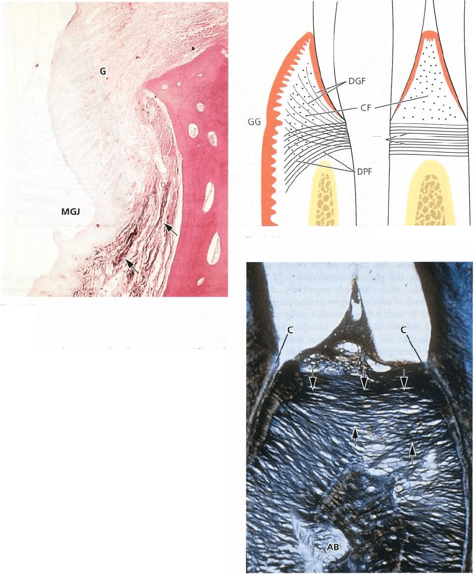
also connect the supra-alveolar cementum (C) with
the crest of the alveolar bone (AB). The four groups of
collagen fiber bundles presented in Fig. 1-46 reinforce
the gingiva and provide the resilience and tone which
is necessary for maintaining its architectural form and
the integrity of the dento-gingival attachment.
Matrix
The matrix of the connective tissue is produced mainly
by the fibroblasts, although some constituents are
produced by mast cells, and other components are
derived from the blood. The matrix is the medium in
which the connective tissue cells are embedded and it
is essential for the maintenance of the normal function
of the connective tissue. Thus, the transportation of
water, electrolytes, nutrients, metabolites, etc., to and
from the individual connective tissue cells occurs
within the matrix. The main constituents of the con-
nective tissue matrix are protein carbohydrate macro-
molecules. These complexes are normally divided
into proteoglycans and glycoproteins. The proteoglycans
contain glycosaminoglycans as the carbohydrate units
(hyaluronan sulfate, heparan sulfate, etc.), which, via
covalent bonds, are attached to one or more protein
chains. The carbohydrate component is always pre-
dominant in the proteoglycans. The glycosaminogly-
can called hyaluronan or "hyaluronic acid" is prob-
ably not bound to protein. The glycoproteins (fi-
bronectin, osteonectin, etc.) also contain polysaccha-
Fig. 1-46.
Fig. 1-47.
rides, but these macromolecules are different from
glycosaminoglycans. The protein component is pre-
dominating in glycoproteins. In the macromolecules,
mono- or oligosaccharides are, via covalent bonds,
connected with one or more protein chains.
Fig. 1-48. Normal function of the connective tissue
depends on the presence of proteoglycans and gly-
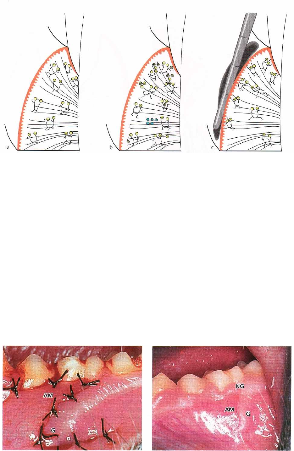
Fig. 1-49. Fig. 1-50.
24 • CHAPTER 1
cosaminoglycans. The carbohydrate moiety of the
proteoglycans, the glycosaminoglycans ( ), are large,
flexible, chain formed, negatively charged molecules,
each of which occupies a rather large space (Fig. 1-
48a). In such a space, smaller molecules, e.g. water
and electrolytes, can be incorporated while larger
molecules are prevented from entering (Fig. 1-48b).
The proteoglycans thereby regulate diffusion and
fluid flow through the matrix and are important
determinants for the fluid content of the tissue and the
maintenance of the osmotic pressure. In other words,
the proteoglycans act as a molecule filter and, in addi-
tion, play an important role in the regulation of cell
migration (movements) in the tissue. Due to their
structure and hydration, the macromolecules exert
resistance towards deformation, thereby serving as
regulators of the consistency of the connective tissue
(Fig. 1-48c). If the gingiva is suppressed, the macro-
molecules become deformed. When the pressure is
eliminated, the macromolecules regain their original
form. Thus, the macromolecules are important for the
resilience of the gingiva.
Epithelial mesenchymal interaction
There are many examples of the fact that during the
embryonic development of various organs, a mutual
inductive influence occurs between the epithelium
and the connective tissue. The development of the
teeth is a characteristic example of such phenomena.
The connective tissue is, on the one hand, a determin-
ing factor for normal development of the tooth bud
while, on the other, the enamel epithelia exert a defi-
nite influence on the development of the mesenchy-
mal components of the teeth.
It has been suggested that tissue differentiation in
the adult organism can be influenced by environ-
mental factors. The skin and mucous membranes, for
instance, often display increased keratinization and
hyperplasia of the epithelium in areas which are ex-
posed to mechanical stimulation. Thus, the tissues
seem to adapt to environmental stimuli. The presence
of keratinized epithelium on the masticatory mucosa
has been considered to represent an adaptation to
mechanical irritation released by mastication. How-
ever, research has demonstrated that the characteristic
Fig. 1-48.
ANATOMY OF THE PERIODONTIUM • 25
Fig. 1-51.
features of the epithelium in such areas are genetically
determined. Some pertinent observations are reported
in the following:
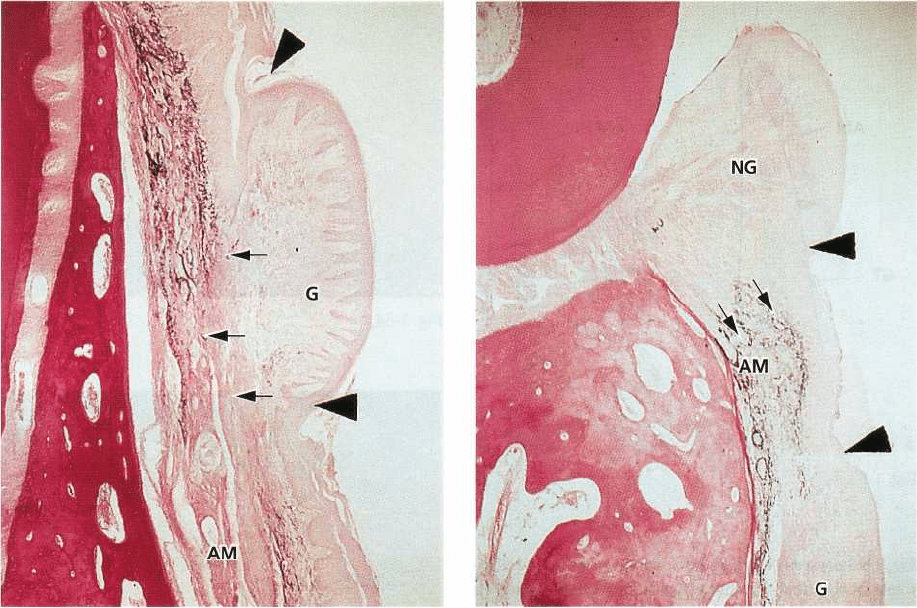
Fig. 1-49 shows an area in a monkey where the gingiva
(G) and the alveolar mucosa (AM) have been trans-
posed by a surgical procedure. The alveolar mucosa is
placed in close contact with the teeth while the gingiva
is positioned in the area of the alveolar mucosa.
Fig. 1-50 shows the same area, as seen in Fig. 1-49, 4
months later. Despite the fact that the transplanted
gingiva (G) is mobile in relation to the underlying
bone, like the alveolar mucosa, it has retained its
characteristic, morphologic features of a masticatory
mucosa. However, a narrow zone of new keratinized
gingiva (NG) has regenerated between the trans-
planted alveolar mucosa (AM) and the teeth.
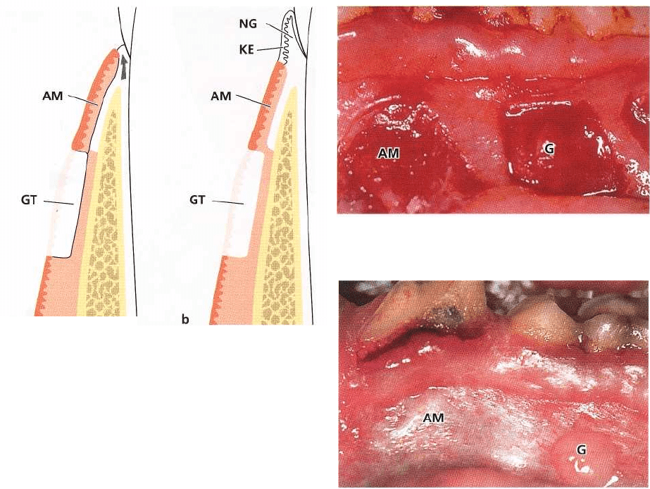
Fig. 1-51 presents a histologic section cut through the
transplanted gingiva seen in Fig. 1-50. Since elastic
fibers are lacking in the gingival connective tissue (G),
but are numerous (small arrows) in the connective
tissue of the alveolar mucosa (AM), the transplanted
gingival tissue can readily be identified. The epithe-
lium covering the transplanted gingival tissue exhib-
its a distinct keratin layer (between large arrows) on
the surface, and also the configuration of the epithe-
lium-connective tissue interface (i.e. rete pegs and
connective tissue papillae) is similar to that of normal
non-transplanted gingiva. Thus, the heterotopically
located gingival tissue has maintained its original
Fig. 1-52.
specificity. This observation demonstrates that the
characteristics of the gingiva are genetically deter-
mined rather than being the result of functional adap-
tation to environmental stimuli.
Fig. 1-52 shows a histologic section cut through the
coronal portion of the area of transplantation (shown
in Fig. 1-50). The transplanted gingival tissue (G)
shown in Fig. 1-51 can be seen in the lower portion of
the photomicrograph. The alveolar mucosa transplant (
AM) is seen between the large arrows in the middle
of the illustration. After surgery, the alveolar mucosa
transplant was positioned in close contact with the
teeth as seen in Fig. 1-49. After healing, a narrow zone
of keratinized gingiva (NG) developed coronal to the
alveolar mucosa transplant (see Fig. 1-50). This new
zone of gingiva (NG), which can be seen in the upper
portion of the histologic section, is covered by kerati-
nized epithelium and the connective tissue contains
no purple-stained elastic fibers. In addition, it is im-
portant to notice that the junction between keratinized
and non-keratinized epithelium (large arrows) corre-
sponds exactly to the junction between "elastic" and "
inelastic" connective tissue (small arrows). The con-
nective tissue of the new gingiva has regenerated from
the connective tissue of the supra-alveolar and peri-
odontal ligament compartments and has separated the
alveolar mucosal transplant (AM) from the tooth (see
Fig. 1-53). However, it is most likely that the
epithelium which covers the new gingiva has mi-
grated from the adjacent epithelium of the alveolar
mucosa.
26 • CHAPTER 1
Fig. 1-53.
Fig. 1-53 presents a schematic drawing of the develop-
ment of the new, narrow zone of keratinized gingiva (
NG) seen in Figs. 1-50 and 1-52.
Fig. 1-53a. Granulation tissue has proliferated
coronally along the root surface (arrow) and has sepa-
rated the alveolar mucosa transplant (AM) from its
original contact with the tooth surface.
Fig. 1-53b. Epithelial cells have migrated from the
alveolar mucosal transplant (AM) onto the newly
formed gingival connective tissue (NG). Thus, the
newly formed gingiva has become covered with a
keratinized epithelium (KE) which has originated
from the non-keratinized epithelium of the alveolar
mucosa (AM). This implies that the newly formed
gingival connective tissue (NG) possesses the ability
to induce changes in the differentiation of the epithe-
lium originating from the alveolar mucosa. This epi-
thelium, which is normally non-keratinized, appar-
ently differentiates to keratinized epithelium because
of stimuli arising from the newly formed gingival
connective tissue (NG). (GT: gingival transplant.)
Fig. 1-54 illustrates a portion of gingival connective
tissue (G) and alveolar mucosal connective tissue (
AM) which, after transplantation, has healed into
wound areas in the alveolar mucosa. Epithelialization
of these transplants can only occur through migration
of epithelial cells from the surrounding alveolar mu-
cosa.
Fig. 1-55 shows the transplanted gingival connective
tissue (G) after re-epithelialization. This tissue portion
has attained an appearance similar to that of the nor-
mal gingiva, indicating that this connective tissue is
now covered by keratinized epithelium. The trans-
Fig. 1-54.
Fig. 1-55.
planted connective tissue from the alveolar mucosa (
AM) is covered by non-keratinized epithelium, and
has the same appearance as the surrounding alveolar
mucosa.
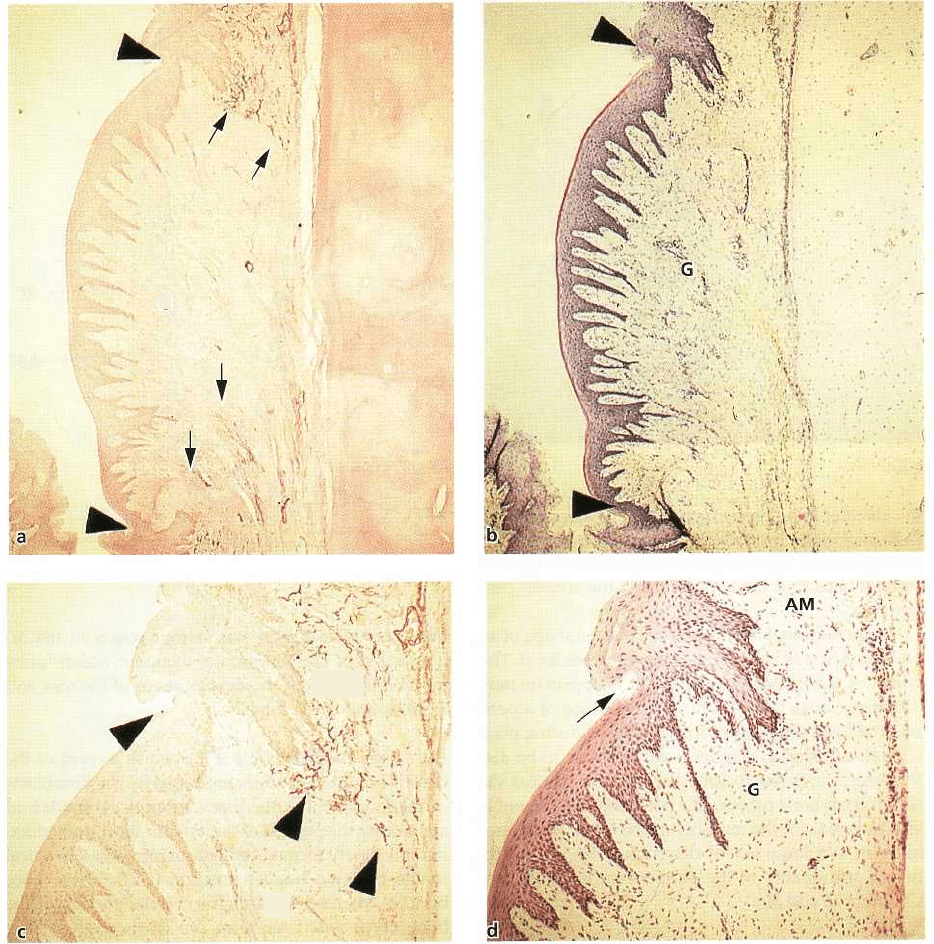
Fig. 1-56 presents two histologic sections through the
area of the transplanted gingival connective tissue.
The section shown in Fig. 1-56a is stained for elastic
fibers (arrows). The tissue in the middle without elas-
tic fibers is the transplanted gingival connective tissue
(G). Fig. 1-56b shows an adjacent section stained with
hematoxylin and eosin. By comparing Figs. 1-56a and
1-56b it can be seen that:
1. the transplanted gingival connective tissue is cov-
ered by keratinized epithelium (between arrow-
heads)
2. the epithelium-connective tissue interface has the
same wavy course (i.e. rete pegs and connective
tissue papillae) as seen in normal gingiva.
The photomicrographs seen in Figs. 1-56c and 1-56d
illustrate, at a higher magnification, the border area
between the alveolar mucosa (AM) and the trans-
planted gingival connective tissue (G). Note the dis-
tinct relationship between keratinized epithelium (ar-
row) and "inelastic" connective tissue (arrowheads),
and between non-keratinized epithelium and "elas-
a
GT
b
ANATOMY OF THE PERIODONTIUM • 27
G
-AM
G
Fig. 1-56.
tic" connective tissue. The establishment of such a
close relationship during healing implies that the
transplanted gingival connective tissue possesses the
ability to alter the differentiation of epithelial cells as
previously suggested (Fig. 1-53). From being non-
keratinizing cells, the cells of the epithelium of the
alveolar mucosa have evidently become keratinizing
cells. This means that the specificity of the gingival
epithelium is determined by genetic factors inherent
in the connective tissue.
PERIODONTAL LIGAMENT
The periodontal ligament is the soft, richly vascular
and cellular connective tissue which surrounds the
roots of the teeth and joins the root cementum with
socket wall. In the coronal direction, the periodontal
ligament is continuous with the lamina propria of the
gingiva and is demarcated from the gingiva by the
collagen fiber bundles which connect the alveolar
bone crest with the root (the alveolar crest fibers).
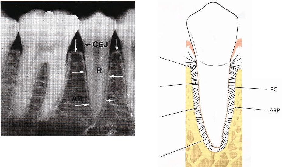
Fig. 1-57 is a radiograph of a mandibular premolar-
molar region. In radiographs, two types of alveolar
bone can be distinguished:
1. The part of the alveolar bone which covers the
alveolus, called "lamina dura" (arrows).
2. The portion of the alveolar process which, in the
radiograph, has the appearance of a meshwork.
This is called the "spongy bone".
The periodontal ligament is situated in the space be-
tween the roots (R) of the teeth and the lamina dura or
28 • CHAPTER 1
Fig. 1-57.
the alveolar bone proper (arrows). The alveolar bone (
AB) surrounds the tooth to a level approximately 1
mm apical to the cemento-enamel junction (CEJ). The
coronal border of the bone is called the alveolar crest (
arrows).
The periodontal ligament space has the shape of an
hourglass and is narrowest at the mid-root level. The
width of the periodontal ligament is approximately 0.
25 mm (range 0.2-0.4 mm). The presence of a peri-
odontal ligament permits forces, elicited during mas-
ticatory function and other tooth contacts, to be dis-
tributed to and resorbed by the alveolar process via
the alveolar bone proper. The periodontal ligament is
also essential for the mobility of the teeth. Tooth mo-
bility is to a large extent determined by the width,
height and quality of the periodontal ligament (see
Chapters 18 and 30).
Fig. 1-58 illustrates in a schematic drawing how the
periodontal ligament is situated between the alveolar
bone proper (ABP) and the root cementum (RC). The
tooth is joined to the bone by bundles of collagen fibers
which can be divided into the following main groups
according to their arrangement:
1. alveolar crest fibers (ACF)
2. horizontal fibers (HF)
3. oblique fibers (OF)
4. apical fibers (APF)
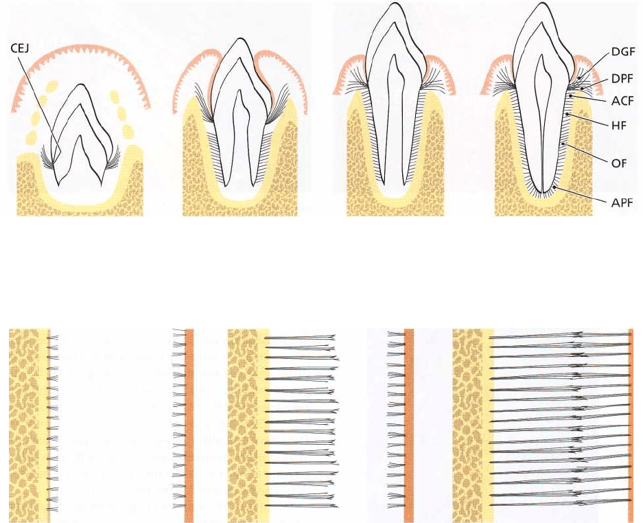
Fig. 1-59. The periodontal ligament and the root ce-
mentum develop from the loose connective tissue (the
follicle) which surrounds the tooth bud. The sche-
ACF
HF
OF
APF
Fig. 1-58.
matic drawing depicts the various stages in the or-
ganization of the periodontal ligament which forms
concomitantly with the development of the root and
the eruption of the tooth.
Fig. 1-59a. The tooth bud is formed in a crypt of the
bone. The collagen fibers produced by the fibroblasts
in the loose connective tissue around the tooth bud
are, during the process of their maturation, embedded
into the newly formed cementum immediately apical
to the cemento-enamel junction (CEJ). These fiber
bundles oriented towards the coronal portion of the
bone crypt will later form the dentogingival fiber
group, the dentoperiosteal fiber group and the
transseptal fiber group which belong to the oriented
fibers of the gingiva (see Fig. 1-46).
Fig. 1-59b. The true periodontal ligament fibers, the
principal fibers, develop in conjunction with the erup-
tion of the tooth. First, fibers can be identified entering
the most marginal portion of the alveolar bone.
Fig. 1-59c. Later, more apically positioned bundles of
oriented collagen fibers are seen.
Fig. 1-59d. The orientation of the collagen fiber bun-
dles alters continuously during the phase of tooth
eruption. First, when the tooth has reached contact in
ANATOMY OF THE PERIODONTIUM • 29
a
b
c
d
Fig. 1-59.
ABP
PL RC
ABP
PL RC
ABP
PL RC
a
b
c
Fig. 1-60.
occlusion and is functioning properly, the fibers of the
periodontal ligament associate into groups of well-
oriented dentoalveolar collagen fibers demonstrated
in Fig. 1-58. These collagen structures undergo con-
stant remodeling (i.e. resorption of old fibers and
formation of new ones).
Fig. 1-60. This schematic drawing illustrates the devel-
opment of the principal fibers of the periodontal liga-
ment. The alveolar bone proper (ABP) is seen to the
left, the periodontal ligament (PL) is depicted in the
center and the root cementum (RC) is seen to the right.
Fig. 1-60a. First, small, fine, brush-like fibrils are de-
tected arising from the root cementum and projecting
into the PL space. The surface of the bone is, at this
stage, covered by osteoblasts. From the surface of the
bone only a small number of radiating, thin collagen
fibrils can be seen.
Fig. 1-60b. Later on, the number and thickness of fibers
entering the bone increase. These fibers radiate to-
wards the loose connective tissue in the mid-portion
of the periodontal ligament area (PL), which contains
more or less randomly oriented collagen fibrils. The
fibers originating from the cementum are still short
while those entering the bone gradually become
longer. The terminal portions of these fibers carry
finger-like projections.
Fig. 1-60c. The fibers originating from the cementum
subsequently increase in length and thickness and
fuse in the periodontal ligament space with the fibers
originating from the alveolar bone. When the tooth,
following eruption, reaches contact in occlusion and
starts to function, the principal fibers become organ-
ized in bundles and run continuously from the bone to
the cementum.
30 • CHAPTER 1
Fig. 1-61a.
Fig. 1-62a.
Fig. 1-62b.
Fig. 1-61b.
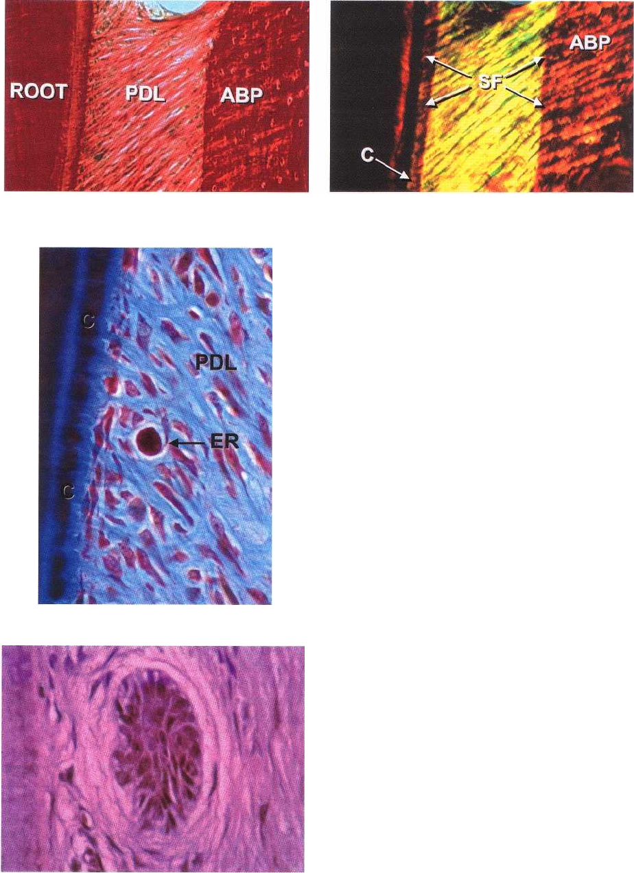
(Sharpey's fibers) have a smaller diameter but are
more numerous than those embedded in the alveolar
bone proper (Sharpey's fibers).
Fig. 1-61b presents a polarized version of Fig. 1-61a. In
this illustration the Sharpey's fibers (SF) can be seen
penetrating not only the cementum (C) but also the
entire width of the alveolar bone proper (ABP). The
periodontal ligament also contains a few elastic fibers
associated with the blood vessels. Oxytalan fibers (see
Fig. 1-44) are also present in the periodontal ligament.
They have a mainly apico-occlusal orientation and are
located in the ligament closer to the tooth than to the
alveolar bone. Very often they insert into the cemen-
tum. Their function has not been determined.
The cells of the periodontal ligament are: fibroblasts,
osteoblasts, cementoblasts, osteoclasts, as well as epithelial
cells and nerve fibers. The fibroblasts are aligned along
the principal fibers, while cementoblasts line the sur-
face of the cementum, and the osteoblasts line the bone
surface.
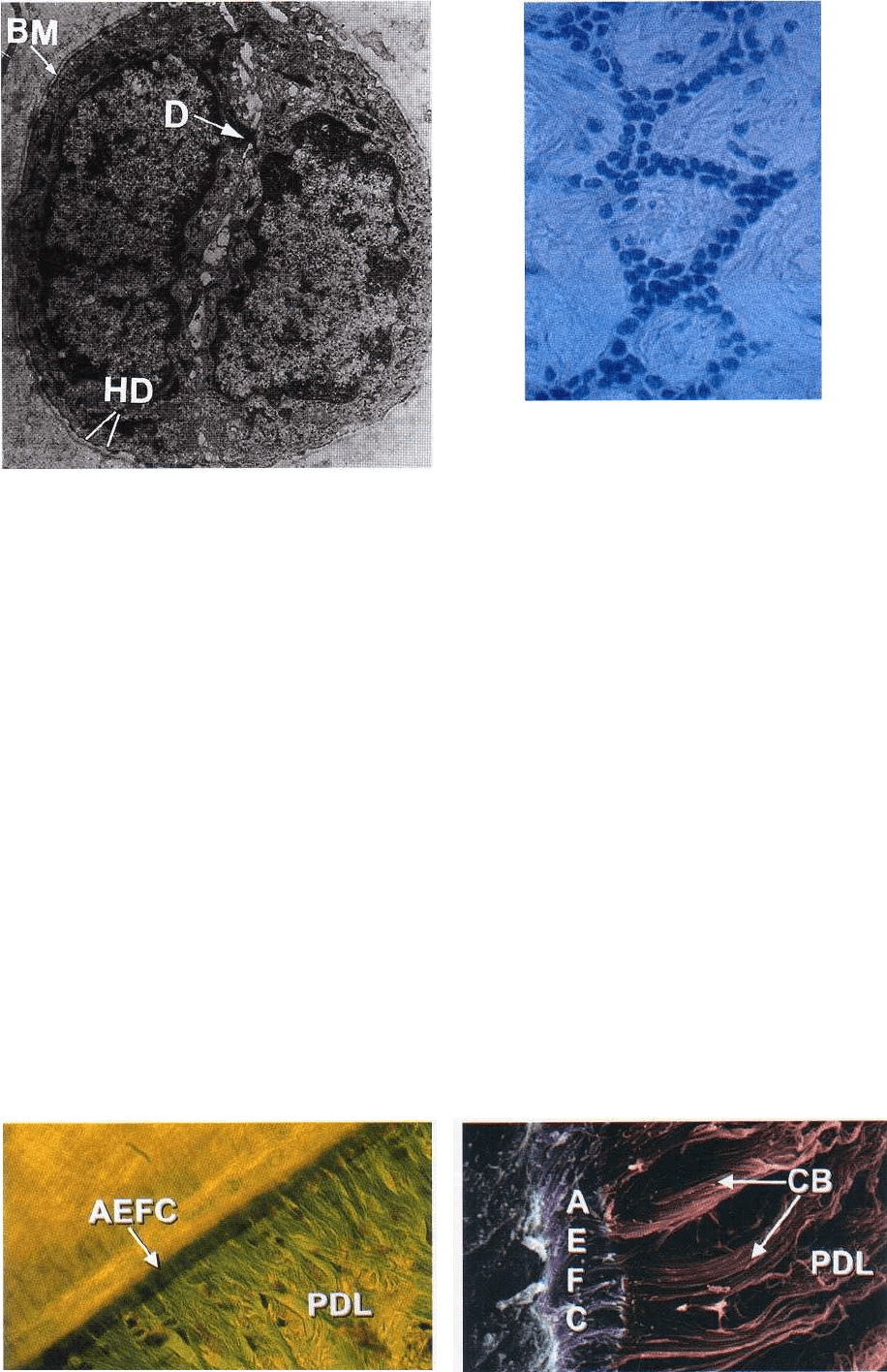
Fig. 1-62a shows the presence of clusters of epithelial
cells (ER) in the periodontal ligament (PDL). These
cells, called the epithelial cell rests of Mallassez, represent
remnants of the Hertwig's epithelial root sheath. The
epithelial cell rests are situated in the periodontal
ligament at a distance of 15-75 µm from the cementum
(C) on the root surface. A group of such epithelial cell
rests is seen in a higher magnification in Fig. 1-62b.
Fig. 1-63. Electron microscopically it can be seen that
the epithelial cell rests are surrounded by a basement
membrane (BM) and that the cell membranes of the
epithelial cells exhibit the presence of desmosomes (D)
as well as hemidesmosomes (HD). The epithelial cells
contain only few mitochondria and have a poorly
developed endoplasmic reticulum. This means that
they are vital, but resting, cells with minute metabo-
lism.
Fig. 1-61a illustrates how the principal fibers of the Fig. 1-64 is a photomicrograph of a periodontal liga-
periodontal ligament (PDL) run continuously from ment removed from an extracted tooth. This specimen the
root cementum to the alveolar bone proper (ABP). prepared tangential to the root surface shows that the The
principal fibers embedded in the cementum epithelial cell rests of Mallassez, which in ordinary
ANATOMY OF THE PERIODONTIUM • 31
Fig. 1-63.
Different forms of cementum have been
described:
Fig. 1-64.
histologic sections appear as isolated groups of epi-
thelial cells, in fact form a continuous network of
epithelial cells surrounding the root. Their function is
at present unknown.
ROOT CEMENTUM
The cementum is a specialized mineralized tissue cov-
ering the root surfaces and, occasionally, small por-
tions of the crown of the teeth. It has many features in
common with bone tissue. However, the cementum
contains no blood or lymph vessels, has no innerva-
tion, does not undergo physiologic resorption or re-
modeling, but is characterized by continuing deposi-
tion throughout life. Like other mineralized tissues, it
contains collagen fibers embedded in an organic ma-
trix. Its mineral content, which is mainly hydroxyapa-
tite, is about 65% by weight; a little more than that of
bone (i.e. 60%). Cementum serves different functions.
It attaches the periodontal ligament fibers to the root
and contributes to the process of repair after damage
to the root surface.
1. Acellular, extrinsic fiber cementum (AEFC) is found in
the coronal and middle portions of the root and
contains mainly bundles of Sharpey's fibers. This
type of cementum is an important part of the at-
tachment apparatus and connects the tooth with the
alveolar bone proper.
2. Cellular, mixed stratified cementum (CMSC) occurs in
the apical third of the roots and in the furcations. It
contains both extrinsic and intrinsic fibers as well
as cementocytes.
3. Cellular, intrinsic fiber cementum (CIFC) is found
mainly in resorption lacunae and it contains intrin-
sic fibers and cementocytes.
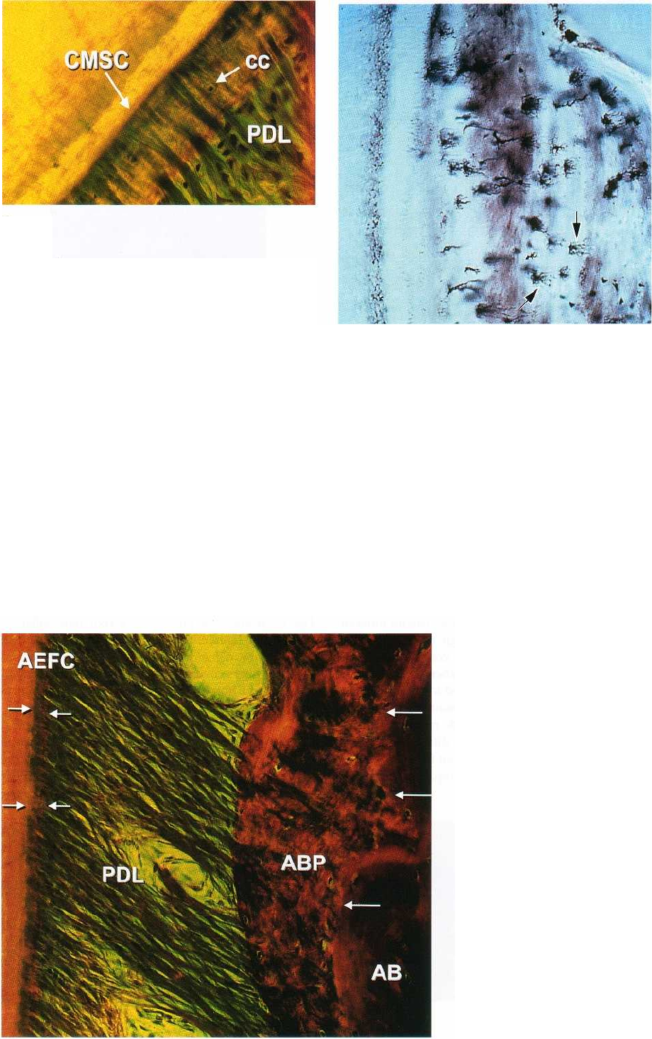
Fig. 1-65a shows a portion of a root with adjacent
periodontal ligament (PDL). A thin layer of acellular,
extrinsic fiber cementum (AEFC) with densely packed
extrinsic fibers covers the peripheral dentin. Cemen-
toblasts and fibroblasts can be observed adjacent to
the cementum.
Fig. 1-65b represents a scanning electron micrograph
of AEFC. Note that the extrinsic fibers attach to the
dentin (left) and are continous with the collagen fiber
bundles (CB) of the periodontal ligament (PDL). The
Fig. 1-65a
Fig. 1-65b.
32 • CHAPTER 1
Fig. 1-66.
AEFC is formed concomitantly with the formation of
the root dentin. At a certain stage during tooth forma
tion, the epithelial sheath of Hertwig, which lines the
newly formed predentin, is fragmented. Cells from
the dental follicle then penetrate the epithelial sheath
of Hertwig and occupy the area next to the predentin.
In this position, the ectomesenchymal cells from the
dental follicle differentiate into cementoblasts and be
-gin to produce collagen fibers at right angles to the
surface. The first cementum is deposited on the highly
mineralized superficial layer of the mantle dentin
called the "hyaline layer" which contains enamel ma-
trix proteins and the initial collagen fibers of the ce-
mentum. Subsequently, cementoblasts drift away
from the surface resulting in increased thickness of the
cementum and incorporation of principal fibers.
Fig. 1-66 demonstrates the structure of cellular, mixed
stratified cementum (CMSC) which, in contrast to
AEFC, contains cells and intrinsic fibers. The CMSC is
laid down throughout the functional period of the
Fig. 1-67.
tooth. The various types of cementum are produced
by cementoblasts or periodontal ligament (PDL) cells
lining the cementum surface. Some of these cells be-
come incorporated into the cementoid, which sub-
sequently mineralizes to form cementum. The cells
which are incorporated in the cementum are called
cementocytes (CC).
Fig. 1-67 illustrates how cementocytes (black cells)
reside in lacunae in CMSC or CIFC. They communi-
cate with each other through a network of cytoplasmic
processes (arrows) running in canaliculi in the cemen-
turn. The cementocytes also, through cytoplasmic
processes, communicate with the cementoblasts on
