Jan Lindhe. Clinical Periodontology
Подождите немного. Документ загружается.

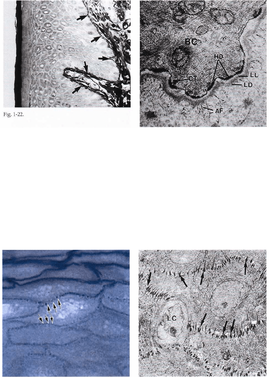
Fig. 1-24. Fig. 1-25.
ANATOMY OF THE PERIODONTIUM • 13
Fig. 1-22.
the cell converge towards such hemidesmosomes. The
hemidesmosomes are involved in the attachment of
the epithelium to the underlying basement mem-
brane.
Fig. 1-24 illustrates an area of stratum spinosum in the
gingival oral epithelium. Stratum spinosum consists
of 10-20 layers of relatively large, polyhedral cells,
equipped with short cytoplasmic processes resem-
bling spines. The cytoplasmic processes (arrows) oc-
cur at regular intervals and give the cells a prickly
appearance. Together with intercellular protein-car-
bohydrate complexes, cohesion between the cells is
provided by numerous "desmosomes" (pairs of
hemidesmosomes) which are located between the cy-
toplasmic processes of adjacent cells.
Fig. 1-23.
Fig. 1-25 shows an area of stratum spinosum in an
electronmicrograph. The dark-stained structures be-
tween the individual epithelial cells represent the des-
mosomes (arrows). A desmosome may be considered
to be two hemidesmosomes facing one another. The
presence of a large number of desmosomes indicates
that the cohesion between the epithelial cells is solid.
The light cell (LC) in the center of the illustration
harbors no hemidesmosomes and is, therefore, not a
keratinocyte but rather a "clear cell" (see also Fig. 1-
19).
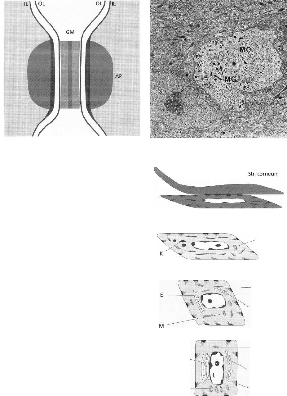
Fig. 1-26 is a schematic drawing describing the com-
position of a desmosome. A desmosome can be con-
14 • CHAPTER 1
sidered to consist of two adjoining hemidesmosomes
separated by a zone containing electron-dense granu-
lated material (GM). Thus, a desmosome comprises
the following structural components: (1) the outer leaf-
lets (OL) of the cell membrane of two adjoining cells, (
2) the thick inner leaflets (IL) of the cell membranes
and (3) the attachment plaques (AP), which represent
granular and fibrillar material in the cytoplasm.
Fig. 1-27. As mentioned previously, the oral epithe-
lium also contains melanocytes, which are responsible
for the production of the pigment melanin. Melano-
cytes are present in individuals with marked pigmen-
tation of the oral mucosa (Indians and Negroes) as
well as in individuals where no clinical signs of pig-
mentation can be seen. In this electronmicrograph a
melanocyte (MC) is present in the lower portion of the
stratum spinosum. In contrast to the keratinocytes,
this cell contains melanin granules (MG) and has no
tonofilaments or hemidesmosomes. Note the large
amount of tonofilaments in the cytoplasm of the adja-
cent keratinocytes.
Fig. 1-28. When traversing the epithelium from the
basal layer to the epithelial surface, the keratinocytes
undergo continuous differentiation and specializa-
tion. The many changes which occur during this proc-
ess are indicated in this diagram of a keratinized
stratified squamous epithelium. From the basal layer
(stratum basale) to the granular layer (stratum granu-
losum) both the number of tonofilaments (F) in the
cytoplasm and the number of desmosomes (D) in-
crease. In contrast, the number of organelles such as
mitochondria (M), lamellae of rough endoplasmic
reticulum (E) and Golgi complexes (G) decrease in the
keratinocytes on their way from the basal layer to-
wards the surface. In the stratum granulosum, elec-
tron dense keratohyalin bodies (K) and clusters of gly-
cogen containing granules start to occur. Such gran-
Str. granulosum
Str. spinosum
F
G
M
Str. basale
Fig. 1-28.
E
M
F
G D
Fig. 1-27.
Fig. 1-26.
ANATOMY OF THE PERIODONTIUM • 15
ules are believed to be related to the synthesis of
keratin.
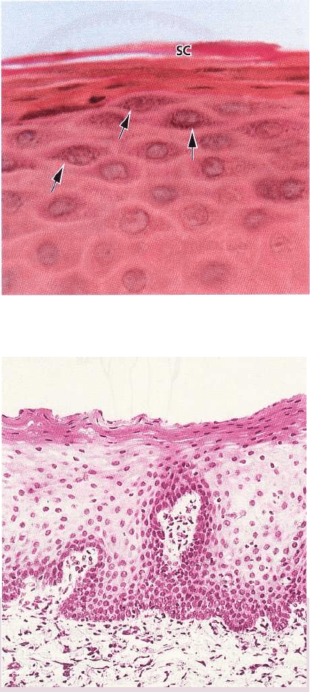
Fig. 1-29 is a photomicrograph of the stratum granu-
losum and stratum corneum. Keratohyalin granules
(arrows) are seen in the stratum granulosum. There is
an abrupt transition of the cells from the stratum
granulosum to the stratum corneum. This is indicative
of a very sudden keratinization of the cytoplasm of the
keratinocyte and its conversion into a horny squame.
The cytoplasm of the cells in the stratum corneum (SC)
is filled with keratin and the entire apparatus for
protein synthesis and energy production, i.e. the nu-
cleus, the mitochondria, the endoplasmic reticulum
and the Golgi complex, is lost. In a parakeratinized
epithelium, however, the cells of the stratum corneum
contain remnants of nuclei. Keratinization is consid-
ered a process of differentiation rather than degenera
tion. It is a process of protein synthesis which
requires energy and is dependent on functional cells,
i.e. cells containing a nucleus and a normal set of
organelles.
Summary
The keratinocyte undergoes continuous differentia-
tion on its way from the basal layer to the surface of
the epithelium. Thus, once the keratinocyte has left the
basement membrane it can no longer divide but main
tains a capacity for production of protein (tonofila-
ments and keratohyalin granules). In the granular
layer, the keratinocyte is deprived of its energy- and
protein-producing apparatus (probably by enzymatic
breakdown) and is abruptly converted into a keratin-
filled cell which via the stratum corneum is shed from
the epithelial surface.
Fig. 1-30 illustrates a portion of the epithelium of the
alveolar (lining) mucosa. In contrast to the epithelium
of the gingiva, the lining mucosa has no stratum cor-
neum. Notice that cells containing nuclei can be iden
tified in all layers, from the basal layer to the
surface of the epithelium.
Fig. 1-29.
Fig. 1-30.
16 • CHAPTER 1
a
RE
b
d
c
Fig. 1-31.
Dento-gingival epithelium
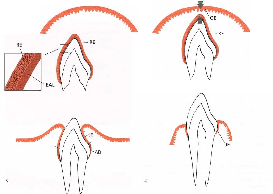
The tissue components of the dento-gingival region
achieve their final structural characteristics in con-
junction with the eruption of the teeth. This is illus-
trated in Fig. 1-31a-d.
Fig. 1-31a. When the enamel of the tooth is fully devel-
oped, the enamel-producing cells (ameloblasts) be-
come reduced in height, produce a basal lamina and
form, together with cells from the outer enamel epi-
thelium, the so-called reduced dental epithelium (RE).
The basal lamina (epithelial attachment lamina: EAL)
lies in direct contact with the enamel. The contact
between this lamina and the epithelial cells is main-
tained by hemidesmosomes. The reduced enamel epi-
thelium surrounds the crown of the tooth from the
moment the enamel is properly mineralized until the
tooth starts to erupt.
Fig. 1-31b. As the erupting tooth approaches the oral
epithelium, the cells of the outer layer of the reduced
dental epithelium (RE), as well as the cells of the basal
layer of the oral epithelium (OE), show increased
mitotic activity (arrows) and start to migrate into the
underlying connective tissue. The migrating epithe-
lium produces an epithelial mass between the oral
epithelium and the reduced dental epithelium so that
the tooth can erupt without bleeding. The former
ameloblasts do not divide.
Fig. 1-31c. When the tooth has penetrated into the oral
cavity, large portions immediately apical to the incisal
area of the enamel are covered by a junctional epithe-
lium (JE) containing only a few layers of cells. The
cervical region of the enamel, however, is still covered
by ameloblasts (AB) and outer cells of the reduced
dental epithelium.
Fig. 1-31d. During the later phases of tooth eruption,
all cells of the reduced enamel epithelium are replaced
ANATOMY OF THE PERIODONTIUM • 17
by a junctional epithelium. This epithelium is continu
ous with the oral epithelium and provides the
attachment between the tooth and the gingiva. If the
free gingiva is excised after the tooth has fully
erupted, a new junctional epithelium,
indistinguishable from that found following tooth
eruption, will develop during healing. The fact that
this new junctional epithelium has developed from
the oral epithelium indicates that the cells of the oral
epithelium possess the ability to differentiate into
cells of junctional epithelium.
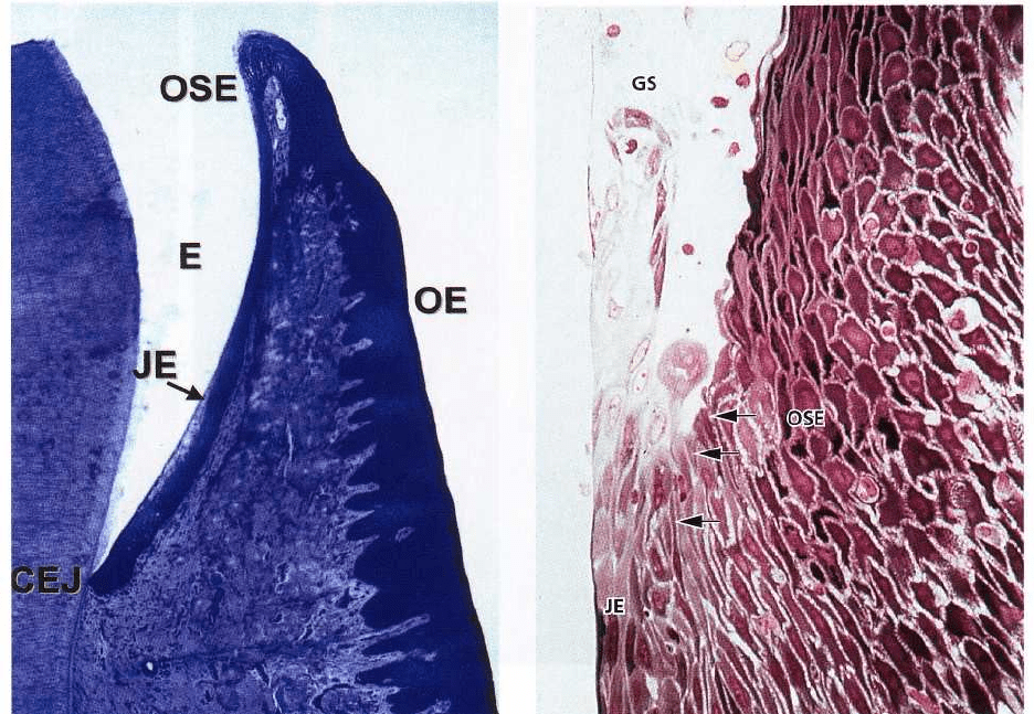
Fig. 1-32 is a histologic section cut through the border
area between the tooth and the gingiva, i.e. the dento-
gingival region. The enamel (E) is to the left. Towards
the right follow the junctional epithelium (JE), the oral
sulcular epithelium (OSE) and the oral epithelium (OE).
The oral sulcular epithelium covers the shallow
groove, the gingival sulcus located between the
enamel and the top of the free gingiva. The junctional
epithelium differs morphologically from the oral sul-
cular epithelium and oral epithelium, while the two
latter are structurally very similar. Although individ-
ual variation may occur, the junctional epithelium is
usually widest in its coronal portion (about 15-20 cell
layers), but becomes thinner (3-4 cells) towards the
cemento-enamel junction (CEJ). The borderline be-
tween the junctional epithelium and the underlying
connective tissue does not present epithelial rete pegs
except when inflamed.
Fig. 1-33. The junctional epithelium has a free surface
at the bottom of the gingival sulcus (GS). Like the oral
sulcular epithelium and the oral epithelium, the junc-
tional epithelium is continuously renewed through
cell division in the basal layer. The cells migrate to the
base of the gingival sulcus from where they are shed.
The border between the junctional epithelium (JE) and
the oral sulcular epithelium (OSE) is indicated by
arrows. The cells of the oral sulcular epithelium are
cuboidal and the surface of this epithelium is kerati-
nized.
Fig. 1-33
Fig. 1-3:
18 • CHAPTER 1
Fig. 1-34.
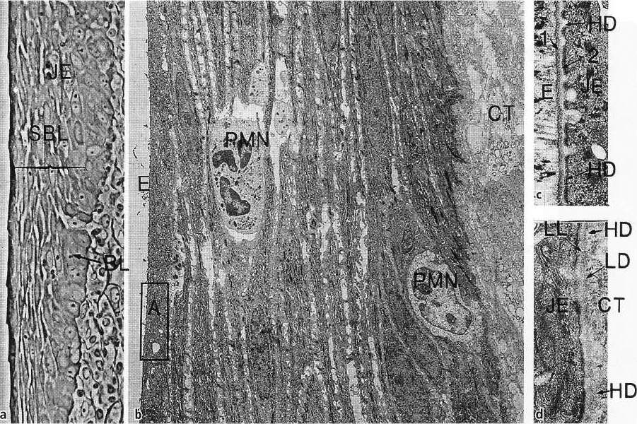
Fig. 1-34 illustrates different characteristics of the junc
tional epithelium. As can be seen in Fig. 1-34a, the
cells of the junctional epithelium (JE) are arranged
into one basal layer (BL) and several suprabasal
layers (SBL). Fig. 1-34b demonstrates that the basal
cells as well as the suprabasal cells are flattened with
their long axis parallel to the tooth surface. (CT =
connective tissue, E = enamel space.)
There are distinct differences between the oral sul-
cular epithelium, the oral epithelium and the junc-
tional epithelium:
1. The size of the cells in the junctional epithelium
is, relative to the tissue volume, larger than in the
oral epithelium.
2. The intercellular space in the junctional
epithelium is, relative to the tissue volume,
comparatively wider than in the oral epithelium.
3. The number of desmosomes is smaller in the junc-
tional epithelium than in the oral epithelium.
Note the comparatively wide intercellular spaces be-
tween the oblong cells of the junctional epithelium,
and the presence of two neutrophilic granulocytes (
PMN) which are traversing the epithelium.
The framed area (A) is shown in a higher magnifi-
cation in Fig. 1-34c, from which it can be seen that the
basal cells of the junctional epithelium are not in direct
contact with the enamel (E). Between the enamel and
the epithelium (JE) one electron-dense zone (1) and
one electron-lucent zone (2) can be seen. The electron
-lucent zone is in contact with the cells of the
junctional
epithelium (JE). These two zones have a structure very
similar to that of the lamina densa (LD) and lamina
lucida (LL) in the basement membrane area (i.e. the
epithelium (JE)-connective tissue (CT) interface) de-
scribed in Fig. 1-23. Furthermore, as seen in Fig. 1-34d,
the cell membrane of the junctional epithelial cells
harbors hemidesmosomes (HD) towards the enamel
as it does towards the connective tissue. Thus, the
interface between the enamel and the junctional epi-
thelium is similar to the interface between the epithe-
lium and the connective tissue.
Fig. 1-35 is a schematic drawing of the most apically
positioned cell in the junctional epithelium. The
enamel (E) is depicted to the left in the drawing. It
can be seen that the electron-dense zone (1) between
the junctional epithelium and the enamel can be
considered a continuation of the lamina densa (LD)
in the basement membrane of the connective tissue
side. Similarly, the electron-lucent zone (2) can be
considered a continuation of the lamina lucida (LL). It
should be noted, however, that at variance with the
epithelium-connective tissue interface, there are no
anchoring fibers (AF) attached to the lamina densa-
like structure (1) adjacent to the enamel. On the other
hand, like the basal cells adjacent to the basement
membrane (at the connective tissue interface), the
cells of the junctional epithelium facing the lamina
lucida-like structure (2) harbor hemidesmosomes.
Thus, the interface between the junctional epithelium
and the enamel is structurally very similar to the
epithelium-connective tissue interface, which means
that the junctional epi-

ANATOMY OF THE PERIODONTIUM • 19
Fig. 1-36.
LL
Fig. 1-35.
thelium is not only in contact with the enamel but is
actually physically attached to the tooth via hemides-
mosomes.
Lamina propria
The predominant tissue component of the gingiva is
the connective tissue (lamina propria). The major
components of the connective tissue are collagen fibers
(around 60% of connective tissue volume), fibroblasts (
around 5%), vessels and nerves (around 35%) which are
embedded in an amorphous ground substance (ma-
trix).
Fig. 1-36. The drawing illustrates a fibroblast (F) resid-
ing in a network of connective tissue fibers (CF). The
intervening space is filled with matrix (M) which
constitutes the "environment" for the cell.
Cells
The different types of cell present in the connective
tissue are: (1) fibroblasts, (2) mast cells, (3) macrophages
and (4) inflammatory cells.
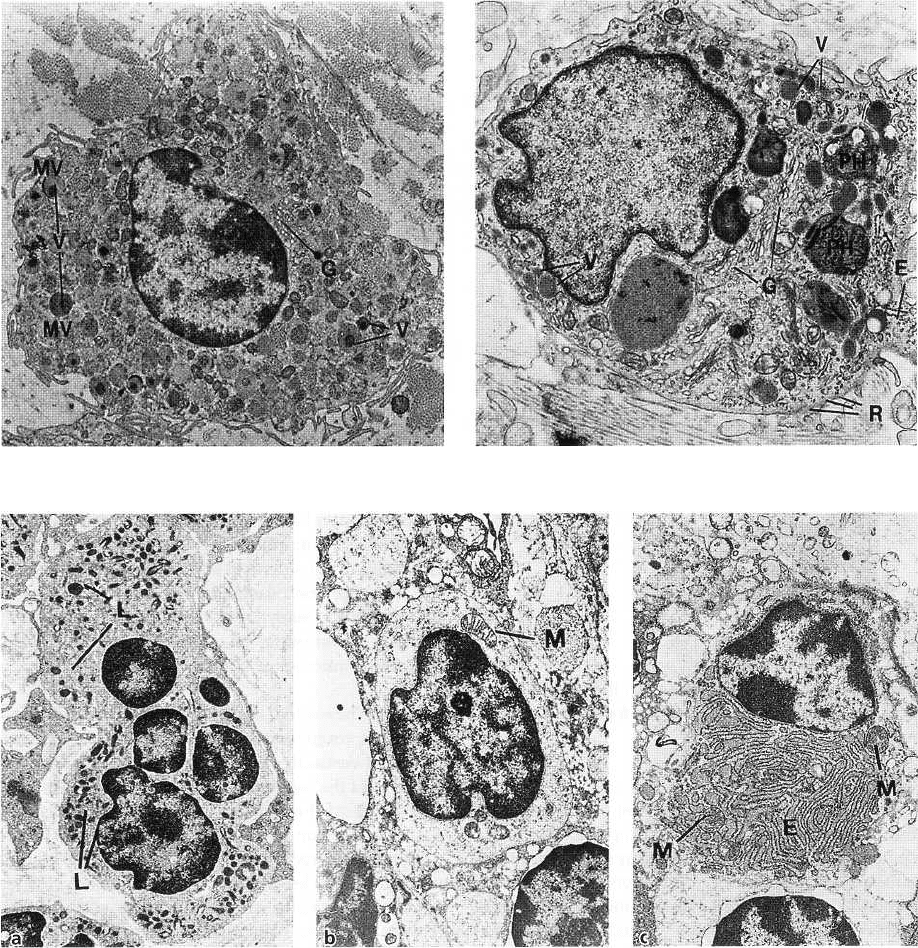
Fig. 1-37. The fibroblast is the most predominant con-
nective tissue cell (65% of the total cell population).
The fibroblast is engaged in the production of various
types of fibers found in the connective tissue, but is
also instrumental in the synthesis of the connective
tissue matrix. The fibroblast is a spindle-shaped or
stellate cell with an oval-shaped nucleus containing
one or more nucleoli. A part of a fibroblast is shown
in electron microscopic magnification. The cytoplasm
contains a well-developed granular endoplasmic
reticulum (E) with ribosomes. The Golgi complex (G)
is usually of considerable size and the mitochondria (
M) are large and numerous. Furthermore, the cyto-
plasm contains many fine tonofilaments (F). Adjacent
to the cell membrane, all along the periphery of the
cell, a large number of vesicles (V) can be found.
Fig. 1-38. The mast cell is responsible for the production
of certain components of the matrix. This cell also
produces vasoactive substances, which can affect the
function of the microvascular system and control the
flow of blood through the tissue. A mast cell is pre-
sented in electron microscopic magnification. The cy-
toplasm is characterized by the presence of a large
number of vesicles (V) of varying size. These vesicles
contain biologically active substances such as pro-
teolytic enzymes, histamine and heparin. The Golgi
Fig. 1-37.
20 • CHAPTER 1
Fig. 1-38. Fig. 1-39.
Fig. 1-40.
complex (G) is well developed, while granular en-
doplasmic reticulum structures are scarce. A large
number of small cytoplasmic projections, i.e. mi-
crovilli (MV), can be seen along the periphery of the
cell.
Fig. 1-39. The macrophage has a number of different
phagocytic and synthetic functions in the tissue. A
macrophage is shown in electron microscopic magni-
fication. The nucleus is characterized by numerous
invaginations of varying size. A zone of electron-
dense chromatin condensations can be seen along the
periphery of the nucleus. The Golgi complex (G) is
well developed and numerous vesicles (V) of varying
size are present in the cytoplasm. Granular endoplas-
mic reticulum (E) is scarce, but a certain number of free
ribosomes (R) are evenly distributed in the cytoplasm.
Remnants of phagocytosed material are often found
in lysosomal vesicles: phagosomes (PH). In the pe-
riphery of the cell, a large number of microvilli of
varying size can be seen. Macrophages are particu-
larly numerous in inflamed tissue. They are derived
from circulating blood monocytes which migrate into
the tissue.
Fig. 1-40. Besides fibroblasts, mast cells and macro-

ANATOMY OF THE PERIODONTIUM • 21
Fig. 1-41.
CF
Fig. 1-42.
phages, the connective tissue also harbors inflamma-
tory cells of various types, for example neutrophilic
granulocytes, lymphocytes and plasma cells.
The neutrophilic granulocytes, also called polymor-
phonuclear leukocytes, have a characteristic appearance
(Fig. 1-40a). The nucleus is lobulate and numerous
lysosomes (L), containing lysosomal enzymes, are
found in the cytoplasm.
The lymphocytes (Fig. 1-40b) are characterized by an
oval to spherical nucleus containing localized areas of
electron-dense chromatin. The narrow border of cyto-
plasm surrounding the nucleus contains numerous
free ribosomes, a few mitochondria (M) and, in local-
ized areas, endoplasmic reticulum with fixed ribo-
somes. Lysosomes are also present in the cytoplasm.
The plasma cells (Fig. 1-40c) contain an eccentrically
located spherical nucleus with radially deployed elec-
tron-dense chromatin. Endoplasmic reticulum (E)
with numerous ribosomes is found randomly distrib-
uted in the cytoplasm. In addition, the cytoplasm
contains numerous mitochondria (M) and a well-de-
veloped Golgi complex.
Fibers
The connective tissue fibers are produced by the fi-
broblasts and can be divided into: (1) collagen fibers, (2)
reticulin fibers, (3) oxytalan fibers and (4) elastic fibers.
Fig. 1-41. The collagen fibers predominate in the gingi-
val connective tissue and constitute the most essential
components of the periodontium. The electronmi-
crograph shows cross- and longitudinal sections of
collagen fibers. The collagen fibers have a charac-
teristic cross-banding with a periodicity of 700 A be-
tween the individual dark bands.
Fig. 1-42 illustrates some important features of the
synthesis and the composition of collagen fibers pro-
duced by fibroblasts (F). The smallest unit, the colla-
gen molecule, is often referred to as tropocollagen. A
tropocollagen molecule (TC) which is seen in the up-
per portion of the drawing is approximately 3000 A
long and has a diameter of 15 A. It consists of three
polypeptide chains intertwined to form a helix. Each
chain contains about 1000 amino acids. One third of
these are glycine and about 20% proline and hy-
droxyproline, the latter being found practically only
in collagen. Tropocollagen synthesis takes place inside
the fibroblast from which the tropocollagen molecule
is secreted into the extracellular space. Thus, the po-
lymerization of tropocollagen molecules to collagen
fibers takes place in the extracellular compartment.
First, tropocollagen molecules are aggregated longitu-
dinally to protofibrils (PF), which are subsequently
laterally aggregated parallel to collagen fibrils (CFR),
with an overlapping of the tropocollagen molecules
by about 25% of their length. Due to the fact that
special refraction conditions develop after staining at
the sites where the tropocollagen molecules adjoin, a
cross-banding with a periodicity of approximately 700
A occurs under light microscopy. The collagen fibers (
CF) are bundles of collagen fibrils, aligned in such a
way that the fibers also exhibit a cross-banding with a
periodicity of 700 A. In the tissue, the fibers are
usually arranged in bundles. As the collagen fibers
mature, covalent crosslinks are formed between the
tropocollagen molecules, resulting in an age-related
reduction in collagen solubility.
Cementoblasts and osteoblasts are cells which also
possess the ability to produce collagen.
22 • CHAPTER 1
Fig. 1-43.
Fig. 1-44.
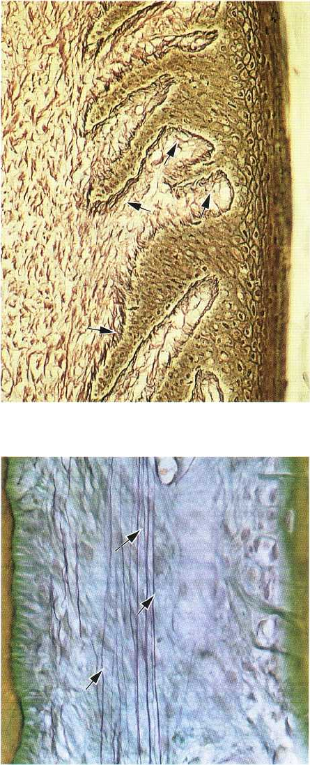
Fig. 1-43. Reticulin fibers — as seen in this photomicro-
graph — exhibit argyrophilic staining properties and
are numerous in the tissue adjacent to the basement
membrane (arrows). However, reticulin fibers also
occur in large numbers in the loose connective tissue
surrounding the blood vessels. Thus, reticulin fibers
are present at the epithelium-connective tissue and the
endothelium-connective tissue interfaces.
Fig 1-44. Oxytalan fibers are scarce in the gingiva but
numerous in the periodontal ligament. They are com-
posed of long thin fibrils with a diameter of approxi-
mately 150 A. These connective tissue fibers can be
demonstrated light microscopically only after pre-
vious oxidation with peracetic acid. The photomicro-
graph illustrates oxytalan fibers (arrows) in the peri-
odontal ligament, where they have a course mainly
parallel to the long axis of the tooth. The function of
these fibers is as yet unknown. The cementum is seen
to the left and the alveolar bone to the right.
Fig. 1-45. Elastic fibers in the connective tissue of the
gingiva and periodontal ligament are only present in
association with blood vessels. However, as seen in
this photomicrograph, the lamina propria and submu-
cosa of the alveolar (lining) mucosa contain numerous
elastic fibers (arrows). The gingiva (G) seen coronal to
the mucogingival junction (MGJ) contains no elastic
fibers except in association with the blood vessels.
Fig. 1-46. Although many of the collagen fibers in the
gingiva and the periodontal ligament are irregularly
or randomly distributed, most tend to be arranged in
groups of bundles with a distinct orientation. Accord-
ing to their insertion and course in the tissue, the
oriented bundles in the gingiva can be divided into the
following groups:
1. Circular fibers (CF) are fiber bundles which run their
course in the free gingiva and encircle the tooth in a
cuff- or ring-like fashion.
2. Dentogingival fibers (DGF) are embedded in the ce-
mentum of the supra-alveolar portion of the root
and project from the cementum in a fan-like con-
figuration out into the free gingival tissue of the
facial, lingual and interproximal surfaces.
3. Dentoperiosteal fibers (DPF) are embedded in the
same portion of the cementum as the dentogingival
fibers, but run their course apically over the ves-
tibular and lingual bone crest and terminate in the
tissue of the attached gingiva. In the border area
between the free and attached gingiva, the epithe-
lium often lacks support by underlying oriented
collagen fiber bundles. In this area the free gingival
groove (GG) is often present.
4. Transseptal fibers (TF), seen on the drawing to the
right, extend between the supra-alveolar cemen-
tum of approximating teeth. The transseptal fibers
run straight across the interdental septum and are
embedded in the cementum of adjacent teeth.
Fig. 1-47 illustrates in a histologic section the orienta-
tion of the transseptal fiber bundles (arrows) in the
supra-alveolar portion of the interdental area. It
should be observed that, besides connecting the ce-
mentum (C) of adjacent teeth, the transseptal fibers
