Jan Lindhe. Clinical Periodontology
Подождите немного. Документ загружается.

TRAUMA FROM OCCLUSION • 353
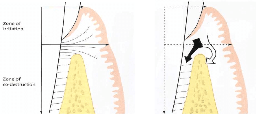
Fig. 15-1. Schematic drawing of the zone of irritation
and the zone of co-destruction according to
Glickman.
vertical pocket formation associated with one or a
varying number of teeth" (Stones 1938). The experi-
ments by Box and Stones, however, have been criti-
cized because they lacked proper controls and because
the experimental design of the studies did not justify
the conclusions drawn.
The interaction between
trauma from occlusion
and
plaque-associated periodontal disease in humans was
in the period 1955-70 frequently discussed in connec
-
tion with "report of a case", "in my opinion state-
ments", etc. Even if such anecdotal data may have
some value in clinical dentistry, it is obvious that
conclusions drawn from research findings are much
more pertinent. The research-based conclusions are
not always indisputable but they invite the reader to
a critique which anecdotal data do not. In this chapter,
therefore, the presentation will be limited to findings
collected from research endeavors involving: (1) hu-
man autopsy material, (2) clinical trials and (3) animal
experiments.
Analysis of human autopsy material
Results reported from carefully performed research
efforts involving examinations of human autopsy ma-
terial have been difficult to interpret. In the specimens
examined (1) the histopathology of the lesions in the
periodontium have been described, as well as (2) the
presence and apical extension of microbial deposits at
adjacent root surfaces, (3) the mobility of the teeth
involved and (4) "the occlusion" of the sites under
scrutiny. It is obvious that assessments made in speci
mens from cadavers have a limited to questionable
value when "cause-effect" relationships between oc-
clusion, plaque and periodontal lesions are to be de-
Fig. 15-2. The inflammatory lesion in the zone of irrita
tion can, in teeth not subjected to trauma, propagate
into the alveolar bone (open arrow), while in teeth also
subjected to trauma from occlusion, the inflammatory
infiltrate spreads directly into periodontal ligament
(
filled arrow).
scribed. It is not surprising, therefore, that
conclusions
drawn from this type of research can be controversial.
This can best be illustrated if "Glickman's concept" is
compared with "Waerhaug's concept" of what aut-
opsy studies have revealed regarding trauma from
occlusion and periodontal disease.
Glickman's
concept
Glickman (1965, 1967) claimed that the pathway of the
spread of a plaque-associated gingival lesion can be
changed if forces of an abnormal magnitude are acting
on teeth harboring subgingival plaque. This would
imply that the character of the progressive tissue de-
struction of the periodontium at a "traumatized
tooth" will be different from that characterizing a
"
non-traumatized" tooth. Instead of an even destruc-
tion of the periodontium and alveolar bone (su-
prabony pockets and horizontal bone loss), which
according to Glickman occurs at sites with uncompli-
cated plaque-associated lesions, sites which are also
exposed to abnormal occlusal force will develop an-
gular bony defects and infrabony pockets.
Since the concept of Glickman regarding the effect
of trauma from occlusion
on the spread of the plaque-
associated lesion is often cited, a more detailed pres-
entation of his theory seems pertinent:
The periodontal structures can be divided into two
zones:
1.
the
zone of irritation
and
2.
the
zone of co-destruction
(Fig. 15-1).
The
zone of irritation
includes the marginal and inter-
dental gingiva. The soft tissue of this zone is bordered
by hard tissue (the tooth) only on one side and is not
affected by forces of occlusion. This means that gingi-
354 • CHAPTER 15
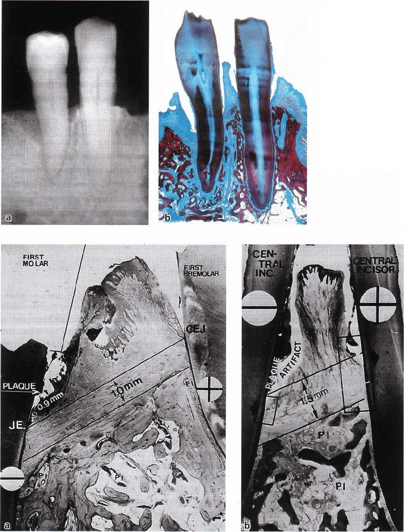
Fig. 15-3. A radiograph of a
mandibular premolar-canine re-
gion. Note the angular bony de-
fect at the distal aspect of the pre-
molar. (b) Histologic mesiodistal
section of the specimen illustrated
in (a). Note the infrabony pocket
at the distal aspect of the premo-
lar. From Glickman & Smulow
(
1965).
Figs. 15-4a,b. Microphotographs illustrating two interproximal areas with angular bony defects. "-" denotes a tooth
not
subjected and "+" denotes a tooth subjected to trauma from occlusion. In categories "-" and "+" the distance be
tween
the apical cells of the junctional epithelium and the supporting alveolar bone is about 1-1.5 mm, and the dis
tance
between the apical extension of plaque and the apical cells of the junctional epithelium about 1 mm. Since the
apical
cells of the junctional epithelium and the subgingival plaque are located at different levels on the two adja-
cent teeth, the outline of the bone crest becomes oblique. A radiograph from such a site would disclose the presence
of an angular bony defect at a nontraumatized C-") tooth.
val inflammation cannot be induced by
trauma from
occlusion
but is the result of irritation from microbial
plaque. The plaque-associated lesion at a "non-trau-
matized" tooth propagates in apical direction by first
involving the alveolar bone and only later the peri-
odontal ligament area. The progression of this lesion
results in an even (horizontal) bone destruction.
The zone of
co-destruction
includes the periodontal
ligament, the root cementum and the alveolar bone and
is coronally demarcated by the transseptal (inter-
dental)
and the dentoalveolar collagen fiber bundles (Fig. 15-1).
The tissue in this zone may become the seat of a lesion
caused by
trauma from occlusion.
The fiber bundles which separate the zone of co-de-
TRAUMA FROM OCCLUSION • 355
struction from the zone of irritation can be affected
from two different directions:
1. from the inflammatory lesion maintained by plaque
in the zone of irritation
2. from trauma-induced changes in the zone of co-de-
struction.
Through this exposure from two different directions
the fiber bundles may become dissolved and/or ori-
ented in a direction parallel to the root surface. The
spread of an inflammatory lesion from the zone of
irritation directly down into the periodontal ligament (
i.e. not via the interdental bone) may hereby be facili-
tated (Fig. 15-2). This alteration of the "normal" path-
way of spread of the plaque-associated inflammatory
lesion results in the development of angular bony
defects. Glickman (1967) in a review paper stated that
trauma from occlusion is an etiologic factor (co-destruc-
tive factor) of importance in situations where angular
bony defects combined with infrabony pockets are
found at one or several teeth (Fig. 15-3).
Waerhaug's concept
Waerhaug (1979) examined autopsy specimens (Fig.
15-4) similar to Glickman's, but measured in addition
the distance between the subgingival plaque and (1)
the periphery of the associated inflammatory cell in-
filtrate in the gingiva and (2) the surface of the adjacent
alveolar bone. He concluded from his analysis that
angular bony defects and infrabony pockets occur
equally often at periodontal sites of teeth which are
not affected by trauma from occlusion as in trauma-
tized teeth. In other words, he refuted the hypothesis
that trauma from occlusion played a role in the spread
of a gingival lesion into the "zone of co-destruction".
The loss of connective attachment and the resorption
of bone around teeth are, according to Waerhaug,
exclusively the result of inflammatory lesions associ-
ated with subgingival plaque. Waerhaug concluded
that angular bony defects and infrabony pockets occur
when the subgingival plaque of one tooth has reached
a more apical level than the microbiota on the neigh-
boring tooth, and when the volume of the alveolar
bone surrounding the roots is comparatively large.
Waerhaug's observations support findings presented
by Prichard (1965) and Manson (1976) which imply
that the pattern of loss of supporting structures is the
result of an interplay between the form and volume of
the alveolar bone and the apical extension of the mi-
crobial plaque on the adjacent root surfaces.
It is obvious, as stated above, that examinations of
autopsy material have a limited value when "cause-
effect" relationships with respect to trauma and pro-
gressive periodontitis are to be determined. As a con-
sequence, the conclusions drawn from this field of
research have not been generally accepted. A number
of authors tend to accept Glickman's conclusions that
trauma from occlusion is an aggravating factor in
periodontal disease (e.g. Macapanpan & Weinmann
1954, Posselt & Emslie 1959, Glickman & Smulow
1962, 1965) while others accept Waerhaug's concept, i.
e. that there is no relationship between occlusal
trauma and the degree of periodontal tissue break-
down (e.g. Lovdahl et al. 1959, Belting & Gupta 1961,
Baer et al. 1963, Waerhaug 1979).
Clinical
trials
In addition to the presence of angular bony defects
and infrabony pockets, increased tooth mobility is fre-
quently listed as an important sign of occlusal trauma.
For details regarding tooth mobility, see Chapter 30 "
Occlusal Therapy". Conflicting data have been re-
ported also regarding the periodontal conditions of
mobile teeth. In one clinical study by Rosling et al. (
1976) patients with advanced periodontal disease
associated with multiple angular bony defects and
mobile teeth were exposed to antimicrobial therapy (i.
e. subgingival scaling after flap elevation). Healing
was evaluated by probing attachment level measure-
ments and radiographic monitoring. The authors re-
ported that "the infrabony pocket located at hypermo-
bile teeth exhibited the same degree of healing as those
adjacent to firm teeth".
In another study, however, Fleszar et al. (1980) re-
ported on the influence of tooth mobility on healing
following periodontal therapy including both root
debridement and occlusal adjustment. They con-
cluded that "pockets of clinically mobile teeth do not
respond as well to periodontal treatment as do those
of firm teeth exhibiting the same disease severity".
A third study (Pihlstrom et al. 1986) studied the
association between trauma from occlusion and perio-
dontitis by assessing a series of clinical and radio-
graphic features at maxillary first molars. Parameters
included in this study were: probing depth, probing
attachment level, tooth mobility, wear facets, plaque
and calculus, bone height, widened periodontal space,
etc. Pihlstrom and his associates concluded from their
measurements and examinations that teeth with
increased mobility and widened periodontal ligament
space had, in fact, deeper pockets, more attachment
loss and less bone support than teeth with-out these
symptoms.
Burgett et al. (1992) studied the effect of occlusal
adjustment in the treatment of periodontitis. Fifty
subjects with periodontitis were examined at baseline
and subsequently treated for their periodontal condi-
tion with root debridement ± flap surgery. Twenty-two
out of the 50 patients, in addition, received compre-
hensive occlusal therapy. Reexaminations performed
2 years later disclosed that probing attachment gain
was on the average about 0.5 mm larger in patients
who received the combined treatment, i.e. scaling and
occlusal adjustment, than in patients in whom the
occlusal adjustment was not included.
The findings by Fleszar, Pihlstrom and Burgett and
co-workers lend some support to the concept that
356 • CHAPTER 15
Tipping movement
Bodily movement
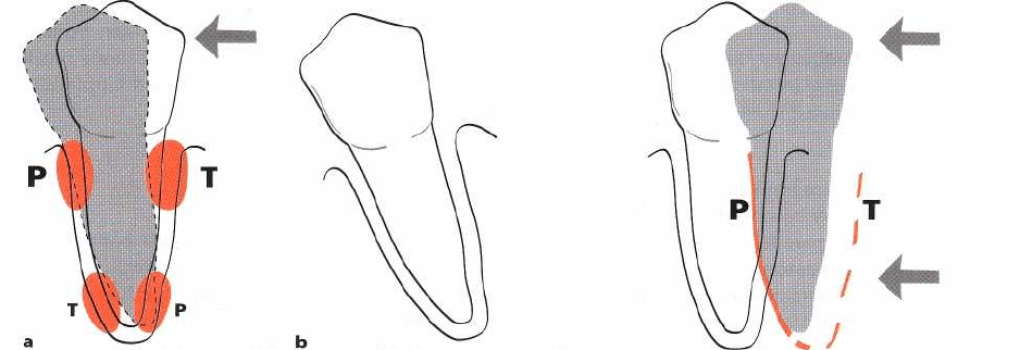
Figs. 15-5. If the crown of a tooth is exposed to exces-
sive, horizontally directed forces (arrow), pressure (P)
and tension (T) zones will develop within the marginal
and apical parts of the periodontium (a). The su-
praalveolar connective tissue remains unaffected by
force application. Within the pressure and tension
zones tissue alterations take place which eventually al
-
low the tooth to tilt in the direction of the force. When
the tooth has escaped the trauma, complete regenera-
tion of the periodontal tissues takes place (b). There is
no apical downgrowth of the dentogingival epithelium.
Fig. 15-6. When a tooth is exposed to forces which pro-
duce
"
bodily tooth movement", e.g. in orthodontic ther-
apy, the pressure (P) and tension (T) zones, depending
on the direction of the force, are extended over the en-
tire tooth surface. The supraalveolar connective tissue
is not affected in conjunction either with tipping or
with bodily movements of teeth. Forces of this kind,
therefore, will not induce inflammatory reactions in the
gingiva. No apical downgrowth of the dentogingival
epithelium occurs.
trauma from occlusion (and increased tooth mobility)
may have a detrimental effect on the periodontium.
Neiderud et al. (1992), however, in a beagle dog study
demonstrated that tissue alterations which occur at
mobile teeth with clinically healthy gingivae (and
normal height of the tissue attachment) may reduce the
resistance offered by the periodontal tissues to
probing. In other words, if the probing depth at two
otherwise similar teeth – one non-mobile and one
hypermobile – is recorded, the tip of the probe will
penetrate 0.5 mm deeper at the mobile than at the
non-mobile tooth. This finding must be taken into
consideration when the above clinical data are inter-
preted.
Since neither analysis of autopsy material nor data
from clinical trials can be used to properly determine
the role
trauma from occlusion
may play in periodontal
pathology, it is necessary to describe the contributions
made by means of animal research in this particular
field. Results from such experiments, describing the
reactions of the normal and subsequently the diseased
periodontium to occlusal forces, are presented below.
Animal experiments
Orthodontic type trauma
The reaction of the periodontal tissues to traumatic
forces initiated by occlusion has been studied princi-
pally in animal experiments. In early experiments the
reaction of the normal periodontium was studied fol-
lowing the application of forces which were inflicted
on teeth in one direction only. Biopsies including tooth
and periodontium were harvested after varying ex-
perimental time intervals and prepared for histologic
examinations. Analysis of the tissue sections (Haupl
&
Psansky 1938, Reitan 1951, Muhlemann & Herzog
1961, Ewen & Stahl 1962, Waerhaug & Hansen 1966,
Karring et al. 1982) revealed the following: when a
tooth is exposed to unilateral forces of a magnitude,
frequency or duration that its periodontal tissues are
unable to withstand and distribute while maintaining
the stability of the tooth, certain well-defined reactions
develop in the periodontal ligament, eventually re-
sulting in an adaptation of the periodontal structures
to the altered functional demand. If the crown of a
tooth is affected by such horizontally directed forces,
the tooth tends to tilt (tip) in the direction of the force
(
Fig. 15-5). This tilting force results in the development
of
pressure
and
tension zones
within the marginal and
apical parts of the periodontium. The tissue reactions
which develop in the
pressure zone
are characterized by
increased vascularization, increased vascular per-
meability, vascular thrombosis, and disorganization
of
cells and collagen fiber bundles. If the magnitude of
forces is within certain limits, allowing the mainte-
nance of the vitality of the periodontal ligament cells,
bone-resorbing osteoclasts soon appear on the bone
surface of the alveolus in the
pressure zone.
A process
of
bone resorption is initiated. This phenomenon is
called
"direct bone resorption".
If the force applied is of higher magnitude, the
result
may be necrosis of the periodontal ligament
tissue in
the
pressure zone,
i.e. decomposition of cells,
TRAUMA FROM OCCLUSION • 357
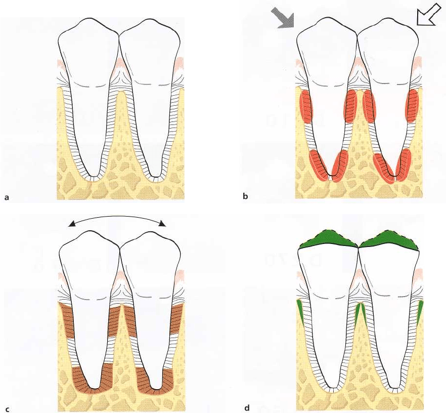
Pig. 15-7. Two mandibular premolars with normal periodontal tissues (a) are exposed to jiggling torces (e) as illus-
trated by the two arrows. The combined tension and pressure zones (encircled areas) are characterized by signs of
acute inflammation including collagen resorption, bone resorption and cementum resorption. As a result of bone re
-
sorption the periodontal ligament space gradually increases in size on both sides of the teeth as well as in the pe-
riapical region. When the effect of the force applied has been compensated for by the increased width of the peri-
odontal ligament space (c), the ligament tissue shows no sign of inflammation. The supraalveolar connective tissue
is
not affected by the jiggling forces and there is no apical downgrowth of the dentogingival epithelium. After oc
clusal
adjustment the width of the periodontal ligament becomes normalized (d) and the teeth are stabilized.
vessels, matrix and fibers
(hyalinization).
"Direct bone
resorption" therefore cannot occur. Instead, osteo
clasts
appear in marrow spaces within the adjacent
bone
tissue where the stress concentration is lower
than in the
periodontal ligament and a process of
undermining or
"
indirect bone resorption" is
initiated. Through this
reaction the surrounding bone is resorbed until there is
a breakthrough to the hyalinized
tissue within the
pressure zone.
This breakthrough re
sults in a reduction
of the stress in this area, and cells
from the neighboring
bone or adjacent areas of the periodontal ligament can
proliferate into the
pressure zone
and replace the
previously hyalinized tissue, thereby reestablishing
prerequisites for "direct bone
resorption". Irrespective of
whether the bone resorp-
tion is of a direct or an indirect nature the tooth moves
(
tilts) further in the direction of the force.
Concomitant with the tissue alterations in the
pres-
sure zone,
apposition of bone occurs in the
tension zone
in
order to maintain the normal width of the periodon
tal
ligament in this area. Because of the tissue reactions
in
the
pressure
and
tension
zones the tooth becomes,
temporarily, hypermobile. When the tooth has moved
(
tilted) to a position where the effect of the forces is
nullified, healing of the periodontal tissues takes place
in
both the
pressure
and the
tension zones
and the tooth
becomes stable in its new position. In orthodontic
tilting
(tipping) movements, neither gingival inflam
mation nor
loss of connective tissue attachment will
occur in a
healthy periodontium and – as long as the
tooth is not
moved through the envelope of the alveo-
358 • CHAPTER 15
D210
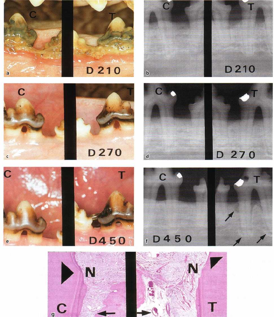
Fig. 15-8. Dogs were allowed to accumulate plaque and calculus in the mandibular premolar regions over a 210-
day period (a). When around 40-50% of the periodontal tissue support had been lost (b) the animals were treated
by scaling, root planing and pocket elimination. During surgery, a notch was prepared in the root at the level of the
bone crest. The dogs were subsequently placed on a plaque control program and 2 months later (Day 270) all ex-
perimental teeth (the lower fourth premolars; 4P and P4) were surrounded by a healthy periodontium with re-
duced height (c and d). The mandibular
left
fourth premolar (T) was exposed to jiggling forces (e). As a conse-
quence, a widened periodontal ligament and increased tooth mobility resulted (f). This increase in tooth mobility
and the development of widened periodontal ligament space did not, however, result in apical downgrowth of the
dentogingival epithelium (g). Arrowheads indicates the apical extension of the junctional epithelium which coin-
cides with the apical border of the notch (N), prepared in the root surface prior to jiggling. C= control tooth. T= test
tooth.
lar process – there is no apical migration of the den-tissue (the tooth) on one side (in the direction of the
togingival epithelium. In other words, since the su-
force), this structure remains unaffected by this type
praalveolar connective tissue is only bordered by hard
of force.
TRAUMA FROM OCCLUSION • 359
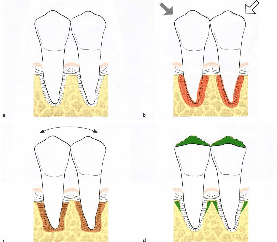
Fig. 15-9. Two mandibular premolars are surrounded by a healthy periodontium with reduced height (a). If such
premolars are subjected to traumatizing forces of the jiggling type (b) a series of alterations occurs in the periodon
-
tal ligament tissue. These alterations result in a widened periodontal ligament space (c) and in an increased tooth
mobility but do not lead to further loss of connective tissue attachment. After occlusal adjustment (d) the width of
the periodontal ligament is normalized and the teeth stabilized.
These tissue reactions do not differ fundamentally
from those which occur as a consequence of
bodily
tooth movement
in orthodontic therapy (Reitan 1951).
The main difference is that the
pressure
and
tension
zones,
depending on the direction of the force, are more
extended in an apical-coronal direction along the root
surface than in conjunction with tipping movement
(
Fig. 15-6). Neither in conjunction with tipping nor in
conjunction with bodily movements of the tooth is the
supraalveolar connective tissue affected by the
force.
Unilateral forces directed to the crown of teeth, there-
fore, will not induce inflammatory reactions in the
gingiva or cause loss of connective tissue attachment.
Studies (Steiner et al. 1981, Wennstrom et al. 1987)
have demonstrated, however, that orthodontic forces
producing bodily (or tipping) movement of teeth may
result in gingival recession and loss of connective
tissue attachment. This breakdown of the attachment
apparatus occurred at sites with gingivitis when, in
addition, the tooth was moved through the envelope
of
the alveolar process. At such sites a bone dehiscence
becomes established and, if the covering soft tissue is
thin (in the direction of the movement of the tooth),
recession (attachment loss) may occur.
Criticism has been directed, however, at experi-
ments in which only unilateral trauma is exerted on
teeth (Wentz et al. 1958). It has been suggested that in
humans, unlike in the animal experiments described
above, the occlusal forces act alternately in one and
then in the opposite direction. Such forces have been
termed
jiggling forces.
Jiggling-type trauma
Healthy periodontium with normal height
Experiments have been reported in which traumatic
forces were exerted on the crowns of the teeth, alter-
nately in buccal and lingual or mesial and distal direc-
360 • CHAPTER
15
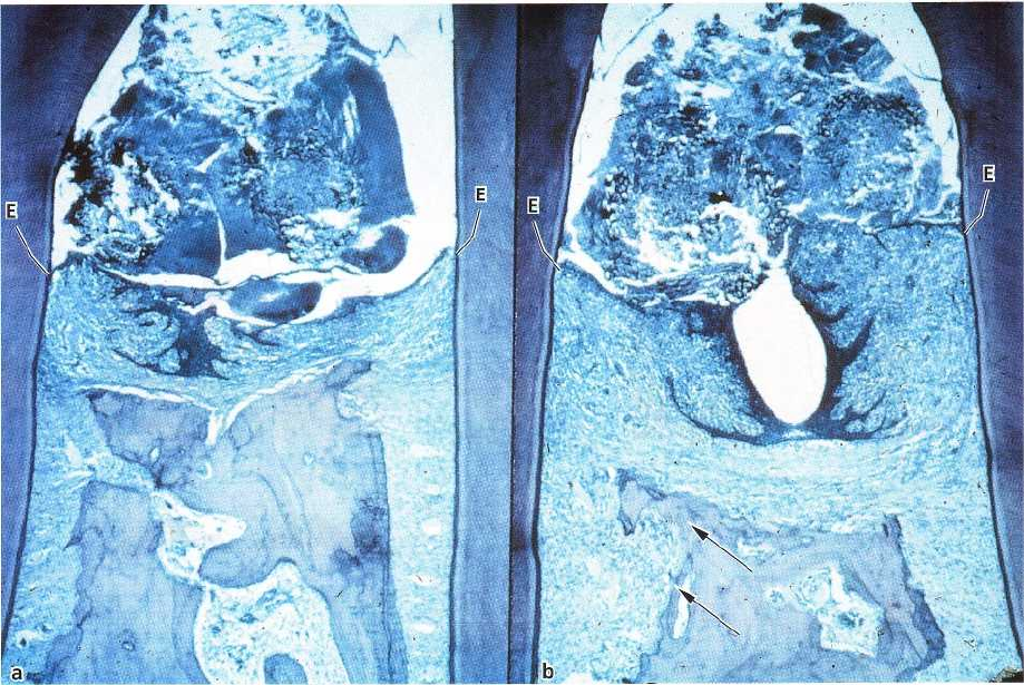
Fig. 15-10. A composite photomicrograph illustrating the interdental space between two pairs of teeth. The teeth
have been subjected to experimental, ligature-induced periodontitis and in (b) also to repetitive mechanical injury.
In (b), there is considerable loss of alveolar bone and an angular widening of the periodontal ligament space (ar
-
rows). However, the apical downgrowth of the dentogingival epithelium in the two areas (a) and( b) is similar. E in
-
dicates the apical level of the dentogingival epithelium. Courtesy of Dr. Meitner.
tions, and in which the teeth were not allowed to move
away from the force (e.g. Wentz et al. 1958, Glickman
& Smulow 1968, Svanberg & Lindhe 1973, Meitner
1975, Ericsson & Lindhe 1982). In conjunction with
"
jiggling-type trauma"
no clearcut
pressure
and
tension
zones
can be identified but rather there is a combina-
tion of pressure and tension on both sides of the
jiggled tooth (Fig. 15-7).
The tissue reactions in the periodontal ligament
provoked by the combined
pressure
and
tension
forces
were found to be similar, however, to those reported
for the pressure zone at orthodontically moved teeth,
with the one difference that the periodontal ligament
space at jiggling gradually increased in width on both
sides of the tooth. During the phase when the peri-
odontal space gradually increased in width (1) inflam-
matory changes were present in the ligament tissue,
(
2) active bone resorption occurred, and (3) the tooth
displayed signs of gradually increasing
(progressive)
mobility When the effect of the forces applied had
been compensated for by the increased width of the
periodontal ligament space, the ligament tissue
showed no signs of increased vascularity or exuda-
tion. The tooth was hypermobile but the mobility was
no longer
progressive
in character. Distinction should
thus be made between
progressive
and
increased
tooth
mobility.
In
"jiggling-type trauma"
experiments, performed
on
animals with a normal periodontium, the su-
praalveolar connective tissue was not influenced by
the occlusal forces, the reason being that this tissue
compartment is bordered by hard tissue on one side
only. This means that a gingiva which was nonin-
flamed at the start of the experiment remained nonin
-
flamed, but also that an overt inflammatory lesion
residing in the supraalveolar connective tissue was
not aggravated by the jiggling forces.
Healthy periodontium with reduced height
Progressive periodontal disease is characterized by
gingival inflammation and a gradually developing
loss
of connective tissue attachment and alveolar
bone.
Treatment of periodontal disease, i.e. removal of
plaque
and calculus and elimination of pathologically
deepened pockets, will result in the reestablishment
of a healthy periodontium but with reduced height.
The question is whether a healthy periodontium with
reduced height has a capacity similar to that of the
normal periodontium to adapt to traumatizing oc-
clusal forces (secondary occlusal trauma).
This problem has also been examined in animal
experiments (Ericsson & Lindhe 1977). Destructive
periodontal disease was initiated in dogs by allowing
the animals to accumulate plaque and calculus for a
TRAUMA FROM OCCLUSION • 361
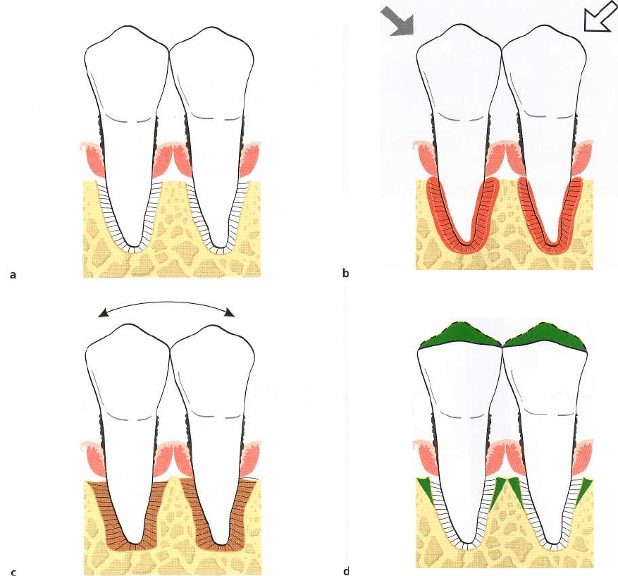
Fig. 15-11. Two mandibular premolars with supra- and subgingival plaque, advanced bone loss and periodontal
pockets of a suprabony character (a). Note the connective tissue infiltrate (shadowed areas) and the
noninflamed
connective tissue between the alveolar bone and the apical portion of the infiltrate. If these teeth
are subjected to
traumatizing forces of the jiggling type (b), pathologic and adaptive alterations occur within the
periodontal liga
ment space. These tissue alterations, which include bone resorption, result in a widened
periodontal ligament
space and increased tooth mobility but no further loss of connective tissue attachment (c). Occlusal adjustment re
-
sults in a reduction of the width of the periodontal ligament (d) and in less mobile teeth.
period of 6 months (Fig. 15-8). When around 50% of
the periodontal tissue support had been lost (Fig.
15-
8a,b), the progressive disease was subjected to
treatment by scaling, root planing and pocket elimi-
nation (Fig. 15-8c). During a subsequent 8-month pe-
riod, the animals were enrolled in a careful plaque
control program. During this period certain premolars
were exposed to traumatizing jiggling forces. The
periodontal tissues in the combined
pressure
and
ten-
sion zones
reacted to the forces by vascular prolifera-
tion, exudation and thrombosis, as well as by bone
resorption. In radiographs, widened periodontal liga-
ments (Fig. 15-8d) could be found around the trauma-
tized teeth, which at clinical examination displayed
signs of
progressive
tooth mobility. The gradual in-
crease in the width of the periodontal ligament and
the
resulting progressive increase in tooth mobility
took place during a period of several weeks but even-
tually terminated. The active bone resorption ceased
and the markedly widened periodontal ligament tissue
regained its normal composition; healing had oc-
curred (Fig. 15-8e). The teeth were hypermobile but
surrounded by periodontal structures which had
adapted to the altered functional demands.
During the entire experimental period the su-
praalveolar connective tissue remained unaffected by
the jiggling forces. There was no further loss of con-
nective tissue attachment and no further downgrowth
of dentogingival epithelium (Fig. 15-8e). The results
from this study clearly reveal that within certain limits
a healthy periodontium with reduced height has a
capacity similar to that of a periodontium with normal
height to adapt to altered functional demands (Fig.
15-
9).
362 • CHAPTER
15
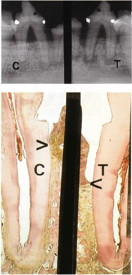
Fig. 15-12. Radiographic appearance of one test tooth
(
T) and one control tooth (C) at the termination of an
experiment in which periodontitis was induced by liga
-
ture placement and plaque accumulation and in which
trauma of the jiggling type was induced. Note angular
bone loss particularly around the mesial root of the
mandibular premolar (T) and the absence of such a de
-
fect at the mandibular premolar (C). From Lindhe &
Svanberg (1974).
Fig. 15-13. Microphotographs from one control (C) and
one test (T) tooth after 240 days of experimental peri-
odontal tissue breakdown and 180 days of trauma
from occlusion of the jiggling type (T). The arrowheads
denote the apical position of the dentogingival epithe-
lium. The attachment loss is more pronounced in T
than in C. From Lindhe & Svanberg (1974).
Plaque-associated periodontal disease
Experiments carried out on humans and animals have
demonstrated that
trauma from occlusion
cannot induce
pathologic alterations in the supraalveolar connective
tissue, i.e. cannot produce inflammatory lesions in a
normal gingiva or aggravate a gingival lesion associ-
ated with plaque and cannot induce loss of connective
tissue attachment. The question remains if abnormal
occlusal forces can influence the spread of the plaque-
associated lesion and enhance the rate of tissue de-
struction in periodontal disease. This has been studied
in animal experiments (Lindhe & Svanberg 1974,
Meitner 1975, Nyman et al. 1978, Ericsson & Lindhe
1982, Poison & Zander 1983). In these experiments
progressive and destructive periodontal disease was
first initiated in dogs or monkeys by allowing the
animals to accumulate plaque and calculus. Teeth thus
involved in a progressive periodontal disease process
were also subjected to trauma from occlusion.
"Traumatizing" jiggling forces (Lindhe & Svanberg
1974) were exerted on premolars and were found to
induce certain tissue reactions in the combined
pres-
sure/tension zones.
The periodontal ligament tissue in
these zones, within a few days of the onset of the
jiggling forces, displayed signs of inflammation, had
increased numbers of vessels, showed increased vas-
cular permeability and exudation, thrombosis, as well
as retention of neutrophils and macrophages. On the
adjacent bone surfaces there were a large number of
osteoclasts. Since the teeth could not orthodontically
move away from the jiggling forces, the periodontal
ligament of both sides of the tooth gradually increased
in width, the teeth became hypermobile
(progressive
tooth mobility) and angular bony defects could be
detected in the radiographs. The forces were eventu-
ally nullified by the increased width of the periodontal
ligament.
If the forces applied were of a magnitude to which