Iozzo Renato V. Proteoglycan Protocols
Подождите немного. Документ загружается.


Phage Display Antibodies to Heparan Sulfate 531
9. Wash the wells 6 times with PBS containing 0.1% (v/v) Tween-20.
10. Incubate the wells with 100 µL of alkaline phosphatase-conjugated rabbit anti-mouse
IgG antibody solution for 60 min.
11. Wash the wells 4 times with PBS containing 0.1% (v/v) Tween-20.
12. Wash the wells 2 times with 0.9% (v/v) NaCl.
13. Incubate the wells with 100 µL of substrate solution (in the dark) until color development
is optimal.
14. Read absorbance at 405 nm.
Method 2
1. Add 6 µg of test molecule to 150 µl antibody solution and incubate overnight at room
temperature.
2. Transfer 100 µl of this solution to a well coated with heparan sulfate and incubate for 60 min.
3. Proceed as under Method 1, starting with step 7.
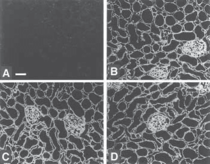
Fig. 3. Specificity of anti-heparan sulfate antibody HS3G8. Rat kidney cryosections were
treated with heparinase III (A), heparinase III incubation buffer (B), chondroitinase ABC (C),
and chondroitinase ABC incubation buffer (D). Next, sections were incubated with periplasmic
fraction containing the antibody. Bound antibodies were visualized using mouse anti-cMyc IgG
followed by Alexa 488-conjugated goat anti-mouse IgG. Bar= 50 µm. Source: from ref. 5.

532 van Kuppevelt et al.
4. Notes
1. E. coli TG1 is a T–phage resistant strain that harbors a mutated tRNA gene. The mutated
tRNA will suppress the UAG amber (stop) codon. A glutamine will be substituted for the
amber codon allowing the expression of scFv-pIII fusion protein on the phage tip.
2. VCS-M13 helper phages provide phage coat proteins and enzymes necessary for
phage rescue.
3. Except for the scFv antibody selection procedure, Marvel can be substituted with bovine
serum abumin (BSA) in the same concentrations.
4. The 9E10 hybridoma cell line is available from the ATCC (American Type Culture Col-
lection). Alternatively, a polyclonal rabbit anti c-Myc antibody (A14, Santa Cruz Bio-
technology) can be used.
5. M13 phages infect F
+
E. coli via sex pili. For production of sex pili, E. coli needs to be
grown into the log phase (absorbance at 600 nm of 0.4–0.5) at 37°C. When grown to a
higher density, sex pili are lost very rapidly. A log phase culture can be kept on ice for no
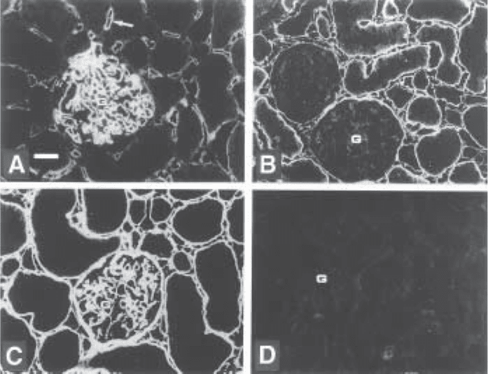
Fig. 4. Immunostaining of rat kidney with three different anti-heparan sulfate scFv antibod-
ies. Cryosections were incubated with periplasmatic fractions of bacteria expressing antibody
HS4C3 (A), HS4D10 (B), HS3G8 (C), and anti-filaggrin (D)(control; filaggrin is not present in
the kidney). Bound antibodies were visualized using mouse anti-cMyc IgG followed by Alexa
488-conjugated goat anti-mouse IgG. Bar=25 µm. All three anti-heparan sulfate antibodies
stain differently, indicating reactivity with different heparan sulfate species. G, glomerulus.
Arrow in a: peritubular capillary. Source from ref. 5.
Phage Display Antibodies to Heparan Sulfate 533
longer than 30 min. Have the eluted and neutralized phages (step 5, Subheading 3.1.3.)
ready for immediate infection.
6. Take the remaining 40 mL of the culture, spin it down and resuspend the pellet in 1 mL
2XTY. Spread it on a 245 ↔ 245 ↔ 25 Nunclon TC dish containing TYE , 100 µg
ampicillin/mL and 1% (w/v) glucose, and grow overnight at 37°C. Harvest the cells in
1–2 mL of ice-cold 2XTY containing 15% (v/v) glycerol and store this stock in 50-µL
aliquots (about 10
8
clones) at –70°C for other selections.
7. Glucose represses transcription of the scFv-pIII fusion protein through the lac operon in
the phagemid.
8. Besides concentrating the phages, this step is also necessary for removing any soluble
antibodies, since the TG1 suppression of the amber codon is never complete.
9. After each selection round an increase in titer is expected, indicating enrichment of
binding clones.
10. The glycerol stocks are used as a backup. When a subsequent selection round fails, use
50 µl of stock for a new round of selection.
11. In this step, 0.1% (w/v) glucose is added to suppress the expression of scFv antibodies
until a sufficient number of cells is produced for large-scale antibody production. The
total amount of glucose will be metabolized at an absorbance at 600 nm of 0.9.
12. In case no suitable apparatus is available for measering absorbance, grow for 3 h, while
shaking, at 37°C before adding IPTG. Bacterial growth should be clearly visible.
13. Include negative controls (e.g., supernatant without scFv antibodies, omission of culture
supernatant).
14. Clones that are weakly positive in both the immunoblot assay and in the ELISA may still
be clones of interest. They may react weakly in ELISA because of poor scFv antibody
production.
15. Non–full-length clones should be ignored, since they are notoriously unspecific binders.
16. Fingerprinting is a quick method for looking for clone diversity. This method is very
useful when a large number of positive clones is to be examined.
17. Next to storage of bacteria, it is recommended to store phagemid DNA of a selected clone
in 70% (v/v) ethanol at –70°C. You can analyze the DNA sequence for the V
H
- gene
number using the Sanger center’s germline query (http://www.sanger.ac.uk/DataSearch/
gq_search.shtml). With the gene number the V
H
family can be identified using a paper by
Tomlinson et al. (4).
18. The stability of scFv antibodies is variable. Some antibodies can be stored at 4°C for
weeks to months, whereas others will stay immunoreactive only for a couple of days.
Most antibodies can be stored at –70°C for months up to years. Bacterial supernatants
containing scFv antibodies are suitable for ELISA, but are generally not suitable for
immunohistochemistry. Periplasmic fractions, in which the antibodies are more concen-
trated, are suitable for both. The use of the HB2151 nonsuppressor strain of E. coli gen-
erally gives a higher yield of (soluble) antibodies.
19. To analyze the HS specificity of the antibody, tissue sections are pretreated with
heparinase III, which digests all heparan sulfates. As a control, pretreatment with
chondroitinase ABC, which digests chondroitin sulfates and dermatan sulfates, is
performed.
20. As a control to verify effectiveness of heparinase III treatment, an antibody (3G10,
Seikagaku) directed against heparan sulfate stubs, generated by the enzyme, may be used.
21. The stained tissue sections can be kept for up to 6 mo. The fluorescent tag (Alexa 488) is
very stable, and fading of the signal hardly occurs. Store frozen or at –20°C. Background
534 van Kuppevelt et al.
staining can often be eliminated by additional blocking steps with 1–2% (w/v) BSA or
with serum (1–5% v/v) from the same species in which the tertiary antibody is raised.
Extra washing steps can also lower the background signals.
References
1. Borrebaeck, C. A. K., ed. (1995) Antibody engineering. Oxford University Press, New
York, NY.
2. Kay, K. B., Winter, J., and McCafferty, J. (1996) Phage Display of Peptides and Proteins.
Academic, San Diego, CA.
3. Nissim, A., Hoogenboom, H. R., Tomlinson, I. M., Flynn, G., Midgley, C., Lane, D., and
Winter, G. (1994) Antibody fragments from a ‘single’ pot, phage display library as immu-
nochemical reagents. EMBO J. 13, 692–698.
4. Tomlinson, I. M., Walter, G., Marks, J. D., Llewelyn, M. B., and Winter, G. (1992) The
repertoire of human germline V
H
sequences reveals about fifty groups of V
H
segments
with different hypervariable loops. J. Mol. Biol. 227, 776–798.
5. Van Kuppevelt, T. H., Dennissen, M. A. B. A., Van Venrooij, W. J., Hoet, R. M. A., and
Veerkamp, J. H. (1998) Generation and application of type-specific anti-heparan sulfate
antibodies using phage display technology. J. Biol. Chem. 273, 12,960–12,966.

Localization of FGF-specific HSPGs 535
535
From:
Methods in Molecular Biology, Vol. 171: Proteoglycan Protocols
Edited by: R. V. Iozzo © Humana Press Inc., Totowa, NJ
51
Tissue-Specific Binding by FGF and FGF Receptors
to Endogenous Heparan Sulfates
Andreas Friedl, Mark Filla, and Alan C. Rapraeger
1. Introduction
Heparan sulfate proteoglycans (HSPGs) bind via their heparan sulfate (HS) gly-
cosaminoglycan chains to a large variety of extracellular ligands. These ligands include
components of the extracellular matrix, other cell surface receptors, viruses, proteases
and their inhibitors, and growth factors. The interaction of growth factors with HS has
been proposed to affect growth factor function, if by no means other than limiting growth
factor diffusion. For certain growth factors, however, work over the past decade has
shown that HS binding has a more direct role in signaling. It is proposed that the HS
chain participates directly in the assembly of these growth factors with their signaling
receptor and may even act as a regulator of the signaling (1,2). This role for HSPGs has
been shown for epidermal growth factor family members, most notably heparin-binding
EGF and the heregulins (3), hepatocyte growth factor/scatter factor (4), and for the mem-
bers of the fibroblast growth factor (FGF) family (5,6).
The role of HSPGs as regulators of growth factor action is best characterized for
the FGF family, and this family will be the focus of this chapter. HSPGs regulate FGF
signaling by participating in the formation of a ternary signaling complex comprised
of the FGF ligand, FGF receptor tyrosine kinase (RTK), and HS (7,8). HS-binding
sites on both the FGF ligand (9) and the RTK (10) facilitate assembly of this signaling
complex. Because the HS structure is known to have considerable variation, particu-
larly in its sulfation pattern, it becomes an important question as to whether this varia-
tion is specific and serves to regulate the formation of these signaling complexes. The
19 FGF family members currently known signal through RTKs that are encoded by
four genes (FGFR1–4). In addition, all FGFRs with the exception of FGFR4 are sub-
ject to RNA splice variation with profound effects on ligand specificity [reviewed in
(11)]. HSPGs have been shown to be both promoters and inhibitors of FGF signaling
536 Friedl et al.
(12,13). This dual role can be explained by differential binding of domains within the
HS chain to FGF ligand and its RTK. For example, a HS that contains a specific
sulfation sequence for both the FGF and the RTK is predicted to behave as a stimula-
tor of FGF signal transduction, whereas a sulfation sequence that binds the FGF ligand
but fails to recognize the RTK is postulated to be a competitive inhibitor. Specificity
in the binding interactions between HS and the other signaling partners is made pos-
sible by the remarkable diversity of HS polysaccharide chains. In fact, the information
density present in HS chains exceeds that of nucleic acids or polypeptides (14).
Experimental evidence is also accumulating in support of the notion that specific
HSPG core proteins bear specific FGF-modulating HS chains (15,16). It is unclear
whether this is a uniform behavior of a specific core protein, since literature reports
have attributed FGF stimulatory and inhibitory activities to basically all existing
HSPG classes, or whether this depends on the cell type that expresses the HSPG.
Divergent reports on the activity of specific HSPGs are likely to reflect differences in
the cell type of origin and metabolic state of the experimental model. In vivo, HSPGs
are widely distributed, but show remarkable tissue- and cell type-specific patterns of
expression. It is also becoming increasingly clear that HSPGs are not passive FGF
co-receptors, but are themselves dynamically regulated with dramatic effects on FGF
signal transduction (17).
An important question regarding HSPG regulation of FGF signaling, therefore, is
whether the HS chains are expressed with specific regulatory properties and whether
this specific expression occurs within a specific tissue or cell type. Much of the
information on HS regulation of FGF signaling has been acquired using heparan sul-
fate isolated in bulk from tissues. These extracts would of course represent a mixture
of HSPGs stemming from multiple cellular and extracellular sources, and any loca-
tion-specific information would inescapably be lost. Although the question can also
be addressed by extracting HSPGs from cells in culture and examining the effects of
the isolated HSPG preparations on FGF signaling, the clear disadvantage of this
approach is that there is no reason to believe that cell lines that have escaped normal
growth control would still be equipped with HSPGs equivalent to their counterparts in
vivo. Therefore, we have developed different assays that allow us to localize HS chains
in situ and to explore their abilities to assemble a ternary complex with FGF and RTK,
thereby providing direct information of their potential role in FGF signaling (18,19).
The strategy that will be described is to reconstitute the signaling complex in situ in
a stepwise fashion by adding each of its components as a binding probe (illustrated
schematically in Fig. 1). As a first step, the HS chains in the tissue need to be local-
ized. This can be achieved by using an antibody directed against the “stubs” remaining
on HSPGs after heparitinase digestion (anti-delta-HS antibody 3G10) (20). This local-
ization is important for establishing whether all of the HS participates in formation of
a signaling complex, or whether this is limited to only a specific HS population. Since
monoclonal antibody 3G10 reacts with HS sugar moieties rather than the protein, it
detects all HSPGs regardless of the core protein identity. Next, HS capable of binding
the FGF under investigation needs to be identified. This is done by incubating the
tissue sections with the FGF and detecting bound growth factor (see Fig. 1A). Due to

Localization of FGF-specific HSPGs 537
the abundant expression of HS is tissues, this can be performed relatively easily using
either frozen and paraffin-embedded tissue sections. Finally, the ability of the FGF/
HS complexes to bind FGF RTK is tested by adding a soluble tagged receptor construct
(see Fig. 1B). The binding pattern of this construct to the FGF provides a detailed picture
of which HS populations are able to bind the FGF and promote its binding to RTK.
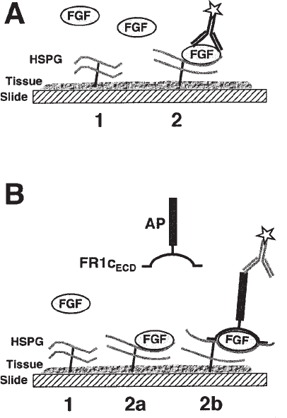
Fig. 1. Schematic representation of the in situ binding assays. (A) Assay to detect growth
factor binding to tissue HSPGs. Tissue sections are incubated with growth factor and bound
growth factor is detected with antibody directed against the growth factor protein or a biotin tag.
This assay distinguishes the following classes of HSPGs: (1) HSPGs that do not bind FGF and
are expected not to play a role in signaling; (2) HSPGs that bind FGF and may be positive or
negative regulators of signaling. (B) Assay to distinguish positive and negative regulators of
growth factor signaling. This assay allows the differentiation of the following classes of HSPGs:
(1) HSPGs that do not bind FGF; (2a) HSPGs that bind FGF, but the HSPG/growth factor
complex does not bind soluble receptor (These HSPGs are expected to sequester FGF and
therefore not to promote signaling.); (2b) HSPGs that not only bind FGF, but assemble the
complete signaling complex (These HSPGs are expected to promote signaling.). AP, alkaline
phosphatase; FR1c
ECD
, FGFR-1c extracellular domain.
538 Friedl et al.
2. Materials
1.1. Preparation of Biotinylated FGF
1. Growth factors: FGF is commercially available from a number of companies. These are
usually expressed in bacteria.
2. Heparin-agarose beads: commercially available from Sigma (St. Louis, MO).
3. PBS: 140 mM NaCl, 2.7 mM KCl, 10 mM Na
2
HPO
4
, 1.8 mM KH
2
PO
4
, pH 7.4.
4. Reaction buffer: 0.2 M sodium bicarbonate in double distilled water (pH 8.1).
5. Bead wash buffer: 20 mM HEPES, 200 mM NaCl, pH 7.4.
6. Elution buffer: PBS, 2.5 M NaCl (sodium chloride concentration may vary depending on
affinity of FGF for heparin).
7. Biotinylation reagent: Sulfo-NHS-biotin (Pierce, Rockford, IL, cat. no. 21217ZZ) dis-
solved in double-distilled H2O as stock at 100 mg/mL (225 mM).
2.2. Purification of FR1c-AP–Binding Probe
1. COS-7 selection medium: DME culture medium (500 mL) containing 10% calf serum to
which is added 10 mL of freshly thawed stocks of 4 mM L-glutamine, 5 mL 1% penicil-
lin/streptomycin, and 500 µg/mL Geneticin (G418 sulfate).
2. COS-7 cell culture medium: Same as selection medium, without added Geneticin.
3. Filter: 0.45 µm filter in a Büchner funnel for filtration of conditioned culture medium.
4. Anti-AP affinity column: 1 mL of commercial preparation of monoclonal anti-AP conju-
gated to agarose beads (Sigma, cat. no. A-2080).
5. AP column elution buffer: 0.1 M glycine (pH 2.5).
6. Neutralization buffer: 1 M Tris (pH 8.0).
7. Human placental alkaline phosphatase: Dilute in PBS to use as a standard for comparison
with purified FR1c-AP (Sigma, cat. no. P1391).
8. 2× AP assay substrate: 2 M diethanolamine, 1 mM MgCl
2
(dissolved at 10 mM concentra-
tion in water prior to addition), 20 mM L-homoarginine, 12 mM p-nitrophenylphosphate
(Sigma 104; Sigma cat. no. 1040-1G).
2.3. Preparation of Tissue Sections and General Histology Material
1. Frozen tissue embedding medium, for example, Tissue Tek OCT compound (Sakura,
Torrence, CA, cat. no. 4583 ).
2. Charged histology slides, for example, Fisher “Plus” slides (Fisher Scientific, Pittsburgh,
PA, cat. no. 12-550-15).
3. Cover slips, e.g. Fisher “Finest” (Fisher Scientific, cat. no. 12-548-5P).
4. Slide-staining racks (optional), for example, Shandon Sequenza racks and cover plates
(Shandon, Pittsburgh, PA).
5. Humidified chamber (as an alternative to staining racks).
6. Fixation of frozen sections: 70 vol% ethanol in double-distilled H
2
O: 4% paraformaldehyde
(e.g., EMS, Ft. Washington, PA, cat. no. 15710) in PBS.
7. Autofluorescence reduction reagents: 0.5mg/mL sodium borohydride, prepared in 4°C
double-distilled H
2
O; 0.1 M glycine, in PBS.
8. Blocking buffer: Bovine serum albumin (2%) in TBS, or 10% serum (e.g., normal swine
serum, Dako, Carpinteria, CA, X0901)
9. Washing buffer TBS: 150 mM NaCl, 10 mM Tris-HCl, pH 7.4.
10. Fluorescently labeled secondary antibody: Alexa-conjugated secondary antibody (e.g.,
Alexa-546-goat-anti-mouse, Molecular Probes, Eugene, OR, cat. no. A-11003).
Localization of FGF-specific HSPGs 539
11. Detection system for brightfield microscopy, for example, Dako EnVision+ system, HRP
(DAB), cat. no. K4006, or Dako LSAB+ peroxidase, Dako, cat. no. K0679.
12. Nuclear counterstain, optional (applied after staining or binding reaction), for example,
Hoechst 33258 for fluorescence microscopy, Meyer’s hematoxylin (e.g., Dako, cat. no.
S3309) for bright-field microscopy.
2.4. Localization of Heparan Sulfate
1. Anti-heparan sulfate antibody: Mouse monoclonal antibody, clone 3G10, Seikagaku,
cat. no. 370260 (Seikagaku-USA [now Cape Cod], Ijamsville, MA).
2. Heparitinase enzyme: Heparitinase (mixture of heparitinase I and II, e.g., Seikagaku,
cat. no. 100703).
3. Heparitinase buffer: 50 mM HEPES, 50 mM NaOAc, 150 mM NaCl, 9 mM CaCl
2
, 0.1%
BSA, pH 6.5.
2.5. Binding of FGF and FR1c-AP to Tissue Sections
1. FGF: Biotinylated (see Subheading 2.1.) or native growth factor.
2. Anti-biotin antibody, for example, mouse monoclonal (Jackson, Cat. # 200-002-096, Jack-
son ImmunoResearch, West Grove, PA) or goat polyclonal (Vector, cat. no. SP-3000,
Vector Laboratories, Burlingame, CA).
3. Anti-placental alkaline phosphatase antibody, for example, mouse monoclonal (Sigma,
clone 8B6, cat. no. A2951) or rabbit polyclonal (Biomeda, cat. no. A67, Foster City, CA).
4. Heparitinase enzyme: Heparitinase (mixture of heparitinase I and II, e.g. Seikagaku,
cat.no. 100703), and heparitinase II, ICN, cat. no. 190102, Costa Mesa, CA.
5. Heparitinase buffer: 50 mM HEPES, 50 mM NaOAc, 150 mM NaCl, 9 mM CaCl
2
, 0.1%
BSA, pH 6.5.
Methods
3.1. Preparation of Biotinylated FGF
The in situ assays require growth factor ligand and soluble receptor-binding probes.
Our experiments have been performed using human recombinant FGFs expressed in
yeast or bacteria. We have used both biotinylated and native FGFs in binding experi-
ments. Biotinylated FGFs offer the advantage that their biotin tag allows detection
without potential background signaling from endogenous FGFs. However, using a
chemically modified FGF introduces potential variability in labeling efficiency and
growth factor inactivation during the biotinylation reaction. In our experience, high
labeling efficiency leads to some loss of activity, which may be of concern. An alter-
native is to use native FGF and observe its localization using high-quality antibodies.
The obvious caveat to this approach is that endogenous FGFs may potentially also be
detected. In practice, this possibility has not been a problem, perhaps because tissue
fixation destroys immunoreactivity of endogenous FGFs sufficiently to prevent their
facile detection. In our routine work, sections consistently lack any endogenous sig-
nal if the incubation step with FGF ligand is omitted before the detection step. In some
embryonic tissues, endogenous FGF can be detected, but its staining is often minor
and can be easily accounted for. For some FGFs and other heparan sulfate-binding
540 Friedl et al.
growth factors, high-quality antibodies allowing in situ localization may not be avail-
able. In this case, biotinylation of ligand may be necessary. Alternatively, the growth
factor could be expressed with a short epitope tag peptide sequence, presumably with-
out affecting its activity (see Note 1).
1. Lyophilized growth factor is dissolved in PBS at 1 mg/mL concentration, aliquoted into
50-µL aliquots, and snap-frozen in liquid nitrogen. It is important to store and handle the
growth factor in polypropylene containers, as it binds avidly to glass.
2. One FGF aliquot (50 µg) is thawed and diluted to 0.5 mL in reaction buffer.
3. Heparin-agaose beads (e.g. 200 µL slurry equaling 100 µL bead volume) are prewashed 1
time with 1 mL elution buffer and 3 times with 1 mL bead wash buffer by centrifugation
using 1.5-mL polypropylene centrifuge tubes. The beads are then equilibrated in 1 mL of
reaction buffer for 10 min.
4. The prewashed beads are combined with the FGF solution and incubated at room tempera-
ture for 10 min, resuspending the beads several times during the binding reaction. The FGF
binds quantitatively to the beads.
5. Sulfo-NHS-biotin stock solution is added to a final concentration of 20 mM. The beads are
incubated in biotinylation reagent at room temperature for 5 min.
6. The reaction is terminated by washing the beads 6 times with bead washing buffer.
7. Biotinylated growth factor is batch-eluted in 200 µL of elution buffer containing NaCl at a
concentration appropriate for the FGF used (e.g., 2.5 M for FGF-2, 1.0 M for FGF-7).
The 2-min incubation is followed by centrifugation, and the process is repeated.
8. The concentration of the biotinylated FGF pooled from the two elution washes is deter-
mined by antibody binding (Westerns blots, ELISA) and comparison with native FGF
standards. The activity of the FGF is compared with native FGF standards in mitogenesis
assays.
3.2. Expression and Purification of RTK-Binding Probe
Soluble FGF RTK is expressed as fusion protein containing the extracellular
(ligand-binding) portion of the RTK at the amino terminus and carboxy-terminal
human placental alkaline phosphatase, which serves as a tag (e.g., FR-AP). Fusion
constructs containing the extracellular portion of the FGFR1c and most other FGF
receptors and their splice variants have been designed and generated by D. Ornitz
(Washington University, St. Louis, MO) (21). FR1c-AP has been extensively charac-
terized in binding studies in vitro and will be used as the example here. This soluble
receptor fusion protein shows HS-dependent binding interactions similar to native
full-length transmembrane receptor. The alkaline phosphatase tag offers a means for
facile purification and the advantage that the protein is easily traceable with a simple
enzyme activity assay. Because human placental alkaline phosphatase is a dimer, the
fusion construct also forms a dimer under physiological conditions. Whether this
dimer formation is critical for its binding activity is uncertain.
1. FR1c-AP is expressed in mammalian COS-7 cells. This cell line is easily transducible and
stably transfected clones can be quickly generated. Using the CMV promoter in the expres-
sion vector (e.g., pCDNA3.1, Invitrogen, Carlsbad, CA) assures high-level expression.
Expression in yeast or insect cells would also be possible, but bacterial expression would
likely not produce satisfactory results because RTK binding requires glycosylation and
disulfide bridges.
