Iozzo Renato V. Proteoglycan Protocols
Подождите немного. Документ загружается.

Phage Display Antibodies to Heparan Sulfate 521
18. Parafilm (American National Can).
19. Polyoxyethylenesorbitan monolaurate (Tween-20, Sigma).
20. PBS containing 0.1% (v/v) Tween-20.
21. 100 mM triethylamine (Merck): add 700 µL (7.18 M) to 50 mL of H
2
O (prepare on the
day of use).
22. 1 M Tris-HCl, pH 7. 4.
23. Sterile 50-mL tubes (Greiner).
24. TYE: 1.5% (w/v) Bacto-Agar, 0.8% (w/v) NaCl, 1.0% (w/v) Pepton, and 0.5% (w/v)
Bacto-Yeast extract.
25. TYE containing 100 µg of ampicillin/mL and 1% (w/v) glucose.
26. Nunclon TC dish 245 × 245 × 25 mm (Nunc); 94/15 Petri dish (Greiner).
27. Glycerol (Sigma).
28. 2XTY containing 15% (v/v) glycerol.
2.2. Screening for Bacterial Clones Expressing Heparan
Sulfate-Binding Antibodies Using ELISA
1. 96-well flat-bottom and 96-well round-bottom sterile Cellstar plates (Greiner).
2. Sterile toothpicks.
3. 2XTY containing 100 µg of ampicillin/mL and 1% (w/v) glucose.
4. 2XTY containing 100 µg of ampicillin/mL and 0.1% (w/v) glucose.
5. Isopropylthio-β-D-galactoside (IPTG, Gibco BRL).
6. 2XTY containing 100 µg of ampicillin/mL and 9 mM IPTG.
7. Glycerol (Gibco BRL).
8. 96-well Microlon ELISA plates, nonsterile (Greiner).
9. PBS.
10. PBS containing 2% (w/v) Marvel (see Note 3).
11. PBS containing 4% (w/v) Marvel.
12. PBS containing 0.1% (v/v) Tween-20.
13. Anti-c-Myc antibody; hybridoma culture supernatant (clone 9E10, mouse IgG; see Note 4).
14. PBS containing 2% (w/v) Marvel and 0.2% (v/v) Tween-20.
15. Alkaline phosphatase-conjugated rabbit anti-mouse IgG antibodies (DAKO).
16. PBS containing 1% (w/v) Marvel and 0.1 % (v/v) Tween-20.
17. 0. 9% (w/v) NaCl.
18. 1 M diethanolamine (Fluka) containing 0.5 mM MgCl
2
, pH 9.8.
19. 4-Nitrophenyl phosphate disodium salt (hexahydrate) (P-NPP, Merck).
2.3. Detection of Antibodies Expressed by Bacterial Clones Using
an Immunoblot Assay
1. 0.45-µm nitrocellulose filter (Schleicher & Schuell).
2. Whattman 3 MM paper.
3. PBS.
4. PBS containing 3% (w/v) Marvel and 1% (v/v) Tween-20.
5. PBS containing 2% (w/v) Marvel and 0.2% (v/v) Tween-20.
6. Anti-cMyc antibody; hybridoma culture supernatant (clone 9E10, mouse IgG; see Note 4).
7. PBS containing 0.1% (v/v) Tween-20.
8. Alkaline phosphatase-conjugated rabbit anti-mouse IgG antibodies (DAKO).
9. PBS containing 1% (w/v) Marvel and 0.1% (v/v) Tween-20.
10. 1 M diethanolamine containing 0.5 mM MgCl
2
, pH 9.8.
522 van Kuppevelt et al.
11. p-Nitro blue tetrazolium chloride (NBT, Merck), and 5-bromo-4-chloro-3-indolyl sulfate
p-toluidine salt (BCIP, Research Organics). Add to 10 mL of 1 M diethanolamine con-
taining 0.5 mM MgCl
2
(pH 9.8): 45 µL of NBT (stock: 75 mg/mL 70% [v/v] dimeth-
ylformamide) and 35 µL of BCIP (stock 50 mg/mL dimethylformamide).
2.4. Screening for “Full-Length” Inserts Using Polymerase Chain
Reaction (PCR) and for V
H
Gene Diversity Using DNA Fingerprinting
1. TYE containing 100 µg of ampicillin/mL and 1% (w/v) glucose.
2. 94/15 Petri dishes.
3. Sterile toothpicks.
4. 5 mM dNTP, Taq DNA polymerase (5 U/µL), and 10 × PCR buffer containing 15 mM
MgCl
2
(Promega).
5. Primers: LMB3 (5'-CAGGAAACAGCTATGAC-3'), fd-SEQ1 (5'-GAATTTTCTGTATGAGG-3').
6. Mineral oil (Sigma).
7. Restriction enzyme BstNI (10 U/µL), NEBuffer 2 (New England Biolabs).
8. DNA marker: φX174/HaeIII Molecular Weight Marker 4 (Eurogentec).
9. SeaKem agarose, NuSieve 3:1 agarose (FMC Bioproducts).
10. 10 mg ethidium bromide (Sigma)/mL H
2
O.
11. 10× TBE buffer: 12.1% (w/v) Tris, 5.1% (w/v) boric acid, and 3.7% (w/v) EDTA.
12. 5× DNA sample buffer: 0.25% (w/v) bromophenol blue, 0.25% (w/v) xylene cyanol FF,
and 40% (w/v) sucrose.
13. QIAprep Spin Miniprep Kit (Qiagen), ABI PRISM
TM
Big Dye Terminator Cycle
Sequencing Ready Reaction Kit (Perkin Elmer).
14. Sequencing primer: (5'- GCCACCTCCGCCTGAACC- 3'), annealing temperature: 65°C.
15. 2XTY containing 100 µg of ampicillin/mL and 1% (w/v) glucose.
16. 2XTY containing 15% (v/v) glycerol.
17. Sterile cryovials (Greiner).
2.5. Production of Culture Supernatant Containing Antibodies
1. Glycerol stock of a bacteria producing an anti-heparan sulfate scFv antibody, stored
at –80°C.
2. TYE containing 100 µg of ampicillin/mL and 1% (w/v) glucose.
3. 94/15 petri dish.
4. 2XTY containing 100 µg of ampicillin/mL and 1% (w/v) glucose.
5. 2XTY containing 100 µg of ampicilin/mL and 0.1% (w/v) glucose.
6. IPTG (Gibco BRL).
7. 10× protease inhibitor mix: 0.1 M EDTA, 250 mM iodacetamid, 1 M N-ethylmaleimide,
1% (w/v) NaN
3
, 1.5 mTIU aprotinin/mL, 0.1% (w/v) pepstatin A, and 1 mM
phenylmethylsulfofluoride in H
2
O.
2.6. Production of Periplasmic Fraction Containing Antibodies
1. See Subheadings 2.5.1. – 2.5.7.
2. Periplasmic fraction buffer (PPF): adjust 0.5 M boric acid (Gibco BRL) to pH 8.0 with
0.5 M sodium borate (Sigma). Take 20 mL of the adjusted solution and add 1.6 mL of 5 M
NaCl, 0.25 mL of 0.2 M EDTA, and adjust the volume to 50 mL with H
2
O.
3. 0.2 µm disposable filter holder.
4. Dialyzing membrane (Spectra/Por, cutoff value 10 kDa).
5. PBS.
Phage Display Antibodies to Heparan Sulfate 523
2.7. Evaluation of Specificity of Antibodies Using
Immunofluorescence Analysis
1. Tissue specimens, snap-frozen in liquid isopentane cooled with liquid nitrogen, and stored
at –70°C.
2. 5-µm tissue cryosections (stored at –20 or –80°C).
3. Heparinase III (0.006 units/mL, Sigma) in incubation buffer (50 mM NaAc and 50 mM
Ca(Ac)
2
, pH 7.0).
4. Chondroitinase ABC (1 unit/mL, Seikagaku) in incubation buffer (25 mM Tris-HCl, pH 8.0).
5. Slides and cover slips.
6. PBS.
7. Blocking solution: PBS containing 1% (w/v) BSA .
8. Washing solution: PBS.
9. Primary antibody solution: add 1 volume of periplasmic fraction of an anti-heparan sul-
fate antibody to 1 volume of blocking solution.
10. Anti-cMyc (9E10) antibody (hybridoma culture supernatant) diluted 1/1 with blocking
solution (see Note 4).
11. Fluorophore (Alexa-488)-conjugated anti-mouse IgG antibody (Molecular Probes) solu-
tion: Make a dilution (1/200–1/500) in PBS containing 0.5% (w/v) BSA.
12. Mowiol (Hoechst) embedding solution.
2.8. Evaluation of Specificity of Antibodies by ELISA
1. ELISA plates (Microlon, Greiner).
2. PBS.
3. Washing solution: PBS containing 0.1% (v/v) Tween-20.
4. Blocking solution: PBS containing 1% (w/v) BSA.
5. Anti-heparan sulfate (primary) antibody solution: add 1 volume of a bacterial culture
supernatant containing an anti-heparan sulfate antibody to 1 volume of blocking solution,
or use diluted periplasmic fraction.
6. Anti-cMyc (9E10) antibody (hybridoma culture supernatant) diluted 1/1 with blocking
solution (see Note 4).
7. Alkaline phosphatase-conjugated rabbit anti-mouse IgG antibodies diluted 1/1000 in PBS
containing 0.5% (w/v) BSA.
8. Substrate solution: 1 mg of p-nitrophenyl phosphate/mL diethanolamine solution (1 M
diethanolamine, 0.5 mM MgCl
2
, pH 9.8).
9. A number of molecules necessary for evaluation of crossreactivity of the antibodies. These
may include heparan sulfate from various sources, heparin, dermatan sulfate, chondroitin
4 and 6-sulfate, keratan sulfate, dextran sulfate, hyaluronate, and DNA.
3. Methods
3.1. Selection of Phages Displaying Antibodies Reactive
with Heparan Sulfate Using Biopanning
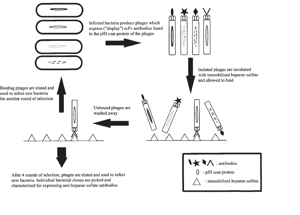
For a schematic outline of the selection procedure, see Fig. 1.
3.1.1. Growth of Antibody Phage Display Library
1. Inoculate 50 mL of 2XTY containing 100 µg of ampicillin/mL and 1% (w/v) glucose with
about 5 × 10
8
bacteria from a glycerol stock of the (semi)-synthetic scFv Library #1 (3).

524 van Kuppevelt et al.
Fig. 1. Schematic representation of the selection of phage display-derived anti-heparan sulfate antibodies by
biopanning.
524
Phage Display Antibodies to Heparan Sulfate 525
2. Grow the culture, while shaking, at 37°C until an absorbance at 600 nm of 0.5 is reached
(see Note 5).
3. Pipet 10 mL of the culture into a sterile 50-mL tube and add about 4 × 10
10
VCS-M13
helper phages (see Note 6).
4. Incubate in a water bath, without shaking, at 37°C for 30 min.
5. Spin the infected culture at 3000g for 10 min at room temperature. Decant the supernatant
and resuspend the pellet in 30 mL of 2XTY containing 100 µg of ampicillin and 25 µg of
kanamycin/mL.
6. Add the 30 mL of bacterial suspension to 270 mL of prewarmed (30°C) 2XTY contain-
ing 100 µg of ampicillin and 25 µg of kanamycin/mL (No glucose! see Note 7). Incu-
bate, while shaking, at 30°C for 16–20 h to allow for large-scale (antibody-displaying)
phage production.
7. Inoculate 5 mL of 2XTY with a single E. coli TG1 colony from a minimal medium
plate and grow, while shaking, at 37°C for 16–20 h. This culture will be used in
Subheading 3.1.3, step 16.
3.1.2. Isolation of Phages
1. Spin the culture from step 6 under Subheading 3.1.1 at 10,000g for 10 min at 4°C to
remove bacteria. Decant the supernatant containing the phages into another sterile bucket.
2. Add 60 mL of ice-cold PEG/NaCl to the supernatant, mix well by inverting the bucket at
least 30 times, and leave the bucket on ice for 1 h. In this step (and step 3) phages are
precipitated (see Note 8).
3. Spin the phages at 10,000g for 30 min at 4°C. Resuspend the pellet in 40 mL of ice-cold,
sterile milli-Q water. Transfer the suspension to a 50-mL tube and add 8 mL of ice-cold
PEG/NaCl. Mix well (as in step 2) and leave for 30 min on ice.
4. Spin the mixture at 3000g for 30 min at 4°C. Decant the supernatant and remove the
remains with a pipet. Respin briefly and remove residual PEG/NaCl. Invert the tube on a
piece of paper tissue, and leave for 30 min to drain any remaining PEG/NaCl.
5. Resuspend the pellet in sterile PBS and spin at 3000g for 10 min at 4°C to remove any
remaining bacterial debris. Decant the supernatant containing the phages into a sterile
tube and store at 4°C until use.
3.1.3. Selection of Phages Binding to Heparan Sulfate
1. Add 2 mL of a 20-µg heparan sulfate/mL solution to an immunotube for 16 h, 4°C. Wash
the immunotube 3 times with PBS and block the tube with PBS containing 2% (w/v)
Marvel. Fill the tube to the brim, cover it with Parafilm, and incubate for at least 2 h at
room temperature to avoid nonspecific binding of phages to the surface of the tube. This
step should be performed early in the day, so the immunotube will be ready when the
phages used for biopanning (step 5, Subheading 3.1.2.) are obtained.
2. Wash the blocked tube 3 times with PBS. Add 2 mL of PBS containing 4% (w/v) Marvel
and 2 mL of phage supernatant (step 5, Subheading 3.1.2.) to the tube, cover with
Parafilm, and incubate for 30 min on an under-and-over turntable (room temperature),
followed by standing for 90 min (room temperature).
3. Discard the phage suspension and wash the tube 20 times with PBS containing 0.1% (v/v)
Tween-20, followed by 20 washes with PBS.
4. Remove the last remains of PBS and elute the bound phages with 1 mL of 100 mM tri-
ethylamine. Cover the tube with Parafilm and rotate for 10 min on an under-and-over
turntable at room temperature.
526 van Kuppevelt et al.
5. Add the 1 mL of eluted phages to a 50-mL tube containing 0.5 mL of 1 M Tris-HCl (pH 7.4)
for pH neutralization. Also add 200 µL of 1 M Tris-HCl (pH 7.4) to the remaining phages in
the immunotube. At this point phages can be stored at 4°C for a short period of time (up to
2 d), or used the same day to infect E. coli TG1 cells. The latter is recommended.
6. Add 1 mL of the eluted phages from the 50-mL tube (Subheading 3.1.3., step 5) to 9 mL of
exponentially growing E. coli TG1 cells in a 50-mL tube. Add 4 mL of the TG1 culture to
the remaining phages in the immunotube (step 5). Incubate both cultures for 30 min at 37°C in
a water bath, without shaking, to allow for infection. Exponentially growing TG1 culture is
prepared as follows: Inoculate 50 mL of 2XTY with 0.5 mL of overnight culture from Sub-
heading 3.1.1., step 7. Grow the culture, while shaking, at 37°C until an absorbance at 600
nm of 0.4–0.5 is reached. The culture obtained is ready for infection (see Note 5).
7. Pool both infected TG1 cultures. Take 100 µL of the pooled culture and make 4 serial
dilutions (1/10
2
, 1/10
4
, 1/10
6
, and 1/10
8
) in 2XTY containing 100 µg of ampicillin/mL
and 1%(w/v) glucose. Plate 100 µl of these dilutions on 94/15 TYE plates containing
100 µg of ampicillin/mL and 1% (w/v) glucose, and grow for 16–20 h at 37°C. Calculate
the titer from these dilutions (see Note 9).
8. Spin the rest of the pooled culture at 3000g for 10 min at room temperature. Decant the
supernatant and resuspend the pellet in 1 mL of 2XTY. Spread the cell suspension on a
Nunclon TC plate with TYE containing 100 µg of ampicillin/mL and 1% (w/v) glucose
and grow for 16–20 h at 37°C.
9. Add 5 mL of ice-cold 2XTY containing 15% (v/v) glycerol to the Nunclon TC dish and scrape
the bacterial cells from the plate with a glass spreader. Take 50 µL of the bacterial suspension
and use it for inoculation of 50 mL 2XTY containing 100 µg of ampicillin/mL and 1% (w/v)
glucose as in Subheading 3.1.1., step 1. Store the remaining bacteria at –70°C (see Note 10).
Repeat the selection procedure for another 3–4 rounds (all steps Subheading 3.1.1.–3.1.3.).
3.2. Screening for Bacterial Clones Expressing Heparan
Sulfate-Binding Antibodies Using ELISA
1. Pick individual bacterial clones, using sterile toothpicks, from the serial dilution plates of
the last 2 selection rounds (see Subheading 3.1.3., step 6) and inoculate 100 µL of 2XTY
containing 100 µg of ampicillin/mL and 1% (w/v) glucose (one clone per well!) in sterile
96-well flat-bottom tissue culture plates. Secure the lid with tape and grow for 16–20 h at
37°C while gentle shaking. These plates will be the master plates.
2. Transfer 2 µL of bacterial culture (step 1) to the corresponding wells of sterile 96-well
round-bottom tissue culture plates with 200 µL 2XTY containing 100 µg of ampicillin/
mL and 0.1%(w/v) glucose (see Note 11). Secure the lid with tape and grow at 37°C,
while shaking, until an absorbance at 600 nm of 0.9 is reached (see Note 12). Add to each
well 25 µL of 2XTY containing 100 µg of ampicillin/mL and 9 mM IPTG. Incubate the
plates, while shaking gently, at 30°C for 16–20 h. Add glycerol to the master plates to a
final concentration of 15% (v/v), mix well, and store the plates at –70°C until further use.
3. Incubate wells from ELISA plates with 100 µl of a 10-µg heparan sulfate/mL solution for
16 h at 4°C. Wash 3 times with PBS and block with PBS containing 2% (w/v) Marvel for
1 h at 37°C. Wash the plates 3 times with PBS containing 0.1% (v/v) Tween-20.
4. Spin the 96-well round-bottom plates (step 2) at 1800g for 10 min at room temperature.
5. Add 60 µL of the culture supernatant (step 4) to 60 µl of PBS containing 4% Marvel, transfer
100 µl to wells of ELISA plates, and incubate for 1 h at room temperature. Use 50 µL of the
culture supernatant in an immunoblot assay used for evaluation of antibody production (see
Subheading 3.3.). The immunoblot assay can be performed simultaneously with the ELISA.
Phage Display Antibodies to Heparan Sulfate 527
6. Discard the culture supernatant and wash the ELISA plates 6 times with PBS containing
0.1% (v/v) Tween-20.
7. Add 100 µL of 9E10 hybridoma supernatant diluted 1/1 with PBS containing 2% (w/v)
Marvel and 0.2% (v/v) Tween-20 to the wells and incubate for 1 h at room temperature.
8. Discard the 9E10 solution and wash the ELISA plates 6 times with PBS containing
0.1% (w/v) Tween-20.
9. Add 100 µL of alkaline phosphatase-conjugated rabbit-anti-mouse IgG antibodies, diluted
1/1000 diluted in PBS containing 1% (w/v) Marvel and 0.1% (v/v) Tween-20 to the wells
and incubate for 1 h at room temperature.
10. Discard the fluid and wash the ELISA plates 5 times with PBS containing 0.1% (v/v)
Tween-20 followed by 1 wash with 0.9% (w/v) NaCl.
11. Add 100 µL 1 M diethanolamine containing 1-mg/mL P-NPP and 0.5 mM MgCl
2
(pH 9.8) to
the wells and incubate, in the dark, at room temperature until color development is optimal.
12. Read the absorbance at 405 nm (see Note 13).
3.3. Detection of Antibody Expression
by Bacterial Clones Using an Immunoblot Assay
1. Cut two Whattman 3 MM papers and one 0.45-µm nitrocellulose filter to the size required
for use in a 96-well dot-blot apparatus.
2. Soak the papers and the filter in PBS for 10 min. Apply the papers and filter to the dot-
blot apparatus (nitrocellulose filter on top). Make sure to remove all air bubbles.
3. Transfer 50 µL of culture supernatant from step 4 under Subheading 3.2. to the dot-blot
apparatus and pull the fluid through the filter by vacuum suction.
4. Remove the nitrocellulose filter from the apparatus and air-dry the filter for 20 min at
room temperature.
5. Block the filter in a container with PBS containing 3% (w/v) Marvel and 1% (v/v) Tween-
20 for 1 h at room temperature, while shaking. The volume of fluids used in this step, as
well as in the following steps, depends on the size of the container used. Make sure the
filter is sufficiently covered with fluid.
6. Discard the blocking solution and add 9E10 hybridoma supernatant diluted 1/1 with PBS
containing 2% (w/v) Marvel and 0.2% (v/v) Tween-20. Incubate, while shaking, for 1 h at
room temperature.
7. Discard the solution and wash 3 times for 10 min, while shaking, with PBS containing
0.1% (v/v) Tween-20.
8. Add alkaline phosphatase-conjugated rabbit anti-mouse IgG antibodies diluted 1/1000 in
PBS containing 1% (w/v) Marvel and 0.1% (v/v) Tween-20. Incubate, while shaking, for
1 h at room temperature.
9. Discard the antibody solution and wash, while shaking, 2 times with PBS containing
0.1% (v/v) Tween-20 for 10 min, 1 time with PBS for 5 min, and 1 time with 1 M
diethanolamine containing 0.5 mM MgCl
2
(pH 9.8) for 5 min.
10. Add NBT/BCIP substrate solution and incubate, while shaking, at room temperature until
the color develops. Discard the substrate solution and wash the filter a couple of times
with water (see Note 14).
3.4. Screening for “Full-Length” Inserts Using PCR
and for V
H
Gene Diversity Using DNA Fingerprinting
In this procedure, the positive clones will be analyzed for the presence of DNA
(about 1 kbp) encoding the scFv antibody. In addition, as an initial screening for the
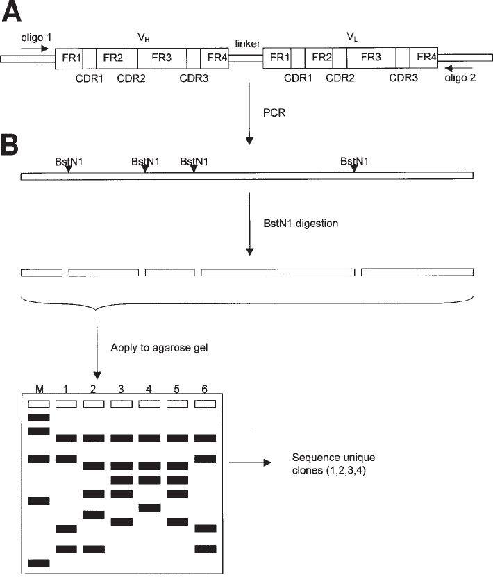
diversity of the clones, restriction enzyme analysis will be performed. (Also see Fig. 2).

528 van Kuppevelt et al.
Fig. 2. Restriction fragment analysis of clones expressing anti-heparan sulfate antibodies.
Polymerase chain reaction (PCR) is preformed to amplify the region encoding the scFv anti-
body (A), using a set of primers flanking the V
H
and V
L
segments. The resulting (full-length)
PCR fragments (B) harbor a unique pattern of BstN1 cleavage sites (CC*
A
/
T
GG), serving as a
fingerprint. Following digestion with restriction enzyme BstN1 (C), the fragments are sepa-
rated on an agarose gel and the restriction patterns of each clone are compared (D). Clones with
unique restriction patterns are selected and sequenced to establish the V
H
family, germ line
segment (DP number, see ref. 4) and V
H
-CDR3 sequence (randomized in the library used; see
ref. 3).FR, framework region; CDR, complementary determining regions; M, DNA marker.
Phage Display Antibodies to Heparan Sulfate 529
1. Select heparan sulfate-positive clones (as detected by ELISA) from the corresponding
master plate (see Subheading 3.2.). Plate bacteria on 94/15 dishes with TYE containing
100 µg of ampicillin/mL and 1% (w/v) glucose to obtain single colonies.
2. Pick a single colony of each clone (mark the colony on the back of the Petri dish) with a
sterile toothpick and transfer the cells into the following PCR-mixture: 34.5 µL H
2
O, 5.0 µL
10X PCR buffer, 2.5 µL 20X dNTP (5 mM each), 2.5 µL LMB3 primer (10 pmol/µL),
2.5 µL fd-SEQ1 primer (10 pmol/µL), 0.5 µL Taq polymerase (5 U/µL). Overlay the PCR
mixture with a droplet of mineral oil and use the following PCR program: 10 min at 94°C,
30 cycles of 1 min at 94°C, 1 min at 60°C, 2 min at 72°C, 10 min at 72°C, cool to 4°C.
3. Take 4 µL of the PCR-mixture and add 1 µL of 5X DNA sample buffer. Run the samples
on a 1% (w/v) Seakem agarose gel (add 3.0 µL ethidium bromide [10 mg/mL] to 75 mL
of agarose solution). Include DNA marker (250 ng) in one of the lanes. Run the gel at
50 V in an appropriate volume of 1X TBE buffer. Analyze the gel on a UV transillumina-
tor. PCR products of about 1000 base pairs indicate full-length clones (see Note 15).
4. For DNA fingerprinting take 20 µL of the PCR-mixture and add 20 µL of the following
restriction enzyme mix: 17.8 µL H
2
O, 2.0 µL 10X NEbuffer 2, 0.2 µL BstNI (10 U/µL).
Overlay with mineral oil and incubate at 60°C for 3 h. Take 8 µL and add 2 µL 5X DNA
sample buffer. Run the restriction mixtures on a 4% (w/v) 3/1 NuSieve agarose gel as
described in step 3, Subheading 3.4., Differences in banding pattern indicate different
clones (see Note 16 and Fig. 2).
5. Take with a sterile toothpick unique clones from the plate with marked colonies (step 2,
Subheading 3.4.) and add to 10 mL 2XTY containing 100 µg ampicillin/mL and 1% (w/v)
glucose. Grow for 16–20 h, while shaking, at 37°C.
6. Take 1.5 mL of the bacterial culture (step 5, Subheading 3.4.) for preparation of phagemid
DNA, used for sequencing and for long-term storage (see Note 17). For phagemid isolation
and sequencing we use materials described in steps 13 and 14, Subheading 2.4.
7. Spin the rest of the culture at 3000g for 10 min at 4°C. Decant the supernatant and resus-
pend the pellet in 1 mL of ice-cold 2XTY containing 15% (v/v) glycerol. Aliquot the
bacterial suspension into several sterile cryovials and store at –70°C.
3.5. Production of Culture Supernatant Containing Antibodies
1. Inoculate 5 mL of 2XTY containing 100 µg ampicillin/mL and 1% (w/v) glucose with a
single colony from a 94/15 dish with TYE containing 100 µg ampicillin/mL and 1% (w/v)
glucose, and derived from a glycerol stock. Grow for 16–20 h, while shaking, at 37°C.
2. Inoculate 500 mL of 2XTY containing 100 µg ampicillin/mL and 0.1% (w/v) glucose
with 5 mL of culture (step 1, Subheading 3.5.) and grow, while shaking, at 37°C until an
absorbance at 600 nm of 0.5 –0.8 is reached.
3. Add IPTG to a final concentration of 1 mM and grow the culture, while shaking, at 30°C
for 16–20 h.
4. Put the culture on ice for 20 min.
5. Spin the culture at 3000g for 10 min at 4°C. Add 0.1 volume of 10X protease inhibitor
mix (see step 7, Subheading 2.5.) to the supernatant and store in aliquots at 4°C, if used
directly, or at –70°C (see Note 18).
3.6. Production of Periplasmic Fraction Containing Antibodies
1. See step 1, Subheading 3.5.
2. See step 2, Subheading 3.5.
3. Add IPTG to a final concentration of 1 mM and grow the culture, while shaking, at
30°C for 3 h.
530 van Kuppevelt et al.
4. Put the culture on ice for 20 min.
5. Spin the culture at 3000g for 10 min at 4°C. Decant the supernatant and resuspend the
bacterial pellet in 5 mL of ice-cold “periplasmic fraction” (PPF) buffer (see step 2,
Subheading 2.6.).
6. Vortex the bacterial suspension vigorously for 10 s and spin at 48,000g for 30 min at 4°C.
7. Filter the supernatant (periplasmatic fraction) through a 0.2-µm disposable filter.
8. Dialyze the periplasmatic fraction for 16–20 h against 5 L of PBS at 4°C.
9. Add 0.1 volume of 10X protease inhibitors (see step 7, Subheading 2.5.). Store the
periplasmatic fraction at 4°C, if used directly, or at –70°C (see Note 18).
3.7. Evaluation of Specificity of
Antibodies Using Immunofluorescence Analysis
1. All incubation steps are carried out in a humid atmosphere at room temperature.
2. Incubate cryosections with heparinase III or chondroitinase ABC overnight at 37°C. As a
control for enzyme reactivity, incubation buffer without enzyme is used (see Notes 19
and 20).
3. Rinse 3 times in PBS and block in PBS containing 1% (w/v) BSA for 30 min.
4. Incubate cryosections with primary antibody solution (anti-heparan sulfate antibodies)
for 60 min.
4. Remove primary antibody solution carefully and rinse once and wash 3 times (10 min)
with PBS.
5. Incubate cryosections with mouse anti-cMyc antibody solution for 60 min.
6. Remove antibody solution carefully and rinse once and wash 3 times (10 min) with PBS.
7. Incubate cryosections with fluorophore-conjugated anti-mouse IgG antibody solution for
60 min.
8. Remove antibody solution carefully and rinse once and wash 3 times (10 min) with PBS.
9. Incubate cryosections in 100% methanol for 10 s for dehydration.
10. Air-dry sections and use mowiol for embedding.
11. Analyze staining patterns by fluorescence microscopy (see Note 21). Figures 3 and 4 are
examples of staining patterns.
3.8. Evaluation of Specificity of Antibodies Using ELISA
The reactivity of the anti-heparan sulfate antibodies with other molecules can be
analyzed by ELISA in two ways: (1) by application of antibodies to wells of microtiter
plates coated with the test molecule, or (2) by an inhibition assay in which the antibod-
ies are incubated with the test molecule.
Method 1
1. All incubation steps are carried out at room temperature.
2. Coat wells with test molecules (see step 9, Subheading 2.8.) by incubation with 10 µg of
test molecules/mL solution for 16 h at 4°C.
3. Wash the wells 6 times with PBS containing 0.1% (v/v) Tween-20.
4. Block the free binding sites with 200 µL of blocking solution for 1 h.
5. Wash the wells 6 times with PBS containing 0.1% (v/v) Tween-20.
6. Incubate the wells with 100 µL of anti-heparan sulfate (primary) antibody solution for 60 min.
7. Wash the wells 6 times with PBS containing 0.1% (v/v) Tween-20.
8. Incubate the wells with 100 µL of mouse anti-cMyc antibody solution for 60 min.
