Iozzo Renato V. Proteoglycan Protocols
Подождите немного. Документ загружается.


Core Proteins Detection 329
329
From:
Methods in Molecular Biology, Vol. 171: Proteoglycan Protocols
Edited by: R. V. Iozzo © Humana Press Inc., Totowa, NJ
32
Detection of Proteoglycan Core Proteins
with Glycosaminoglycan Lyases and Antibodies
John R. Couchman and Pairath Tapanadechopone
1. Introduction
Proteoglycans are quite abundant components of many extracellular matrices, while
most cell surfaces also bear these macromolecules. Frequently the profiles are com-
plex. For example, several members of the syndecan and glypican families of cell
surface heparan sulfate proteoglycans may be present on a single cell type (1,2). Some
extracellular matrices, e.g., from brain, may also contain a variety of proteoglycans
including several members of the hyalectans or aggregating proteoglycans such as
neurocan, brevican, and versican (3). Frequently it is useful to monitor the nature and
variety of proteoglycans in a pool from tissues or cell cultures in a simple manner,
before moving on to further purification steps, use of core protein-specific antibodies,
or pursuit of a potentially novel core protein.
While proteoglycans require some specialized techniques for analysis, advantage can
be taken of their glycanation to identify core proteins even when their precise character-
istics remain unresolved. Specific enzymes are readily available, first from bacterial
sources but more recently of recombinant origin, which selectively degrade glycosami-
noglycans. Chondroitinase ABC will degrade virtually all chondroitin and dermatan
sulfates, while leaving heparan and keratan sulfate chains intact. Conversely, heparitinase
enzymes will degrade nearly all forms of heparan sulfate, but are unable to degrade
chondroitin, dermatan, or keratan sulfate (see Fig. 1). Further, the consequences of
glycosaminoglycan removal can be monitored by sodium dodecyl sulfate-polyacryla-
mide gel electrophoresis (SDS-PAGE). The heterogeneous nature of proteoglycans
ensues largely from the variable number, length and charge of glycosaminoglycans in a
pool of a single core protein (e.g., aggrecan from cartilage, or perlecan from a basement
membrane preparation). Proteoglycans are frequently seen as broad smears, or
sometimes, when large, may not even penetrate a 3% resolving gel (see Fig. 2A). Once
glycosaminoglycan lyases have removed most of the chains, the core proteins become
much more readily resolved by SDS-PAGE as discrete polypeptides (see Fig. 2).

330 Couchman and Tapanadechopone
Chondroitinases and heparitinases are eliminases, so that the remaining core pro-
teins have serine (usually) residues bearing not only the stem oligosaccharide (xylose-
galactose-galactose-uronic acid) but also a disaccharide or larger oligosaccharide with
a terminal unsaturated uronic acid residue. This, it turns out, is quite antigenic, and
monoclonal (4,5) as well as polyclonal antibodies have been raised (6) which recog-
nize the carbohydrate “stubs” remaining after chondroitinase or heparitinase treat-
ments. They are also very specific. An antibody recognizing a heparan sulfate “stub”
with a terminal unsaturated uronic acid residue will not recognize the equivalent “stub”
generated by a chondroitinase enzyme, and vice versa. Therefore, the combined use of
enzymes and antibodies can be used, for example in Western blotting, to estimate the
sizes of core proteins, and the type of glycosaminoglycan present. This can be particu-
larly useful where a particular core protein, e.g., perlecan, can be substituted with
heparan and/or chondroitin sulfate chains (see Fig. 1). It can provide evidence of hybrid
proteoglycans that bear more than one glycosaminoglycan type. Further, since the
antibodies do not recognize core protein epitopes, a mixed population of heparan and/
or chondroitin and dermatan sulfate proteoglycan can be quickly analyzed for their
number, size, and glycanation profiles. Such evidence can be supported by more tradi-
tional metabolic labeling methods, combined with chemical or enzymatic degradation
techniques followed by gel filtration analysis.
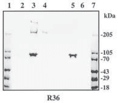
Fig. 1. Detection of chondroitin/dermatan sulfate proteoglycans core proteins from EHS
tumor with R36, a polyclonal antibody recognizing chondroitin/dermatan sulfate stubs remain-
ing after chondroitinase ABC treatment. Lanes 1 and 7 are standards whose molecular weight
in kilodaltons is indicated. Lane 2, untreated sample; lane 3, sample treated with chondroitinase
ABC and heparinase II and III; lane 4, sample treated with chondroitinase ABC only; lane 5
contains heparinase II and III only, while lane 6 contains chondroitinase ABC alone. The com-
mon polypeptide seen in lanes 3 and 5 is present in the heparinase II preparation. The data show
that a CS/DS proteoglycan with a core protein of Mr ~ 200,000 (in lanes 3 and 4) is accompa-
nied by a second, large core protein that is revealed only after additional heparinase treatment.
This, therefore, represents a hybrid form of perlecan bearing both HS and CS/DS chains.

Core Proteins Detection 331
2. Materials
1. Samples for analysis dissolved in suitable buffers (see Note 1).
2. Heparitinase buffer: 0.1 M sodium acetate, 0.1 mM calcium acetate, pH 7.0.
3. Chondroitinase buffer: 50 mM Tris-HCl, 30 mM sodium acetate, 20 mM ethyleme-
diamintetraacetic acid (EDTA), 10 mM NEM, 0.2 mM phenylmethyl sulfonyl flouride
(PMSF), and 0.02 sodium azide, pH 8.0 (see Note 1).
4. Protease inhibitor for all heparitinase treatments (see Note 2): 10× trypsin inhibitor
type III-0 (ovomucoid 100 µg/mL, Sigma).
5. Glycosaminoglycan lyases (Seikagaku or Sigma):
a. Heparan sulfate: heparinase III (EC 4.2.2.8). This enzyme is also known as heparitinase
and heparitinase I.
b. Chondroitin sulfate and dermatan sulfate: chondroitin ABC lyase (EC 4.2.2.4).
c. Chondroitin sulfate: chondroitin AC II lyase (EC 4.2.2.5).
d. Dermatan sulfate: chondroitin B lyase (no EC number).
6. Glycosaminoglycan carriers (optional): chondroitin sulfate type A (chondroitin 4-sulfate)
or type C (chondroitin 6-sulfate), heparan sulfate (Sigma).
7. 2× SDS-PAGE sample buffer (Sigma), with or without reducing agent (e.g., 40 mM
dithioerythreitol).
8. Prestained protein molecular-weight standards (Sigma or Bio-Rad).
9. SDS-PAGE gels. If a wide size range of core proteins is suspected, 3–15% gradient gels
can be useful.
10. Electroblotting and transfer buffers and apparatus.
11. Transfer membrane: 0.45-µm nitrocellulose (Bio-Rad Trans-Blot
®
transfer medium or
Schleicher & Schuell #BA85), PVDF (Millipore Immobilon P), or positively charged nylon
(Bio-Rad Zetabind) membranes.
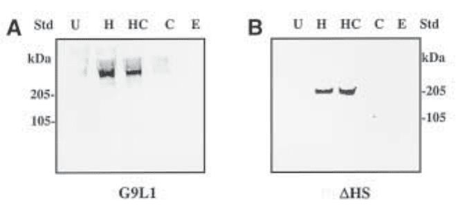
Fig. 2. Detection of intact murine perlecan (A) and recombinant domain IV-V of mouse
perlecan transfected into COS-7 cells (B) with a rat monoclonal antibody specific to perlecan
core protein (A) and monoclonal ∆-heparan sulfate antibody recognizing HS stub after
heparinase III (heparitinase) treatments (B). (A) shows that the intact perlecan core protein is
not clearly visible until the HS chains have been removed, while (B) shows HS substitution on
the recombinant perlecan. In each blot, the proteoglycans are untreated (U), heparitinase-pre-
treated (H), heparitinase- and chondroitinase-pretreated (HC), or chondroitinase-pretreated (C).
Lanes E contain heparitinase and chondroitinase enzymes only.
332 Couchman and Tapanadechopone
12. Blocking buffer: 5% nonfat dried milk in 0.1% Tween 20 is optional in phospate-buffered
saline, PBS (TPBS).
13. Diluting buffer: 1% nonfat dried milk, 0.1% bovine serum albumin, and 0.1% (v/v) Tween 20
in PBS (for monoclonal antibodies) or triphosphate-buffered saline, TBS (for polyclonal
antibodies).
14. Primary antibodies recognizing carbohydrate “stubs” (Seikagaku):
a. Monoclonal ∆-heparan sulfate (for heparan sulfate GAG).
b. Monoclonal anti proteoglycan ∆-di-0S, -4S, -6S (for chondroitin/ dermatan sulfate GAGs;
see Note 3).
These antibodies are also available as biotin conjugates.
c. Equivalent antibodies recognizing protein of interest.
15. Secondary antibodies: horseradish peroxidase- or alkaline phosphatase-anti-Ig conjugate.
Alternately, streptavidin-horseradish peroxidase conjugate should be used where the pri-
mary antibodies are biotin conjugates.
16. Chromogenic and chemiluminescence visualization system, e.g., ECL™ Western blot-
ting detection reagents (Amersham Pharmacia Biotech) for peroxidase conjugates or
alkaline phosphatase-conjugate substrate kit (Bio-Rad).
3. Methods
1. Divide the proteoglycan sample to be analyzed into equal aliquots. The number depends
on the enzyme treatments to be performed. For example, if a proteoglycan pool is sus-
pected to contain chondroitin and heparan sulfate proteoglycans, four aliquots should be
used. One sample is left untreated, while others receive chondroitinase ABC, heparitinase,
or both enzyme treatments. Ideally, each sample should contain 1–10 µg of proteoglycan.
The choice of buffer depends on the enzymes to be used (see Note 1). Further controls
contain buffer with enzyme only (no proteoglycan).
2. Treat samples with appropriate enzymes at 37°C. The amount and duration of enzyme
treatment depend on the proteoglycan concentration. For 1–10 µg of proteoglycan, 2–3 h
of incubation with 0.5–1 mU chondroitinase ABC or 1–2 mU heparitinase in the presence
of protease inhibitor (1× ovomucoid, see Note 2) is suggested. Where concentrations of
proteoglycan are higher, adding a second aliquot of enzyme after 2 h, for a further incuba-
tion can be beneficial. Where proteoglycan concentrations are very low (below 100–200 ng
per sample), adding approximately 0.5 µg of appropriate free glycosaminoglycan can be
added as carrier to aid efficiency and recovery (but see Note 4).
3. If enzyme activity needs to be verified, set up samples of free glycosaminoglycan (approx
0.5 mg/mL) in buffer, to which the enzymes are added, and incubate simultaneously.
Enzyme activity is monitored spectrophotometrically at 232 nm.
4. Enzyme treatments are terminated by adding SDS-PAGE sample buffer (with or without
reducing agent, Subheading 2.) and heating to 100°C, if required. Samples can be frozen
at –20°C or immediately resolved by SDS-PAGE.
5. Samples are applied to SDS-PAGE gels for conventional electrophoresis and transfer to
nitrocellulose or other medium (see Note 5). If a range of core protein masses is sus-
pected, or not known, acrylamide gradients are preferable (e.g., 3–15%).
6. Membranes are blocked conventionally, for example in 5% dried milk powder in phos-
phate-buffered saline for at least 1 h. They are then probed with monoclonal or
polyclonal antibodies recognizing carbohydrate “stubs” created by glycosaminoglycan
lyases. These are available as purified IgG, and sometimes in biotinylated form, and
should be used at 10–25 µg/mL. Incubation can be at 4°C overnight, or shorter periods
at room temperature or 37°C, but for at least 1 h. Constant gentle agitation is advised.
Core Proteins Detection 333
7. Thorough washing is followed by secondary antibody (e.g., affinity purified goat anti-mouse
IgG conjugated to horseradish peroxidase) in the same buffer for 1 h at room temperature.
Antibody concentrations should accord with manufacturer’s instructions. Extensive washes
are then followed by visualization as preferred, such as chemiluminescence.
4. Notes
1. A suitable buffer for chondroitinase ABC or AC II is listed under Subheading 2., as is
one suitable for heparitinase (also known as heparinase III). However, where samples are
to be treated with both enzymes, we have found the heparitinase buffer to be suitable. It
should be noted that chondroitinase B is inhibited by phosphate. Heparinase activity is
increased by the presence of calcium ions, but it is reported that the activity of heparinase
III is not much decreased by its absence.
2. Polysaccharide lyases are primarily of microbial origin. Protease contamination can be
present in the enzyme preparation, especially all heparinase enzymes. This can cause
misleading results. Thus, protease inhibitor should be added in case of heparitinase treat-
ments. Chondroitinase ABC is available in protease-free form.
3. Separate, specific antibodies are available that, while all recognizing the terminal unsaturated
uronic acid residue, as described, have specificity for the presence and position of sulfate on the
adjacent galactosamine residue. The three antibodies can be used combined. At the current time
they are only available separately. Most commonly, the prevalence of sulfation is 4S > 6S > 0S.
4. We have found that the use of chondroitinase ABC and heparinase III together leads to a
less efficient identification of chondroitin sulfate proteoglycan core proteins than the use
of the former enzyme alone. The reasons are not clear, but it may be that products of
heparan sulfate lyases are slightly inhibitory to chondroitinase enzymes. It is known that
heparin will inhibit chondroitinases, and should therefore not be used as a carrier.
5. Intact proteoglycans transfer poorly to nitrocellulose or similar membrane. The more gly-
cosaminoglycan present on a core protein, the more difficult it becomes. This is a result
of high mass as well as charge. Therefore, while decorin with one chain can be quite
efficiently transferred, aggrecan with >100 chains may not. Transfer to cationic mem-
branes can enhance proteoglycan capture.
References
1. Bernfield, M., Götte, M., Park, P. W., Reizes, O., Fitzgerald, M. L., Lincecum, J., and
Zako, M. (1999) Functions of cell surface heparan sulfate proteoglycan. Annu. Rev.
Biochem. 68, 729–777.
2. David, G., Bai, X. M., Van der Schueren, B., Cassiman, J-J, and Van der Berghe, H.
(1992) Developmental changes in heparan sulfate expression: in situ detection with mAbs.
J. Cell Biol. 119, 961–975.
3. Iozzo, R. V. (1998) Matrix proteoglycans: from molecular design to cellular function.
Annu. Rev. Biochem. 67, 609–652.
4. Couchman, J. R., Caterson, B., Christner, J. E., and Baker, J. R. (1984) Mapping by mono-
clonal antibody detection of glycosaminoglycans in connective tissues. Nature 307, 650–652.
5. Tapanadechopone, P., Hassell, J. R., Rigatti, B., and Couchman, J. R. (1999) Localization
of glycosaminoglycan substitution sites on domain V of mouse perlecan. Biochem.
Biophys. Res. Commun. 265, 680–690.
6. Couchman, J. R., Kapoor, R., Sthanam, M., and Wu, R. R. (1996) Perlecan and basement
membrane-chondroitin sulfate proteoglycan (bamacan) are two basement membrane chon-
droitin/dermatan sulfate proteoglycans in the Engelbreth-Holm-Swarm tumor matrix.
J. Biol. Chem. 271, 9595–9602.

Proteoglycan Fragments 335
335
From:
Methods in Molecular Biology, Vol. 171: Proteoglycan Protocols
Edited by: R. V. Iozzo © Humana Press Inc., Totowa, NJ
33
Proteoglycan Core Proteins and Catabolic Fragments
Present in Tissues and Fluids
John D. Sandy
1. Introduction
The full- or partial-length c-DNA and deduced core protein sequence is now avail-
able for at least 38 distinct proteoglycans [for review, see (1)]. Many of these can be
placed into several large family groupings, such as the 10 members of the small leu-
cine-rich repeat proteoglycans (decorin, biglycan, fibromodulin, lumican, keratocan,
PRELP, epiphycan, mimecan, oculoglycan and osteoadherin), the glypicans (GPC-1,
cerebroglycan, OCI-5, K-glypican, GPC-5, and GPC-6), the hyaluronan-binding
proteoglycans (aggrecan, brevican, neurocan, and versican), the syndecans (SYN-1,
fibroglycan, neuroglycan/N-syndecan, and amphiglycan/ryudocan), and the gly-
cosaminoglycan-substituted collagens (types IX, XII, and XVIII). In addition, there is
a group of apparently unrelated species, which includes agrin, perlecan, leprecan,
bamacan, betaglycan, serglycin, phosphacan, NG2, CD44/epican, and testican.
Since most extracellular matrix proteins, and presumably also proteoglycans,
undergo some degree of proteolytic modification during biosynthesis or catabolism,
each of the core species described above probably exists in vivo in both the intact form
and in one or more fragmented forms. Proteolysis may indeed be necessary for the
conversion of the intact proteoglycan to a product, or a series of products, that can
serve one or more functions in the cell or tissue where it is located. Proteolysis will
almost certainly be involved in the removal of proteoglycans from cells or tissues,
whether this be part of the normal turnover process or in pathological states where
degradative pathways may be altered or accelerated. Despite the wealth of knowledge
on core structures, tissue distribution, and function of proteoglycans, there is still very
limited information on the role of proteolysis in the biology of these molecules. On the
other hand, there are now a number of examples of what appear to be physiologically
important proteolytic processing events for proteoglycans, and for aggrecan (2) and
336 Sandy
brevican (3) the precise cleavage sites and the family of proteinases responsible
(ADAMTS) appears to have been established.
A 17-kDa N-terminal fragment of decorin accumulates in human skin with aging
(4,5) and a 20-kDa N-terminal biglycan fragment is generated by bFGF treatment of
bovine aortic endothelial cells (6), but the details of cleavage of these proteoglycans
are unknown in both cases. The ectodomain of syndecan-1 is shed from cell sur-
faces into wound fluids by proteolysis in vivo (7,8), and similar cell -associated pro-
teolysis appears to generate fragments of betaglycan (9), NG2 (10), testican (11), and
perlecan (12), but the structural details have not been reported either. The most detailed
analyses of proteoglycan core protein degradation in vivo have been done with the
family of hyaluronan-binding proteoglycans, aggrecan, brevican, neurocan, and
versican. At least two much-studied HA-binding proteins, hyaluronectin (13) and glial
hyaluronate-binding protein (GHAP) (14) appear to represent the N-terminal globular
domain of versican, although the versican isoform involved, the precise cleavage sites
and the protease(s) responsible for the cleavage(s) are unclear. Neurocan has been
isolated from rat brain in two fragments (15) which are generated by cleavage by an
unknown proteinase near the middle of the core protein (apparently at the methionine
638–leucine 639 bond), and immunostaining with monoclonal antibodies that recog-
nize only the N-terminal fragment (1F6) or the C-terminal fragment (1D1) suggests
that the two fragments have very different functions (16).
Studies on brevican/BEHAB (17) and particularly aggrecan (18–20) have provided
the majority of the molecular details on the pathways and enzymes involved in vivo in
the proteolysis of proteoglycans. Brevican is present in the brain as the full-length
protein (about 145 kDa) and a proteolytic fragment (about 80 kDa), which represents
the C-terminal portion. The sequence at the cleavage responsible for this fragment
(glutamate 400– serine 401) shows a striking similarity to the five cleavage sites that
have been identified in aggrecan degradation in cartilage. The enzyme(s) responsible
(aggrecanases) have now been identified as members of the ADAMTS family of
metalloproteinases. The detailed studies on cartilage aggrecan degradation leading to
the discovery of aggrecanase activity (18) and their cloning as ADAMTS-4 (2) and
ADAMTS-11 (alias ADAMTS-5) (20) have provided some proven methodological
approaches to the study of proteoglycan fragmentation, and these methods would
appear to have general applicability to other proteoglycans. Indeed, the protocols to be
described here have largely been developed (18,21–23) for analysis of small quantities
of aggrecan and fragments (5- to 250-mg glycosaminoglycan [GAG]) such as those
present in small tissue biopsies, biological fluids, and cell or tissue explant culture
medium.
2. Materials
2.1. Proteoglycan and Core Protein Preparation from Fluids, Culture
Medium, and Collagen-Rich Tissues
1. Streptomyces hyaluronidase (Str. hyalurolyticus), chondroitinase ABC (protease-free,
Proteus vulgaris), Endobetagalactosidase (Escherichia freundii), and keratanase
II(Bacillus sp.) (Seikagaku).
2. DE 52 cellulose, preswollen microgranular (Whatman).
Proteoglycan Fragments 337
3. Polyprep chromatography columns (Bio-Rad).
4. Dialysis of small volumes in Slide-a-Lyzer minidialysis units, 10,000 MWCO (Pierce)
and large volumes (greater than 200 µL) in Specta/Por dialysis tubing, 12,000–14,000
MWCO (Fisher).
2.2. Proteoglycan and Core Protein Preparation from Brain
and Spinal Chord
1. 10-mL glass homogenizer with Teflon plunger (Thomas Scientific).
2.3. Preparation of Tissue Proteoglycans with No Glycosaminoglycan
Substitution
1. Eppendorf microfuge (model 5413 or 5415C).
2.4. N-Terminal Sequencing of Proteoglycan Core Proteins
1. Protein assayed by bicinchoninic acid assay kit (Pierce).
2. Pharmacia FPLC Superose 12 (HR-10/30) and a fast desalting column (HR-10/10).
3. PVDF membranes (Hybond PVDF from Amersham or Immobilon from Millipore).
2.5. Western Blot Analysis and Core Identification
1. Sample preparation in 0.6-mL Snap-cap microcentrifuge tubes (Continental Lab Products).
2. 2× Tris-glycine sodium dodecyl sulfate (SDS) sample buffer as a premixed buffer
(Novex).
3. Gel-loading pipet tips (200 µL) (Labsource, Chicago, IL).
4. Mini-Protean II gel assembly (Bio-Rad) and E19001-XCELL II Mini Cell (Novex).
5. Trans-Blot transfer medium (roll of pure nitrocellulose membrane) (0.45 µ, Bio-Rad).
6. Dry nonfat milk, blotting-grade blocker (Bio-Rad), and polyvinyl chloride laboratory
wrap (Fisher).
7. Primary antibodies to most proteoglycans are described in the literature, and aggrecan
antibodies, including those which detect neoepitopes on specific cleavage products, have
recently been reviewed (24). Secondary antibodies for rabbit primary antibodies are
HRP-conjugated goat anti-rabbit IgG (Chemicon), and for mouse monoclonals are gener-
ally HRP-conjugated goat anti-mouse IgG (Sigma), but HRP-conjugated goat anti-mouse
IgM (Sigma) for antibody 3-B-3.
8. Chemiluminescent substrates, ECL (Amersham) or SuperSignal West Pico (Pierce), and
high-performance chemiluminescence film, Hyperfilm ECL (Amersham).
2.6. Image Capture and Quantitation
1. ScanJet 3c/T, with DeskScan II software for PC or Mac (Hewlett Packard.) NIH Image
(obtainable online from wayne@helix.nih.gov(for Mac) or from Scioncorp.com (for Win-
dows). Adobe Photoshop for presentation.
3. Method
3.1. Proteoglycan and Core Protein Preparation from Fluids
and Culture Medium (Note 1)
1. Typically, biological fluids (synovial fluid) or medium from cell or tissue culture, which
contains at least 5 µg of proteoglycan core protein, will be required. A desirable starting
amount for aggrecan analyses is 250 µg of GAG (as chondroitin sulfate [CS] and keratan
sulfate [KS]) or about 25µg of core protein.
338 Sandy
2. If the fluid is viscous due to a high content of hyaluronan (such as is found in synovial
fluid or fibroblast-conditioned medium), it should be predigested as follows: Add 1/5 vol
of 0.5 M ammonium acetate, 20 mM ethylenediaminetetraacetic acid (EDTA), 0.5 mM
4-(2-aminoethyl)benzenesulfonyl flouride (AEBSF), pH 6.0, and 2 TRU of streptomyces
hyaluronidase and incubate at 60°C for 3 h.
3. The digest is dried to remove the ammonium acetate, and may be deglycosylated directly
for Western analysis (Subheading 3.6.) as in steps 5 and 6 to follow. If the sample contains
a high concentration of nonproteoglycan proteins (such as for serum-containing medium),
the dried sample is taken up in about 2 mL of 7 M urea, 50 mM Tris-acetate, pH 8.0, and
applied to a 0.5-mL-bed-volume column of DE 52 cellulose in Poly-Prep chromatography
columns. The DE 52 cellulose is prepared by washing 3 times in distilled water and stored
as a 50% slurry in water at 4°C. The 0.5-mL-bed-volume column is equilibrated in at least
2 mL of 7 M urea, 50 mM Tris-acetate, pH 8.0, before the sample is loaded. After sample
loading and collection of unbound material (flow-through), the column is eluted as follows:
(a) 2 mL of 7 M urea, 50 mM Tris-acetate, pH 8.0, which is added to the flow-through (b) 8
mL of 0.1 M NaCl, 7 M urea, 50 mM Tris-acetate, pH 8.0, (c) 4 mL of of 0.2 M NaCl, 7 M
urea, 50 mM Tris-acetate, pH 8.0, (d) 1.5 mL of 0.8 M NaCl, 7 M urea, 50 mM Tris-acetate,
pH 8.0, and (e) 1.5 mL of 1.5 M NaCl, 7 M urea, 50 mM Tris-acetate, pH 8.0.
4. All fractions (b, c, d, e) are dialyzed exhaustively against distilled water (at least 24 h at
4°C in 4 L of water with multiple changes) and the retentates are dried.
5. If the samples contain chondroitin sulfate/dermatan sulfate (CS/DS), the dried retentates
are dissolved in 50 mM sodium acetate, 50 mM Tris, 10 mM EDTA,pH 7.6, and 25 mU
(per 100 µg GAG) chondroitinase ABC (protease-free) is added, followed by incubation
at 37°C for 1–2 h.
6. If the samples contain KS alone or in addition to CS/DS, the above digestion condi-
tion is adjusted to pH 6.0 with acetic acid and 0.5 mU (per 100 µg GAG) of endo-
betagalactosidase and 0.5 mU (per 100 µg GAG) of Keratanase II are added and incubated
at 37°C for 2–4 h.
3.2. Proteoglycan and Core Protein Preparation from Collagen-Rich
Tissues
1. Typically, a tissue sample containing at least 5 µg of proteoglycan core protein will be required.
2. The tissue (cartilage, tendon, ligament, meniscus, aorta, etc.) is rinsed in cold PBS,
chopped finely, and extracted by rocking at 4°C (15 mL of extractant per gram wet
weight) for 48 h in 4 M guanidine-HCl, 10 mM MES, 50 mM sodium acetate, 5 mM
EDTA, 0.1 mM AEBSF, 5 mM iodoacetic acid, 0.3 M aminohexanoic acid, 15 mM
benzamidine,1µg/mL pepstatin, pH 6.8.
3. A clear extract is obtained after centrifugation to pellet the tissue and the extract is
dialyzed exhaustively against distilled water (at least 24 h at 4°C in 4 L of water with
multiple changes), and the retentate is either dried and deglycosylated for Western
analysis (Subheading 3.6.) as in steps 5 and 6 under Subheading 3.1., or, if purification
is required, it is adjusted to 7 M urea, 50 mM Tris-acetate, pH 8.0, and processed for
analysis as for proteoglycans in steps 3–6 under Subheading 3.1.
3.3. Proteoglycan and Core Protein Preparation from Brain
and Spinal Chord
1. The tissue (typically 50–300 mg wet weight of brain or spinal chord) is finely sliced and
added to ice-cold 0.3 M sucrose, 4 mM HEPES, 0.15 M NaCl, 5 mM EDTA, 0.1 mM
Proteoglycan Fragments 339
AEBSF, 5 mM iodoacetic acid, 0.3 M aminohexanoic acid, 15 mM benzamidine,1 µg/mL
pepstatin, pH 6.8 (about 9 mL of extractant per gram wet weight of tissue).
2. After a brief (3 × 1 min), cold homogenization, the sample is clarified by centrifugation at
maximum speed in an Eppendorf microfuge for 30 min at 4°C and the supernatant is ap-
plied to a 0.5-mL-bed-volume column of DE 52 cellulose in Poly-Prep chromatography
columns which is equilibrated in 50 mM Tris-HCl, 0.15 M NaCl, 0.1% Triton X-100, pH
8.0. After sample loading and collection of unbound material (flow-through) the column is
eluted as follows: (a) 4 mL of 50 mM Tris-HCl, 0.15 M NaCl, 0.1% Triton X-100, pH 8.0,
which is added to the flow-through,(b) 4 mL of 50 mM Tris-HCl, 6 M urea, 0.25 M NaCl,
0.1% Triton X-100, pH 8.0, and (c) 50 mM Tris-HCl, 1.5 M NaCl, 0.5% CHAPS, pH 8.0.
3. The flow-through pool, the 0.25 M NaCl wash and the 1.5 M NaCl wash are each prepared
for Western analysis (Subheading 3.6.) as in steps 4–6 under Subheading 3.1.
3.4. Preparation of Tissue Proteoglycans with No Glycosaminoglycan
Substitution (Notes 2–4)
1. Some proteoglycans, and/or fragments, such as the “free” G1 domains of the different
hyaluronan-binding proteoglycans, are present in tissues without GAG substitution. Solu-
bilization of these proteins for SDS-PAGE and Western analysis may be achieved by
direct extraction in detergent-containing buffer.
2. Tissue (cartilage, tendon, ligament, meniscus, spinal chord, aorta, sclera, cornea, brain, etc.)
is washed in cold PBS, sliced finely, and suspended (about 6 mL of extractant per gram wet
weight tissue) by mild agitation in 1% Triton X-100, 50 mM Tris-HCl, 150 mM NaCl, 5
mM CaCl2, 0.05%(v/v) BRIJ-35, 0.025% NaAzide, 5 mM EDTA, 0.1 mM AEBSF, 5 mM
iodoacetic acid, 15 mM benzamidine, 1 µg/mL pepstatin, pH 6.8. After 30 min at room
temperature the tissue is removed by centrifugation (microfuge at maximum speed) at 4°C
and a portion of the extract taken directly for SDS-PAGE and Western blot. Since
deglycosylation is not used, only proteoglycans without GAG substitution will be detected.
3.5. N-Terminal Sequencing of Proteoglycan Core Proteins (Note 5)
1. Deglycosylated core proteins from step 6 under Subheading 3.1. can be prepared for
N-terminal sequencing as follows: A portion of the deglycosylated product (contain-
ing at least 15 µg of proteoglycan core protein) is fractionated on Superose 12, eluted
in 0.5 M guanidine·HCl at 0.5 mL/min/fraction. The eluant is monitored at 214 nm
and the high molecular-weight-pool (fractions 4–8) and low-molecular-weight pool
(fractions 9–13) are concentrated to 0.5 mL and desalted on a fast-desalting column
run in water at 1 mL/min and monitored at 214 nm. The desalted protein is dried and
taken directly for N-terminal analysis.
2. If multiple N-terminal sequences are obtained, the proteins can be further separated on
SDS-PAGE, electroblotted to PVDF membrane, stained with Coomassie blue, and indi-
vidual stained bands cut out with a scalpel for direct N-terminal analysis.
3.6. Western Blot Analysis and Core Identification (Figs 1–3; Note 6)
1. Dry samples (0.1–5 µg of protein) are dissolved in 20–40 µL of gel sample buffer
(prepared fresh with 12 mg of dithiothreitol, 200 µL of 6 M urea, and 200 µL of 2×
Tris-glycine SDS sample buffer, pH 6.8) heated in a heating block at 100°C for 5–10 min,
followed by centrifugation to spin down the liquid.
2. Samples (up to 40 µL) are applied with gel-loading pipet tips to Novex precast gels
(4–12% or 4–20% Tris-glycine gels, 1.0 mm × 10 wells) and run in electrode buffer
(50 mM Tris base, 384 mM glycine, 0.2% SDS, pH 8.8) at 200 V for about 45 min at 4°C.
