Iozzo Renato V. Proteoglycan Protocols
Подождите немного. Документ загружается.

Heparan Sulfate Degradation 351
7. Desai, U. R., Wang, H.-M., Ampofo, S. A., and Linhardt, L. J. (1993) Oligosaccharide
composition of heparin and low-molecular-weight heparins by capillary electrophoresis.
Anal. Biochem. 213, 120–127.
8. Desai, U. R., Wang, H.-M., Kelly, T. R., and Linhardt, R. J. (1993) Structure elucidation
of a novel acidic tetrasaccharide and hexasaccharide derived from a chemically modified
heparin. Carbohydr. Res. 241, 249–259.
9. Guo, Y. and Conrad, H. E. (1988) Analysis of oligosaccharides from heparin by reversed
phase ion-pairing high pressure liquid chromatography. Anal. Biochem. 168, 54–62.
10. Kitagawa, H., Kinoshita, A., and Sugahara, K. (1995) Microanalysis of glycosami-
noglycan-derived disaccharides labeled with the fluorophore 2-aminoacridone by cap-
illary electrophoresis and high-performance liquid chromatography. Anal. Biochem.
232, 114–121.
11. Liu, J., Shworak, N. W., Fritze, L. M., Edelberg, J. M., and Rosenberg, R. D. (1996)
Purification of heparan sulfate D-glucosaminyl 3-O-sulfotransferase. J. Biol. Chem. 271,
27,072–27,082.
12. Merchant, Z. M., Kim, Y. S., Rice, K. G., and Linhardt, R. J. (1985) Structure of heparin-
derived tetrasaccharides. Biochem. J. 229, 369–377.
13. Murata, K., Murata, A., and Yosida, K. (1995) High-performance liquid chromatographic
identification of eight constitutional disaccharides from heparan sulfate isomers digested
with heparitinases. J. Chromatogr. B 670, 3–10.
14. Pervin, A., Al-Hakim, A., and Linhardt, R. J. (1994) Separation of glycosaminoglycan-
derived oligosaccharides by capillary electrophoresis using reverse polarity. Anal.
Biochem. 221, 182–188.
15. Shively, J. E., and Conrad, H. E. (1976) Formation of anhydrosugars in the chemical
depolymerization of heparin. Biochemistry 15, 3932–3942.
16. Conrad, H. E., James, M. E., and Varboncoeur, E. (1973) Qualitative and quantitative
analysis of reducing carbohydrates by radiochromatography on ion exchange papers. Anal.
Biochem. 51, 486–500.

Heparan Lyases 353
353
From:
Methods in Molecular Biology, Vol. 171: Proteoglycan Protocols
Edited by: R. V. Iozzo © Humana Press Inc., Totowa, NJ
35
Degradation of Heparan Sulfate with Heparin Lyases
Laurie A. LeBrun and Robert J. Linhardt
1. Introduction
Glycosaminoglycan (GAG), heparan sulfate (HS), and heparin are a polydisperse
mixture of linear polysaccharides composed of glucosamine residues 1→ 4 linked
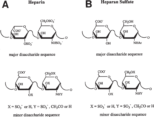
to uronic acid residues. The major repeating unit in heparin is → 4)-α-D-N-sulfoglu-
cosamine-6-sulfate (1→ 4)-α-L-iduronic acid-2-sulfate (1→, corresponds to 75–90%
of its sequence (1) (see Fig. 1A), whereas heparan sulfate consists of 50–75% → 4)-
α-D-N-acetylglucosamine (1→ 4)-β-glucuronic acid (1→ and smaller amounts of
→ 4)-α-D-N-acetylglucosamine-6-sulfate (1→ 4)-β-D-glucuronic acid (1→ and → 4)-
α-D-N-sulfoglucosamine (1→ 4)-β-D-glucuronic acid (1→ (see Fig. 1B). Heparin,
which contains approx 2.7 sulfate groups per disaccharide unit, is more highly
sulfated than HS, which contains less than one sulfate per disaccharide unit.
HS proteoglycans (PGs) are localized on the surface of many mammalian cells and
in the extracellular matrix. HS proteoglycans are important for several different bio-
logical activities such as cell–cell and cell–protein interactions (2). These biological
activities are controlled mainly through the binding of a variety of proteins to the HS
chains. Specific sequences in the HS chain are thought to be responsible for the binding
of growth factors, protease inhibitors, and adhesion molecules. The use of HS-degrading
enzymes can help in separating and identifying biological active oligosaccharides (3).
HS can be degraded enzymatically by using heparin lyases from bacterial sources.
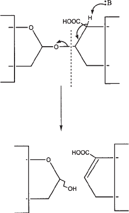
The lyase enzymes degrade GAGs by endolytic cleavage (4–6). The enzymes cut glu-
cosamine–uronate linkage by elimination (see Fig. 2), leaving a C4–C5 unsaturated
bond containing product that can be easily detected by ultraviolet (UV) absorbance. In
contrast, mammalian heparanases cleave this linkage by hydrolysis.
Heparin lyases have been isolated from Flavobacterium heparinum (7), Bacteriodes
species (8), Bacteriodes heparinolyticus (9), and Prevotella heparinolytica (10). Hep-
arin lyases from F. heparinum have been purified to homogeneity, studied extensively
(11), and are available commercially from Sigma and Seikagaku.

354 LeBrun and Linhardt
Three types of heparin lyases have been purified from Flavobacterium: heparin lyase
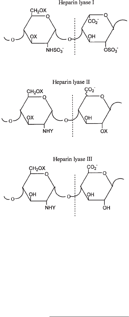
I, heparin lyase II, and heparin lyase III (see Fig. 2 and Note 1). Heparin lyase I acts
primarily on heparin, heparin lyase II cleaves both heparin and HS, and heparin lyase III
is active only on HS (see Table 1). The primary linkages cleaved by these enzymes and
their relative activities toward heparin and HS are shown in Table 1 and Fig. 3.
From both the DNA and amino acid sequences, there is only 15% alignment
between heparin lyase I, II, and III (12). There are certain conserved sequences such as
the heparin-binding sites and the calcium-binding regions in heparin lyase I and III.
Recently, chemical modification studies and site-directed mutagenisis have been used
to help identify critical residues for enzyme activity (13–17). Further studies and the
crystal structures of the heparin lyases are needed to help us understand better the
relationship between function and structure of these enzymes.
This chapter will explain how the heparin lyase enzymes can be used to degrade
both heparin- and HS-containing samples and how to assay the activity of the enzyme.
The heparin lyase enzymes can be used to identify the presence of HS/heparin in
samples or to purify HS oligosaccharides for structural analysis.
2. Materials
2.1. Enzyme Preparation
1. Heparin Lyase I, II, or III. Enzymes can be ordered from Sigma (St. Louis, MO) and
Siekagaku America (Falmouth, MA) (see Notes 1 and 2).
Fig. 1. (A) Structure of the major and minor disaccharide sequences of heparin. (B) Struc-
ture of the major and minor disaccharide sequences of heparan sulfate.

Heparan Lyases 355
2. Reagents required to make the appropriate buffers:
a. Dibasic sodium phosphate (EM Science, Gibbstown, NJ).
b. Phosphoric acid (Fisher Scientific, Fair Lawn, NJ).
c. Sodium chloride (Fisher Scientific).
3. 500-µL polypropylene microcentrifuge tubes.
2.2. Enzyme Assay
1. HS (bovine kidney HS, sodium salt from Siekagaku America).
2. Heparin lyase solution (see Subheading 3.1.).
3. Buffer (see Table 2).
4. UV spectrophotometer.
5. 2 × 1-mL quartz cuvets.
2.3.Sample Digestion
1. HS or heparin samples (see Note 3) or samples containing radiolabeled HS or heparin.
2. Spectropor dialysis membrane (molecular-weight cutoff [MWCO] 1000) (Spectrum, Los Ange-
les, CA) or Centricon (YM3, MWCO 3000) centrifugal filter units (Millipore, Bedford, MA).
3. 500-µL polypropylene microcentrifuge tubes.
4. Heparin lyase solution.
5. Water baths at 30°C and 35°C for enzyme digestion and at 100°C to inactivate the enzyme reaction.
2.4. Product Analysis
High-performance liquid chromatography (HPLC), capillary electrophoresis (CE),
gel-permeation chromatography, or polyacrylamide gel electrophoresis (PAGE) may
be used to purify and analyze oligosaccharides prepared from HS/heparin.
Fig. 2. Eliminative cleavage of GAGs by lyases.

356 LeBrun and Linhardt
3. Methods
3.1. Preparation of Lyases for Use
1. Preparation of buffers: The following buffers can be stored at room temperature for
over 1 mo (see Note 4).
a. For heparin lyase I, prepare 50 mM sodium phosphate buffer containing 100 mM
sodium chloride at pH 7.1. To prepare 1 L of buffer, dissolve 7.1 g of dibasic sodium
phosphate and 5.8 g of sodium chloride into 900 mL of distilled water. Adjust the pH to
7.1 with phosphoric acid and bring the volume up to 1 L with distilled water.
b. For heparin lyase II and III, prepare 50 mM sodium phosphate buffer by dissolving
7.1 g of dibasic sodium phosphate in 900 mL of distilled water. For heparin lyase II
adjust the pH to 7.1 with concentrated phosphoric acid, and adjust to 7.6 for heparin
lyase III. Adjust the final volume to 1 L with distilled water.
2. Aliquot samples:
a. Dissolve 0.1 U of lyophilized enzyme in 100 µL of the appropriate buffer (see Table 2).
b. Store the enzyme in 10-mU aliquots in 500-µL polypropylene tubes at –70°C (see
Note 5).
3.2. Activity Assay for Lyases
1. Add 640 µL of the appropriate buffer (see Table 2) to a 1-mL quartz cuvet. Warm the
cuvet to 30°C in a temperature-controlled UV spectrophotometer (see Note 6).
2. Thaw 10-µL aliquots of the appropriate enzyme solution (see Table 1) at room temperature.
3. Remove 90 µL of warm buffer out of the cuvet and transfer the solution into the tube
containing the enzyme solution. Immediately transfer the entire 100 µL of buffer and en-
zyme back into the cuvet, which is incubating at 30°C.
4. Adjust the baseline of the spectrophotometer to zero at 232 nm.
5. Remove the cuvet from the spectrophotometer and add 50 µL of 20 mg/mL of the appro-
priate substrate HS/heparin (see Table 1) to the cuvet. Cover the cuvet with Parafilm and
Table 1
Activity of Heparin Lyases
Activity and
substrate conversion Heparin lyase I Heparin lyase II Heparin lyase III
Heparin
a
Percent activity
b
100 60 <1
% Conversion
c
60 (80)
d
85 6
Heparan sulfate
e
Percent activity 10 100 100
Percent conversion 20 40 94
a
Porcine mucosal heparin.
b
Percent activity = [initial rate on the substrate examined/initial rate on substrate giving the highest
activity] × (100).
c
Percent conversion = [moles of linkages cleaved/total moles of hexosamine → uronic acid
linkages] × (100).
d
Bovine lung heparin.
e
Bovine kidney heparan sulfate.

Heparan Lyases 357
invert two times to mix. Remove the Parafilm and place the cuvet back into the spectro-
photometer.
6. Within 30 s after the addition of substrate, begin to measure the absorbance continuously
or at 30-s intervals for 2–10 min. Graph absorbance at 232 nm vs time. The initial rate is
determined by measuring the slope of the linear portion of absorbance vs time.
7. Calculate the enzyme activity from the initial rate using the extinction coefficient
(ε = 3800 M
–1
) for the reaction products (see Note 7). Each product formed has an unsat-
urated uronic acid residue at its nonreducing terminus that absorbs at 232 nm. The en-
zyme activity is calculated as
Fig. 3. Primary glycosidic linkages cut by hepain lyases. Abbreviations: X, H or SO
3
–
; Y,
CH
3
CO or SO
3
–
. Heparin lyase II cleaves at either glucuronic or iduronic acid residues.
Enzyme Activity =
(∆Abs 232 nm/ min) (700 µL)
(3800 M
–1
)

358 LeBrun and Linhardt
3.3.Sample Digestion
3.3.1. Complete Heparin Lyase-Catalyzed Depolymerization of a Sample
Containing HS/Heparin
1. Dissolve samples, containing HS/heparin (see Note 8) in buffer and dialyze using a 1000-
MWCO dialysis membrane or a Centricon (YM3, MWCO 3000) centrifugal filter unit
(see Note 9).
2. Thaw 10 µL of the appropriate enzyme solution (see Table 1) at room temperature (assay
if desired as described under Subheading 3.2.) and then add 40 µL of the appropriate
buffer (see Table 1) to a 500-µL polypropylene microcentrifuge tube (see Note 10). Also
add 50 µL of buffer to one tube as a blank control.
3. Add 50 µL of the HS/heparin-containing sample to each tube and mix by gently inverting.
4. Incubate for 8–12 h at the appropriate temperature as indicated in Table 2 (see Note 11).
5. Terminate the reaction by heating the tubes at 100°C for 2–3 min (see Note 12).
6. Product formation can be determined by either UV detection, colormetric assay, or HPLC.
Pure samples containing >10 µg of HS can be measured by absorbance. The C4–C5
unsaturated bond of the oligosaccharide product is a chromophore that can be measured
at a λ
max
= 232 nm with a molar absorptivity of 5500 M
–1
in 30 mM hydrochloric acid
(see Note 13) (18). If the sample contains a high concentration of protein, the Azure A
metachromatic assay (19) should be used. For smaller samples or impure samples, gel-
permeation chromatography using UV or colorimetric detection (20) can be used to mea-
sure the quantity of product (see Note 14).
3.3.2. Complete Heparin Lyase-Catalyzed Depolymerization
of Radiolabeled HS
1. Dissolve GAG sample containing radiolabeled HS in 50 µL of sodium phosphate buffer.
Dialyze sample against sodium phosphate buffer using 1000 MWCO dialysis membrane or
a Centricon (YM3, MWCO 3000) centrifugal filter unit (see Note 9).
2. Thaw 10 µL of heparin lyase III solution at room temperature, immediately prior to use
(see Note 5).
Table 2
Properties of Heparin Lyases and Reaction Conditions
MW T
opt
b
Enzyme Substrate (Da) pI (ºC) Buffer system
Heparin lyase I
a
(EC 4.2.2.7) Heparin 42,800 9.2 30 50 mM NaPO
4
,
100 mM NaCl,
pH 7.1
Heparin lyase II Heparin 84,100 9.0 35 50 mM NaPO
4
,
HS pH 7.1
Heparin lyase III HS 70,800 10 35 50 mM NaPO
4
,
(EC 4.2.2.8) pH 7.6
a
EC is the Enzyme Commission number.
b
T
opt
, optimum temperature for the enzyme.
Heparan Lyases 359
3. Add 30 µL of sodium phosphate buffer to the 500-µL polypropylene microcentrifuge
tube containing the enzyme solution.
4. (Optional)(see Note 15). Add 1.7 µL each of 20-mg/mL chondroitin sulfate A, chon-
droitin sulfate C, and dermatan sulfate substrate solutions (34 µg of each GAG) to the
enzyme in buffer.
5. Add 50 µL of radiolabeled heparan sulfate solution and incubate 8–12 h at 30°C.
6. Heat at 100°C for 2 min to inactivate the enzyme.
7. Analyzed depolymerized radioactive sample by gel-permeation chromatography using
radioisotope detection methods (see Note 14).
3.4. Analysis of Product
The heparin and heparan sulfate oligosaccharides can be analyzed by gradient
PAGE, capillary electrophoresis (CE), or strong-anion-exchange HPLC (21–24).
4. Notes
1. Heparin lyase I from Flavobacterium heparinum is sold as heparinase I by Sigma and as
heparinase by Seikagaku. Heparin lyase II is sold as heparinase II by Sigma and as
heparitinase II by Seikagaku. Heparin lyase III from Flavobacterium heparinum is sold
as heparinase III by Sigma and as heparatinase or heparatinase I by Seikagaku.
2. Often the purchased lyophilized enzyme contains bovine serum albumin (BSA) as a
stabilizer. For example, Sigma samples contain 25% BSA.
3. Samples consisting of tissue, biological fluids, PGs, and GAGs that contain microgram
quantities of HS can often be analyzed directly using heparin lyases without the use of
radioisotopes.
4. Since calcium is an activator for heparin lyase I and III, 20 mM sodium acetate buffer in
the presence of 2 mM calcium acetate can be used with these enzymes. These enzymes
are also compatible with a wide range of other biological buffers.
5. Storage: The lyophilized enzyme is stable at –20°C for at least 2 yr. The dissolved enzyme
is stable when frozen at –20°C for 1 mo and for over a year at –70°C. The heparin lyases
are sensitive to freeze-thawing, especially heparin lyase III. Once heparin lyase III
samples are thawed, they should be used immediately.
6. If a temperature-controlled spectrophotometer is not available, activity can be measured
at room temperature, or samples can be incubated in a water bath and the absorbance can
be measured at fixed time points.
7. One unit is equal to 1 µmol product formed per minute.
8. If the sample is believed to contain HS, heparin lyase III should be used. Samples that
contain heparin should be treated with heparin lyase I. In samples where the identity of
the GAG is unknown or believed to be a mixture of HS and heparin use either heparin
lyase II or an equal unit mixture of heparin lyase I, II, and III to ensure complete depoly-
merization.
9. The presence of metals, detergents, and denaturants can interfere with the activity of the
lyases. Before digesting the samples, detergents should be removed by precipitation with
potassium chloride or by using a detergent-removal column such as Biobeads (Bio-Rad).
Urea and guanidine should be removed by exhaustive dialysis using controlled-pore
dialysis membrane (MWCO 1000).
10. Additional enzyme (10- to 100-fold) may be required to break down small, resistant oli-
gosaccharides (25–26).
360 LeBrun and Linhardt
11. If possible, gently shake the samples during digestion.
12. Following the use of a lyase, residual lyase activity can be destroyed by heating the reaction
mixture to 100°C or by adding denaturants or detergents. Most lyases are cationic pro-
teins and can be removed from anionic oligosaccharide products by passing the reaction
mixture through a small cation-exchange column, such as SP-Sephadex (Sigma), adjusted
to an acidic pH. The oligosaccharide products (void volume) are then recovered, read-
justed to neutral pH, and analyzed. This method can also be used to remove BSA, an
excipient found in many of the commercial enzymes, from the oligosaccharide products.
13. This assay is to be used with relatively pure GAGs. High concentrations of protein inter-
fere with the measure of oligosaccharide production due to UV absorbance of the protein.
14. In gel-permeation chromotography of HS/heparin, following complete depolymerization
using the appropriate heparin lyase, the products should elute close to the column’s total
volume, corresponding to an apparent molecular weight <1500 daltons (confirming the
presence of heparin/HS), while the substrate (control without enzyme) should elute close
to the column’s void volume, corresponding to a molecular weight of >10,000 daltons.
15. When attempting to use heparin lyases to depolymerize radiolabeled samples that contain
very small quantities of heparin or heparan sulfate, it is often useful to add cold substrate
as a carrier so that the activity of heparin lyase can be distinguished from that of trace
amounts of chondrotin lyases that may be present in heparin lyase preparations (27).
References
1. Linhardt, R. J., Ampofo, S. A., Fareed, J. Hoppensteadt, D., Mulliken, J. B., and Folkman,
J. (1992) Characterization of a human heparin. Biochemistry 31, 12,441–12,445.
2. Stringer, S. E. and Gallagher, J. T. (1997) Heparan sulphate. Int. J. Biochem. Cell Biol. 29,
709–714.
3. Toida, T. and Linhardt, R. J. (1998) Structural analysis of heparan sulfate and heparan
oligosacccharides. Trends Glycosci. Glycotechnol. 10, 125–136.
4. Linhardt, R. J., Galliher, P. M., and Cooney, C. L. (1986) Polysaccharide lyases. Appl.
Biochem. Biotechnol. 12, 135–176.
5. Ernst, S., Langer, R., Cooney, C. L., and Sasisekharan, R. (1995) Enzymatic degradation
of glycosaminoglycans. Crit. Rev. Biochem. Mol. Biol. 30, 387–444.
6. Sutherland, I. W. (1995) Polysaccharide lyases. FEMS Microbiol. Rev. 16, 323–347.
7. Payza, A. N. and Korn, E. D. (1956) Bacterial degradation of heparin. Nature 177, 88–89.
8. Ahn, M. Y., Shin, K. H., Kim, D. H., Jung, E. A., Toida, T., Linhardt, R. J., and Kim, Y. S.
(1998) Characterization of a Bacteroides species from human intestine that degrades
glycosaminoglycans. Can. J. of Microbiol. 44, 423–429.
9. Nakamura, T., Shibata, Y., and Fujimura S. (1988) Purification and properties of
Bacteroides heparinolyticus heparinase (heparin lyase, EC 4.2.2.7). J. Clin. Microbiol.
26, 1070–1071.
10. Watanabe, M., Tsuda, H., Yamada, S., Shibuta, Y., Nakamura, T., and Sugahara, K. (1998)
Characterization of heparinase from an oral bacterium Prevotella heparinolytica. J.
Biochem. 123, 283–288.
11. Lohse, D. L. and Linhardt, R. J. (1992) Purification and characterization of heparin lyases
from Flavobacterium heparinum. J. Biol. Chem. 267, 24,347–24,355.
12. Godavarti, R. and Sasisekharan, R. (1996) A comparative analysis of the primary
sequences and characteristics of heparinases I, II, and III from Flavobacterium heparinum.
Biochem. Biophys. Res. Commun. 229, 770–777.
Heparan Lyases 361
13. Shriver, Z., Hu, Y., and Sasisekharan, R. (1998) Heparinase II from Flavobacterium
heparinum. Role of histidine residues in enzymatic activity as probed by chemical modifi-
cation and site-directed mutagenesis. J. Biol. Chem. 273, 10,160–10,167.
14. Shriver, Z., Hu, Y., Pojasek, K., and Sasisekharan, R. (1998) Heparinase II from Fla-
vobacterium heparinum. Role of cysteine in enzymatic activity as probed by chemical
modification and site-directed mutagenesis. J. Biol. Chem. 273, 22,904–22,912.
15. Godavarti, R. and Sasisekharan, R. (1998) Heparinase I from Flavobacterium heparinum.
Role of positive charge in enzymatic activity. J. Biol. Chem. 273, 248–255.
16. Sasisekharan, R., Leckband, D., Godavarti, R., Venkataraman, G., Cooney, C. L., and
Langer, R. (1995) Heparinase I from Flavobacterium heparinum: the role of the cysteine
residue in catalysis as probed by chemical modification and site-directed mutagenesis.
Biochemistry 34, 14,441–14,448.
17. Godavarti, R., Cooney, C. L., Langer, R., and Sasisekharan, R. (1996) Heparinase I from
Flavobacterium heparinum. Identification of a critical histidine residue essential for
catalysis as probed by chemical modification and site-directed mutagenesis. Biochemistry
35, 6846–6852.
18. Linker, A. and Hovingh, P. (1972) Isolation and characterization of oligosaccharides
obtained from heparin by the action of heparinase. Biochemistry 11, 563–568.
19. Grant A. C., Linhardt, R. J., Fitzgerald, G. L., Park, J. J., and Langer R. (1984) Metachro-
matic activity of heparin and heparin fragments. Anal. Biochem. 137, 25–32.
20. Bitter, T. and Muir, H. M. (1962) A modified uronic acid carbozole reaction. Anal.
Biochem. 4, 330–334.
21. Ampofo, S. A., Wang, H. M., and Linhardt, R. J. (1991) Disaccharide compositional
analysis of heparin and heparan sulfate using capillary zone electrophoresis. Anal.
Biochem. 199, 249–255.
22. Linhardt, R. J. and Pervin, A. (1996) Separation of negatively charged carbohydrates by
capillary electrophoresis. J. Chromotogr. A. 720, 323–335.
23. Hileman R. E., Smith, A. E., Toida, T., and Linhardt, R. J. (1997) Preparation and struc-
ture of heparin lyase-derived heparan sulfate oligosaccharides. Glycobiology 7, 231–239.
24. Sugahara, K. Tohno-oka, R., Yamada, S., Khoo, K.-H., Morris, H. R., and Dell, A. (1994)
Structural studies on the oligosaccharides isolated from bovine kidney heparan sulfate and
characterization of bacterial heparitinases used as substrates. Glycobiology 4, 535–544.
25. Rice, K. G. and Linhardt R. J. (1989) Study of defined oligosaccharides substrates of
heparin and heparan monosulfate lyases. Carbohydr. Res. 190, 219–233.
26. Desai, U. R., Wang, H., and Linhardt, R. J. (1993) Substrate specificity of heparin lyases
from Flavobacterium heparinum. Arch. Biochem. Biophys. 306, 461–468.
27. Linhardt, R. J. (1994) Analysis of glycosaminoglycans with polysaccharide lyases, in
Current Protocols in Molecular Biology, Analysis of Glycoconjugates, (Varki, A., ed.),
Wiley-Interscience, Boston, MA, vol. 2, pp.p 17.13.17–17.13.32.
